. 2024 Jan-Dec:53:19160216241248669.
doi: 10.1177/19160216241248669.

Pull-Out Strength of Orthodontic Miniscrews in the Temporal Bone

Affiliations

Pull-Out Strength of Orthodontic Miniscrews in the Temporal Bone

Christian Menke et al. J Otolaryngol Head Neck Surg. 2024 Jan-Dec.

Abstract

Background: Minimally invasive cochlear implant surgery by using a microstereotactic frame demands solid connection to the bone. We aimed to determine the stability of commercially available orthodontic miniscrews to evaluate their feasibility for frame's fixation. In addition, which substitute material most closely resembles the mechanical properties of the human temporal bone was evaluated.

Methods: Pull-out tests were carried out with five different types of orthodontic miniscrews in human temporal bone specimens. Furthermore, short fiber filled epoxy (SFFE), solid rigid polyurethane (SRPU50), bovine femur, and porcine iliac bone were evaluated as substitute materials. In total, 57 tests in human specimens and 180 tests in the substitute materials were performed.

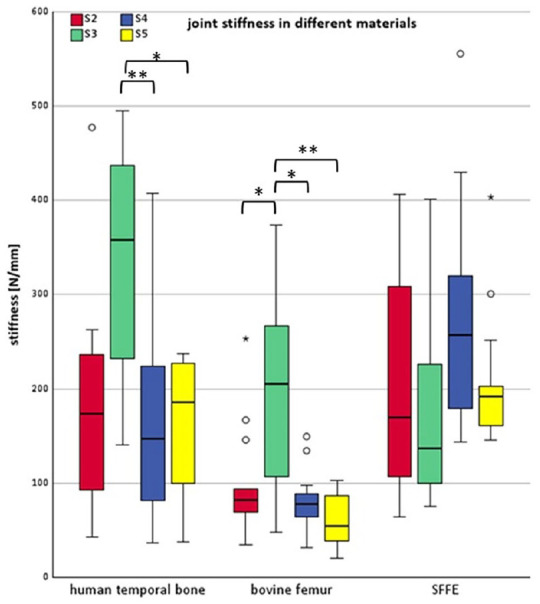
Results: In human temporal bone, average pull-out forces ranged from 220 N to 285 N between screws. Joint stiffness in human temporal bone ranged between 14 N/mm and 358 N/mm. Statistically significant differences between the tested screws were measured in terms of stiffness and elastic energy. One screw type failed insertion due to tip breakage. No significant differences occurred between screws in maximum pull-out force. The average pull-out values of SFFE were 14.1 N higher compared to human specimen.

Conclusion: Orthodontic miniscrews provided rigid fixation when partially inserted in human temporal bone, as evidenced by pull-out forces and joint stiffness. Average values exceeded requirements despite variations between screws. Differences in stiffness and elastic energy indicate screw-specific interface mechanics. With proper insertion, orthodontic miniscrews appear suitable for microstereotactic frame anchoring during minimally invasive cochlear implant surgery. However, testing under more complex loading is needed to better predict clinical performance. For further pull-out tests, the most suitable substitute material is SFFE.

Keywords: bone substitute material; minimally invasive cochlear implant surgery; orthodontic miniscrews; pull-out test.


Conflict of interest statement

Declaration of Conflicting InterestsThe authors declared no potential conflicts of interest with respect to the research, authorship, and/or publication of this article.

Figures

Graphical abstract
Graphical abstract
Figure 1.
Figure 1.
Illustration of the reference frame and fixation with miniscrews.
Figure 2.
Figure 2.
Illustration of the pull-out test setup: 1: baseplate; 2: specimen; 3: screw; 4: spacer; 5: tension ring.
Figure 3.
Figure 3.
(a) Illustration of investigated bone screws. S1: PSM quattro standard (PSM Medical Solutions). S2: Promedia LeForte (Promedia Medizintechnik). S3: KLS maxdrive drill-free (Gebrüder Martin GmbH & Co KG). S4: General implants mini bone screw (General implants Germany GmbH). S5: Promedia dual top (Promedia Medizintechnik). (b) Detailed photograph of the flank at the tip of S3.
Figure 4.
Figure 4.
Example of a force-displacement curve. Y-axis: force in N; X-axis: displacement in mm. Between the two crosses, a linear regression was applied.
Figure 5.
Figure 5.
Overview of results of pull-out tests displayed in boxplots regarding different materials. X-axis: tested material; Y-axis: pull-out forces in N; left: pull-out forces in human temporal bone; middle: pull-out forces in bovine femur; right: pull-out forces in SFFE; S2: red boxplots; S3: green boxplots; S4: blue boxplots; S5: yellow boxplots; SFFE: short fiber filled epoxy. Asterisk brackets show statistic significant difference.
Figure 6.
Figure 6.
Overview of results of joint stiffness displayed in boxplots regarding different materials. X-axis: tested material; Y-axis: joint stiffness in N/mm; left: joint stiffness in human temporal bone; middle: joint stiffness in bovine femur; right: joint stiffness in SFFE; S2: red boxplots; S3: green boxplots; S4: blue boxplots; S5: yellow boxplots; SFFE: short fiber filled epoxy. Asterisk brackets show statistic significant difference.
Figure 7.
Figure 7.
Overview of results of elastic energy displayed in boxplots. X-axis: tested material; Y-axis: elastic energy in Nmm; left: elastic energy in human temporal bone; middle: elastic energy in bovine femur; right: elastic energy in SFFE; S2: red boxplots; S3: green boxplots; S4: blue boxplots; S5: yellow boxplots; SFFE: short fiber filled epoxy. Asterisk brackets show statistic significant difference.
Figure 8.
Figure 8.
Comparison between S1 before drilling into the bone (left) and after the pull-out test (right).

References

    1. Deep NL, Dowling EM, Jethanamest D, Carlson ML. Cochlear implantation: an overview. J Neurol Surg B Skull Base. 2019;80(2):169-177. doi:10.1055/s-0038-1669411 - DOI - PMC - PubMed
    1. Matin F, Artukarslan EN, Illg A, Lesinski-Schiedat A, Lenarz T, Suhling MC. Cochlear implantation in elderly patients with residual hearing. J Clin Med. 2021;10(19):4305. doi:10.3390/jcm10194305.4305 - DOI - PMC - PubMed
    1. Varadarajan VV, Sydlowski SA, Li MM, Anne S, Adunka OF. Evolving criteria for adult and pediatric cochlear implantation. Ear Nose Throat J. 2021;100(1):31-37. doi:10.1177/0145561320947258 - DOI - PubMed
    1. Sargsyan G, Kanaan N, Lenarz T, Lesinski-Schiedat A. Comparison of speech recognition in cochlear implant patients with and without residual hearing: a review of indications. Cochlear Implants Int. 2021;22(5):257-264. doi:10.1080/14670100.2021.1898111 - DOI - PubMed
    1. Peters JPM, van Heteren JAA, Wendrich AW, et al.. Short-term outcomes of cochlear implantation for single-sided deafness compared to bone conduction devices and contralateral routing of sound hearing aids-Results of a Randomised controlled trial (CINGLE-trial). PLoS One. 2021;16(10):e0257447. doi:10.1371/journal.pone.0257447 - DOI - PMC - PubMed

LinkOut - more resources