[Preprint]. 2024 Feb 23:2024.02.22.581453.
doi: 10.1101/2024.02.22.581453.

An egg sabotaging mechanism drives non-Mendelian transmission in mice

Affiliations

An egg sabotaging mechanism drives non-Mendelian transmission in mice

Frances E Clark et al. bioRxiv. .

Update in

Abstract

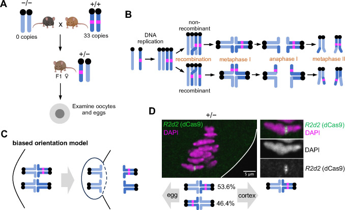
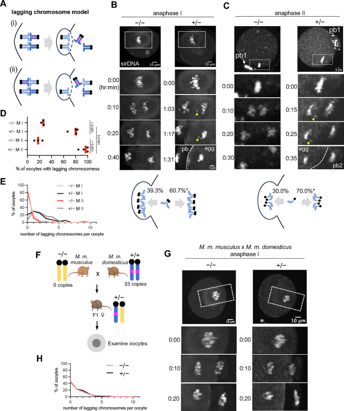
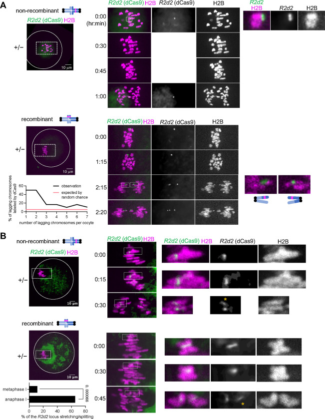
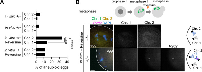
During meiosis, homologous chromosomes segregate so that alleles are transmitted equally to haploid gametes, following Mendel's Law of Segregation. However, some selfish genetic elements drive in meiosis to distort the transmission ratio and increase their representation in gametes. The established paradigms for drive are fundamentally different for female vs male meiosis. In male meiosis, selfish elements typically kill gametes that do not contain them. In female meiosis, killing is predetermined, and selfish elements bias their segregation to the single surviving gamete (i.e., the egg in animal meiosis). Here we show that a selfish element on mouse chromosome 2, R2d2, drives using a hybrid mechanism in female meiosis, incorporating elements of both male and female drivers. If R2d2 is destined for the polar body, it manipulates segregation to sabotage the egg by causing aneuploidy that is subsequently lethal in the embryo, so that surviving progeny preferentially contain R2d2. In heterozygous females, R2d2 orients randomly on the metaphase spindle but lags during anaphase and preferentially remains in the egg, regardless of its initial orientation. Thus, the egg genotype is either euploid with R2d2 or aneuploid with both homologs of chromosome 2, with only the former generating viable embryos. Consistent with this model, R2d2 heterozygous females produce eggs with increased aneuploidy for chromosome 2, increased embryonic lethality, and increased transmission of R2d2. In contrast to a male meiotic driver, which kills its sister gametes produced as daughter cells in the same meiosis, R2d2 eliminates "cousins" produced from meioses in which it should have been excluded from the egg.

Keywords: aneuploidy; chromosome segregation; female meiotic drive; meiosis; mouse oocyte; selfish genetic element.


Conflict of interest statement

Declaration of interests The authors declare no competing interests.

Figures

Figure 1.
Figure 1.. R2d2 randomly orients on the metaphase I spindle in female meiosis.
(A) Female F1 hybrids between two M. m. domesticus strains with different R2d2 copy numbers (R2d2 heterozygous mice, +/−) as experimental systems to study meiotic drive of R2d2. Oocytes and eggs collected from +/− mice (C57BL/6J x WSB/EiJ and BXD19/TyJ x WSB/EiJ hybrids) and −/− mice (the CF1 strain) were used throughout this study unless specified in the figure legend. (B) Schematic showing recombinant and non-recombinant chromosomes, depending on whether there is crossover between R2d2 and the centromere. Non-recombinants can cheat in meiosis I whereas recombinants can cheat in meiosis II. (C) Schematic showing the biased orientation model for R2d2 meiotic drive. (D) R2d2 heterozygous (+/−) oocytes expressing dCas9-EGFP and gRNA targeting the R2d2 sequence were fixed shortly before anaphase I and stained for EGFP. The fraction of oocytes with the R2d2 locus oriented toward either the egg or cortical pole was measured; n = 39 cells. White line, oocyte cortex. Note that we focused only on non-recombinant chromosomes, which can cheat in meiosis I.
Figure 2.
Figure 2.. R2d2 meiotic drive is associated with increased anaphase lagging.
(A) Schematic showing the anaphase lagging model for R2d2 meiotic drive. (B, C) Control (−/−) and R2d2 heterozygous (+/−) meiosis I oocytes (B) and meiosis II eggs (C) were stained with sirDNA to visualize chromosomes and imaged live at anaphase (n = 65 and 80 meiosis I oocytes for control and R2d2 heterozygote, respectively and n = 47 and 49 meiosis II eggs for control and R2d2 heterozygote, respectively). Note that the lagging chromosomes (yellow arrowheads) eventually remained in the egg. The segregation pattern of lagging chromosomes was quantified; chi-square test for goodness of fit was used to examine the deviation from the expected 50:50 ratio (B, n = 145 lagging chromosomes from 76 meiosis I oocytes; *P = 0.01004 and C, n = 40 lagging chromosomes from 17 meiosis II eggs; *P = 0.01141). (D, E) Based on the live imaging data in B and C, the fraction of oocytes with at least one lagging chromosome (D, each dot represents an independent experiment; red line, mean; unpaired two-tailed t-test was performed) and the distribution of the number of lagging chromosomes per oocyte were quantified. (F) Female F1 hybrids between a M. m. domesticus strain with expanded R2d2 (WSB/EiJ) and a M. m. musculus strain without it (PWD/PhJ) show Mendelian segregation of R2d2. (G, H) Control (−/−, PWD/PhJ x C57BL/6J) and R2d2 heterozygous (+/−, PWD/PhJ x WSB/EiJ) oocytes in the M. m. domesticus x M. m. musculus hybrid genetic background were stained with sirDNA to visualize chromosomes and imaged live at anaphase I (G), and the distribution of the number of lagging chromosomes per oocyte was quantified (H, n = 66 and 48 oocytes for control and R2d2 heterozygote, respectively).
Figure 3.
Figure 3.. R2d2 induces its own chromosome to lag in anaphase.
(A) R2d2 heterozygous (+/−) oocytes expressing H2B-mCherry, dCas9-EGFP, and gRNA targeting the R2d2 DNA sequence were imaged live at anaphase I (white line, oocyte cortex). The histogram shows the distribution of the fraction of lagging chromosomes that have dCas9 signals (n = 35 cells). For the non-recombinant chromosome example (top), we imaged the dCas9-EGFP signals only at the first timepoint and at the anaphase onset to minimize photobleaching and phototoxicity. In the recombinant chromosome example (bottom), three dCas9 signals were observed at the 2 hr 20 min timepoint due to the splitting of the R2d2 locus on one of the homologs (also see B). (B) R2d2 heterozygous (+/−) oocytes expressing H2B-mCherry, dCas9-EGFP, and gRNA targeting the R2d2 DNA sequence were imaged live at the metaphase I to anaphase I transition. The fraction of oocytes with the dCas9 signal splitting or stretching was quantified in metaphase I and anaphase I (n = 26 cells); Chi-Square Test of Independence was used to calculate the P values in the graph; white line, oocyte cortex; orange asterisks, stretching R2d2 locus. We used the Denoise.ai software (Nikon) to reduce noise and follow the R2d2 locus better in anaphase I.
Figure 4.
Figure 4.. Chromosome 2 with R2d2 has a higher aneuploid rate.
(A) Quantification of the fraction of the oocytes that are aneuploid for chromosome 1 or 2 at the metaphase II stage (n = 52, 52, 84, 35, 162, 61, 78, and 36 oocytes for in vitro −/− Reversine Chr. 2, in vitro −/− Reversine Chr. 1, in vitro +/− Reversine Chr. 2, in vitro +/− Reversine Chr. 1, in vitro +/− Chr. 2, in vitro +/− Chr. 1, in vivo +/− Chr. 2, and in vivo +/− Chr. 1, Chi-Square Test of Independence was used to calculate the P values in the graph). (B) Top schematic shows the Reversine treatment of mouse oocytes at metaphase I. Control (−/−) and R2d2 heterozygous (+/−) oocytes were matured in vitro in the presence of Reversine and fixed at metaphase II to visualize chromosome 1 and 2 and the R2d2 locus with Oligopaint FISH. White lines, egg cell cortex.

References

    1. Clark F.E., and Akera T. (2021). Unravelling the mystery of female meiotic drive: where we are. Open biology 11, 210074. 10.1098/rsob.210074. - DOI - PMC - PubMed
    1. Chmátal L., Schultz R.M., Black B.E., and Lampson M.A. (2017). Cell Biology of Cheating—Transmission of Centromeres and Other Selfish Elements Through Asymmetric Meiosis. In Centromeres and Kinetochores (Springer, Cham), pp. 377–396. 10.1007/978-3-319-58592-5_16. - DOI - PubMed
    1. Pardo-Manuel de Villena F., and Sapienza C. (2001). Nonrandom segregation during meiosis: the unfairness of females. Mammalian genome : official journal of the International Mammalian Genome Society 12, 331–339. 10.1007/s003350040003. - DOI - PubMed
    1. Kruger A.N., and Mueller J.L. (2021). Mechanisms of meiotic drive in symmetric and asymmetric meiosis. Cellular and molecular life sciences : CMLS 78, 3205–3218. 10.1007/s00018-020-03735-0. - DOI - PMC - PubMed
    1. Courret C., Chang C.-H., Wei K.H.-C., Montchamp-Moreau C., and Larracuente A.M. (2019). Meiotic drive mechanisms: lessons from Drosophila. Proceedings of the Royal Society B: Biological Sciences 286, 20191430. 10.1098/rspb.2019.1430. - DOI - PMC - PubMed

Publication types