Review
. 2024 Jun 6:15:1363032.
doi: 10.3389/fimmu.2024.1363032. eCollection 2024.

State-of-the-art diagnosis of autoimmune blistering diseases

Affiliations
Review

State-of-the-art diagnosis of autoimmune blistering diseases

Nina van Beek et al. Front Immunol. .

Abstract

Autoimmune blistering disorders (AIBDs) are a heterogeneous group of approximately a dozen entities comprising pemphigus and pemphigoid disorders and dermatitis herpetiformis. The exact diagnosis of AIBDs is critical for both prognosis and treatment and is based on the clinical appearance combined with the detection of tissue-bound and circulating autoantibodies. While blisters and erosions on the skin and/or inspectable mucosal surfaces are typical, lesions may be highly variable with erythematous, urticarial, prurigo-like, or eczematous manifestations. While direct immunofluorescence microscopy (IFM) of a perilesional biopsy is still the diagnostic gold standard, the molecular identification of the major target antigens opened novel therapeutic avenues. At present, most AIBDs can be diagnosed by the detection of autoantigen-specific serum antibodies by enzyme-linked immunosorbent assay (ELISA) or indirect IFM when the clinical picture is known. This is achieved by easily available and highly specific and sensitive assays employing recombinant immunodominant fragments of the major target antigens, i.e., desmoglein 1 (for pemphigus foliaceus), desmoglein 3 (for pemphigus vulgaris), envoplakin (for paraneoplastic pemphigus), BP180/type XVII collagen (for bullous pemphigoid, pemphigoid gestationis, and mucous membrane pemphigoid), laminin 332 (for mucous membrane pemphigoid), laminin β4 (for anti-p200 pemphigoid), type VII collagen (for epidermolysis bullosa acquisita and mucous membrane pemphigoid), and transglutaminase 3 (for dermatitis herpetiformis). Indirect IFM on tissue substrates and in-house ELISA and immunoblot tests are required to detect autoantibodies in some AIBD patients including those with linear IgA disease. Here, a straightforward modern approach to diagnosing AIBDs is presented including diagnostic criteria according to national and international guidelines supplemented by long-term in-house expertise.

Keywords: autoantibody; autoimmune bullous disease; dermatitis herpetiformis; epidermolysis; immunofluorescence; linear IgA dermatosis; pemphigoid; pemphigus.


Conflict of interest statement

ES has a scientific cooperation with Euroimmun. NvB and ES have a pending patent together with Euroimmun. ES holds patents together with Euroimmun. The remaining authors declare that the research was conducted in the absence of any commercial or financial relationships that could be construed as a potential conflict of interest. ES declared that they was an editorial board member of Frontiers, at the time of submission. This had no impact on the peer review process and the final decision.

Figures

Figure 1
Figure 1
Clinical presentation of pemphigus. Classical presentation of pemphigus vulgaris with lesions of the oral mucosa, erosions on the soft palate (A), crusts on the lips (B), and erosions of the buccal mucosa (C). Skin involvement shows erosions (D, E) and crusts with postinflammatory hyperpigmentation (E). Pemphigus foliaceus with erythema and erosions predominantly in the seborrheic areas (F). IgA pemphigus with pustules, erosions, and blisters (G, H).
Figure 2
Figure 2
Clinical presentation of bullous pemphigoid (A–E) and pemphigoid gestationis (F). Tense blisters and erythematous plaques on the trunk and arms (A, B), fibrin-covered erosions of the buccal mucosa and palate (C), and eczematous and urticarial lesions in bullous pemphigoid (D, E). Pemphigoid gestationis in a pregnant woman with pruritic plaques and excoriations around the umbilical region (F).
Figure 3
Figure 3
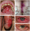
Clinical presentation of mucous membrane pemphigoid. Erosions on the hard palate (A); vulva, perineal, and perianal regions (B); and penis (C). Conjunctival erythema (D) and symblepharon (E) as typical features of ocular involvement.
Figure 4
Figure 4
Clinical presentation of linear IgA disease (LAD), (A–C) anti-p200 pemphigoid, (D, E) and epidermolysis bullosa acquisita (EBA) (F–H). LAD in a 4-year-old child with blisters along the edges of erythematous plaques (➔, “string-of-pearls”, A). Annular pattern of lesions on the legs (B, C). Anti-p200 pemphigoid with pruritic urticarial plaques and tense blisters (D, E). Inflammatory variant of EBA presenting with erosions and postinflammatory hyperpigmentation on the back (F). Mechanobullous form of EBA with high skin fragility and erosions after the removal of Band-Aids (G). IgA EBA manifesting with eczema, excoriated papules, and erythematous plaques (H).
Figure 5
Figure 5
Direct immunofluorescence microscopy of perilesional biopsies. Granular deposits of IgA at the tips of dermal papillae and along the dermal–epidermal junction in dermatitis herpetiformis (×200 magnification) (A). Intercellular deposits of IgG in the epidermis in pemphigus vulgaris (×200 magnification) (B). In pemphigoid diseases, u- and n-serrated patterns of linear deposits at the dermal–epidermal junction can be distinguished. In epidermolysis bullosa acquisita, u-serrated pattern can be seen with arches closed at the bottom (×1,000 magnification) (C). In all other pemphigoid diseases, n-serrated pattern is observed with arches closed at the top (×1,000 magnification) (D).
Figure 6
Figure 6
Diagnostic algorithm for direct immunofluorescence microscopy (IFM). 1, In paraneoplastic pemphigus, a combined pattern with anti-DEJ deposits may be present; 2, in mucosal biopsies, pattern analysis is not possible; 3, so far, in none of the undetermined cases, an EBA or BSLE has been diagnosed based on serum anti-type VII collagen IgG/IgA (64); 4, unless in ocular MMP, where direct IFM can be repeatedly negative; in these cases, diagnosis of MMP can be made based on the clinical picture and the clinical and histopathological exclusion of differential diagnoses (65, 68). BSLE, bullous systemic lupus erythematosus; EBA; epidermolysis bullosa acquisita; DH, dermatitis herpetiformis; DEJ, dermal–epidermal junction; LAD, linear IgA disease; MMP, mucous membrane pemphigoid.
Figure 7
Figure 7
Indirect immunofluorescence on monkey esophagus, rat bladder, and human salt-split skin. Detection of IgG binding with an intercellular pattern in the epithelium of monkey esophagus pemphigus vulgaris (A). IgG reactivity to rat bladder epithelium typical for paraneoplastic pemphigus (B). IgA binding to the endomysium of monkey esophagus in dermatitis herpetiformis (C). Linear IgG binding on monkey esophagus (D) and along the roof of the artificial blister of salt-split human skin (E) in a bullous pemphigoid patient. IgG reactivity along the blister floor of salt-split human skin, which can be seen in anti-laminin 332 mucous membrane pemphigoid, epidermolysis bullosa acquisita, and anti-p200 pemphigoid (F).
Figure 8
Figure 8
Diagnostic algorithm for serum autoantibodies in pemphigoid diseases. 1, Commercial assays are available; 2, with predominant skin lesions; 3, in pregnancy; 4, with predominant mucosal lesions; 5, in-house assays only available in specialized laboratories; 6, in approximately 25% of MMP patients with anti-laminin 32 reactivity, a malignancy was found; 7, on high clinical suspicion of BP, BP180 NC16A-specific ELISA/indirect IFM is recommended; on suspicion of MMP, additional serological tests in particular for IgG reactivity against laminin 332 are recommended; on suspicion of EBA, type VII collagen-specific ELISA/indirect IFM is recommended. BP, bullous pemphigoid; BSLE, bullous systemic lupus erythematosus; EBA; epidermolysis bullosa acquisita; DH, dermatitis herpetiformis; DEJ, dermal–epidermal junction; LAD, linear IgA disease; MMP, mucous membrane pemphigoid; PG, pemphigoid gestationis.
Figure 9
Figure 9
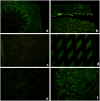
Biochip™ mosaic with six substrates that allow the serological diagnosis of approximately 90% of autoimmune blistering diseases when the clinical picture is known (89). Linear IgG binding on monkey esophagus (A), primate salt-split skin blister roof (B), positive IgG staining on recombinant BP180 NC16A (D), and positive fluorescence of HEK293 cells transfected with BP230 (F), while HEK293 cells transfected with desmogleins 1 (C) and 3 (E) show no fluorescence compatible with bullous pemphigoid.
Figure 10
Figure 10
Biochips™ for serum IgG against laminin 332 and laminin β4. Biochips™ with HEK293 cells transfected with either the heterotrimer of laminin 332 (A, B) or laminin β4 (C, D). IgG reactivity is seen in a patient with anti-laminin 332 mucous membrane pemphigoid (B) and a patient with anti-p200 pemphigoid (D), while the corresponding negative controls show no reactivity (A, C), respectively.
Figure 11
Figure 11
Immunoblotting with dermal extract, extracellular matrix of cultured human keratinocytes, and conditioned concentrated medium of cultured human keratinocytes. Immunoblot with extract of human dermis shows reactivity against the 200-kDa p200 protein in anti-p200 pemphigoid (p200) and the 290-kDa full-length type VII collagen in epidermolysis bullosa acquisita (EBA). IgG4 reactivity with the α3 chain of laminin 332 in mucous membrane pemphigoid (MMP; 220-kDa unprocessed and 165-kDa processed forms) as well as with BP180 (180-kDa full-length and 120-kDa processed forms) and with BP230 (230 kDa) in patients with bullous pemphigoid without reactivity against the immunodominant NC16A domain (BP1 and BP2) by immunoblotting with extracellular matrix of cultured human keratinocytes. Immunoblot with conditioned concentrated medium of cultured human keratinocytes for detection of IgA antibodies against the soluble ectodomain of BP180 [linear IgA disease antigen 1; LAD-1 in linear IgA diseases (LAD)]. Normal human sera (NHS1–3) were used as controls.

References

    1. Beek NV, Zillikens D, Schmidt E. Bullous autoimmune dermatoses-clinical features, diagnostic evaluation, and treatment options. Dtsch Arztebl Int. (2021) 118:413–20. doi: 10.3238/aerztebl.m.2021.0136 - DOI - PMC - PubMed
    1. Kasperkiewicz M, Ellebrecht CT, Takahashi H, Yamagami J, Zillikens D, Payne AS, et al. . Pemphigus. Nat Rev Dis Primers. (2017) 3:17026. doi: 10.1038/nrdp.2017.26 - DOI - PMC - PubMed
    1. Schmidt E, Kasperkiewicz M, Joly P. Pemphigus. Lancet. (2019) 394:882–94. doi: 10.1016/S0140-6736(19)31778-7 - DOI - PubMed
    1. Stanley JR, Amagai M. Pemphigus, bullous impetigo, and the staphylococcal scalded-skin syndrome. N Engl J Med. (2006) 355:1800–10. doi: 10.1056/NEJMra061111 - DOI - PubMed
    1. Amber KT, Murrell DF, Schmidt E, Joly P, Borradori L. Autoimmune subepidermal bullous diseases of the skin and mucosae: clinical features, diagnosis, and management. Clin Rev Allergy Immunol. (2018) 54:26–51. doi: 10.1007/s12016-017-8633-4 - DOI - PubMed

MeSH terms