Review
. 2024 Jun 6:15:1386645.
doi: 10.3389/fphys.2024.1386645. eCollection 2024.

Direct modulation of G protein-gated inwardly rectifying potassium (GIRK) channels

Affiliations
Review

Direct modulation of G protein-gated inwardly rectifying potassium (GIRK) channels

Ha Nguyen et al. Front Physiol. .

Abstract

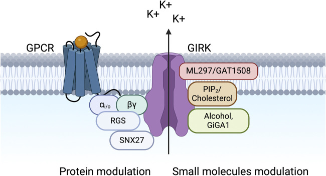
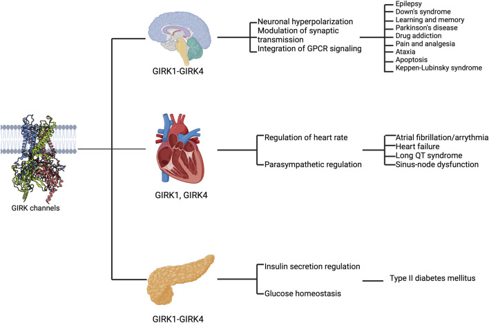
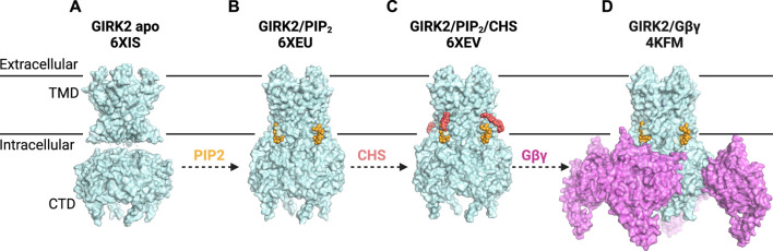
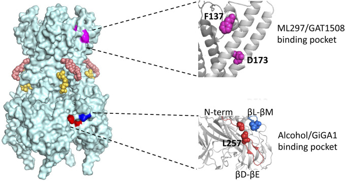
Ion channels play a pivotal role in regulating cellular excitability and signal transduction processes. Among the various ion channels, G-protein-coupled inwardly rectifying potassium (GIRK) channels serve as key mediators of neurotransmission and cellular responses to extracellular signals. GIRK channels are members of the larger family of inwardly-rectifying potassium (Kir) channels. Typically, GIRK channels are activated via the direct binding of G-protein βγ subunits upon the activation of G-protein-coupled receptors (GPCRs). GIRK channel activation requires the presence of the lipid signaling molecule, phosphatidylinositol 4,5-bisphosphate (PIP2). GIRK channels are also modulated by endogenous proteins and other molecules, including RGS proteins, cholesterol, and SNX27 as well as exogenous compounds, such as alcohol. In the last decade or so, several groups have developed novel drugs and small molecules, such as ML297, GAT1508 and GiGA1, that activate GIRK channels in a G-protein independent manner. Here, we aim to provide a comprehensive overview focusing on the direct modulation of GIRK channels by G-proteins, PIP2, cholesterol, and novel modulatory compounds. These studies offer valuable insights into the underlying molecular mechanisms of channel function, and have potential implications for both basic research and therapeutic development.

Keywords: G protein-gated inwardly rectifying potassium channel (GIRK); Kir3; PIP2; alcohol; cholesterol; small molecule modulator.


Conflict of interest statement

The authors declare that the research was conducted in the absence of any commercial or financial relationships that could be construed as a potential conflict of interest.

Figures

FIGURE 1
FIGURE 1
GIRK channels are modulated by various proteins and small molecules. The schematic shows a macromolecular signaling complex that contains a G protein-coupled receptor (GPCR), which couples to pertussis toxin (PTX)-sensitive Gi/o G proteins (Gαi/o and Gβγ), a GIRK channel, a regulator of G protein signaling (RGS) protein, and sorting nexin 27 (SNX27). Small molecule modulators of GIRK channels are also shown, including PIP2, cholesterol, alcohol, and novel compounds (ML297, GAT1508, and GiGA1).
FIGURE 2
FIGURE 2
GIRK channels are expressed in various organs and implicated in disease states. The schematic highlights the expression of GIRK subunits in different regions (the brain, the heart, and the pancreas from top to bottom), their functional roles (first text column) and their implications in diseases (second text column).
FIGURE 3
FIGURE 3
G Protein-Gated Inwardly Rectifying Potassium (GIRK) Channel Structures and Modulatory Sites. (A) structure of GIRK2 apo (PDB: 6XIS) shows the CTD disengaged from the TMD (disordered linkers not shown). (B) structure of GIRK2 in the presence of PIP2 (yellow) (PDB: 6XEU) suggests a portion of GIRK2/PIP2 particles adopt an engaged or docked CTD. (C) structure of GIRK2 in complex with PIP2 (yellow) and CHS (salmon) (PDB: 6XEV) demonstrates that PIP2 binds at its canonical site, while CHS can be docked near the PIP2 binding site. CHS potentially stabilizes the channel-PIP2 interactions and increases the portion of engaged-CTD particles. (D) structure of GIRK2 with four Gβγ subunits (lilac), four PIP2 molecules (yellow) and Na+ ions (not visible) (PDB: 4KFM) reveals the interplay of these modulators in GIRK2 gating.
FIGURE 4
FIGURE 4
Model of the GIRK2 tetramer with four subunits and locations of various modulatory sites. Corresponding PIP2 molecules and cholesterol hemi succinate, CHS, are presented in yellow and salmon, respectively. The region where ML297/GAT1508 potentially bind is highlighted in pink and region where alcohol/GiGA1 bind is highlighted in blue. Expansions of the regions containing key residues implicated in activation by ML297/GAT1508 (top) and alcohol/GiGA1 (bottom) are shown on the right. Amino acids involved in ML297 activation of GIRK1/GIRK2 channels are GIRK1 F137 and D173 (magenta). Key residues forming the alcohol/GiGA1 binding pocket are L257 (red) in the βD-βE loop, I55 (red) in the N-term and L344 (blue) in the βL-βM loop. All residue numbers provided are for mouse GIRK1 and GIRK2 isoforms.

Similar articles

Cited by

References

    1. An X., Cho H. (2023). Increased GIRK channel activity prevents arrhythmia in mice with heart failure by enhancing ventricular repolarization. Sci. Rep. 13 (1), 22479. 10.1038/s41598-023-50088-2 - DOI - PMC - PubMed
    1. Anderson A., Masuho I., Fernandez de Velasco E. M., Nakano A., Lutz B., Martemyanov K. A., et al. (2020). GPCR-dependent biasing of GIRK channel signaling dynamics by RGS6 in mouse sinoatrial nodal cells. Proc. Natl. Acad. Sci. U. S. A. 117 (25), 14522–14531. 10.1073/pnas.2001270117 - DOI - PMC - PubMed
    1. Anderson G. R., Arthur S., Song J. H., Martemyanov K. A. (2007). The membrane anchor R7BP controls the proteolytic stability of the striatal specific RGS protein, RGS9-2. J. Biol. Chem. 282 (7), 4772–4781. 10.1074/jbc.M610518200 - DOI - PubMed
    1. Araya A., Gallegos S., Viveros R., Martin L. S., Muñoz B., Harvey R. J., et al. (2021). Presence of ethanol-sensitive and ethanol-insensitive Glycine receptors in the ventral tegmental area and prefrontal cortex in mice. Br. J. Pharmacol. 178 (23), 4691–4707. 10.1111/bph.15649 - DOI - PMC - PubMed
    1. Arora D., Hearing M., Haluk D. M., Mirkovic K., Fajardo-Serrano A., Wessendorf M. W., et al. (2011). Acute cocaine exposure weakens GABA(B) receptor-dependent G-protein-gated inwardly rectifying K+ signaling in dopamine neurons of the ventral tegmental area. J. Neurosci. Official J. Soc. Neurosci. 31 (34), 12251–12257. 10.1523/JNEUROSCI.0494-11.2011 - DOI - PMC - PubMed

LinkOut - more resources