

Structures and functions of the normal and injured human olfactory epithelium
- PMID: 38903957
- PMCID: PMC11188711
- DOI: 10.3389/fncir.2024.1406218
Structures and functions of the normal and injured human olfactory epithelium
Abstract
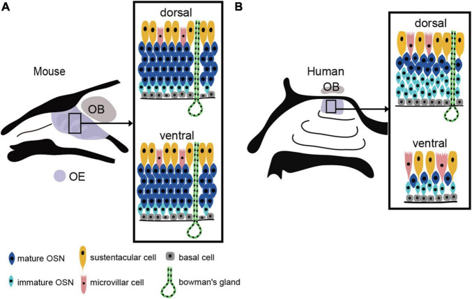
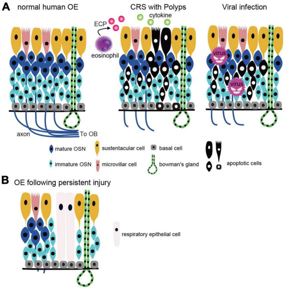
The olfactory epithelium (OE) is directly exposed to environmental agents entering the nasal cavity, leaving OSNs prone to injury and degeneration. The causes of olfactory dysfunction are diverse and include head trauma, neurodegenerative diseases, and aging, but the main causes are chronic rhinosinusitis (CRS) and viral infections. In CRS and viral infections, reduced airflow due to local inflammation, inflammatory cytokine production, release of degranulated proteins from eosinophils, and cell injury lead to decreased olfactory function. It is well known that injury-induced loss of mature OSNs in the adult OE causes massive regeneration of new OSNs within a few months through the proliferation and differentiation of progenitor basal cells that are subsequently incorporated into olfactory neural circuits. Although normal olfactory function returns after injury in most cases, prolonged olfactory impairment and lack of improvement in olfactory function in some cases poses a major clinical problem. Persistent inflammation or severe injury in the OE results in morphological changes in the OE and respiratory epithelium and decreases the number of mature OSNs, resulting in irreversible loss of olfactory function. In this review, we discuss the histological structure and distribution of the human OE, and the pathogenesis of olfactory dysfunction associated with CRS and viral infection.
Keywords: chronic rhinosinusitis; olfactory dysfunction; olfactory epithelium; respiratory metaplasia; viral infection.
Copyright © 2024 Kikuta, Nagayama and Hasegawa-Ishii.
Conflict of interest statement
The authors declare that the research was conducted in the absence of any commercial or financial relationships that could be construed as a potential conflict of interest.
Figures
Similar articles
-
Neuropathology of the olfactory mucosa in chronic rhinosinusitis.Am J Rhinol Allergy. 2010 Mar-Apr;24(2):110-20. doi: 10.2500/ajra.2010.24.3435. Am J Rhinol Allergy. 2010. PMID: 20021743 Free PMC article.
-
Tumor necrosis factor alpha inhibits olfactory regeneration in a transgenic model of chronic rhinosinusitis-associated olfactory loss.Am J Rhinol Allergy. 2010 Sep-Oct;24(5):336-40. doi: 10.2500/ajra.2010.24.3498. Am J Rhinol Allergy. 2010. PMID: 21243089 Free PMC article.
-
The role of TNF-α in inflammatory olfactory loss.Laryngoscope. 2011 Nov;121(11):2481-6. doi: 10.1002/lary.22190. Epub 2011 Aug 31. Laryngoscope. 2011. PMID: 21882204 Free PMC article.
-
[Progress on the mechanisms of olfactory epithelium injury in chronic rhinosinusitis associated olfactory dysfunction].Zhonghua Er Bi Yan Hou Tou Jing Wai Ke Za Zhi. 2025 Apr 7;60(4):470-477. doi: 10.3760/cma.j.cn115330-20240430-00250. Zhonghua Er Bi Yan Hou Tou Jing Wai Ke Za Zhi. 2025. PMID: 40262989 Review. Chinese.
-
Olfactory dysfunction in chronic rhinosinusitis: insights into the underlying mechanisms and treatments.Expert Rev Clin Immunol. 2023 Jul-Dec;19(8):993-1004. doi: 10.1080/1744666X.2023.2235891. Epub 2023 Jul 13. Expert Rev Clin Immunol. 2023. PMID: 37432663 Review.
Cited by
-
A new surgical technique to increase airflow in the olfactory cleft: superior turbinate lateralization procedure.Eur Arch Otorhinolaryngol. 2024 Nov;281(11):5863-5871. doi: 10.1007/s00405-024-08848-x. Epub 2024 Jul 17. Eur Arch Otorhinolaryngol. 2024. PMID: 39017995
References
Publication types
MeSH terms
LinkOut - more resources
Full Text Sources