. 2024 Jul 31;44(7):BSR20240768.
doi: 10.1042/BSR20240768.

Comprehensive analysis of the endothelin system in the kidneys of mice, rats, and humans

Affiliations

Comprehensive analysis of the endothelin system in the kidneys of mice, rats, and humans

Margi Patel et al. Biosci Rep. .

Abstract

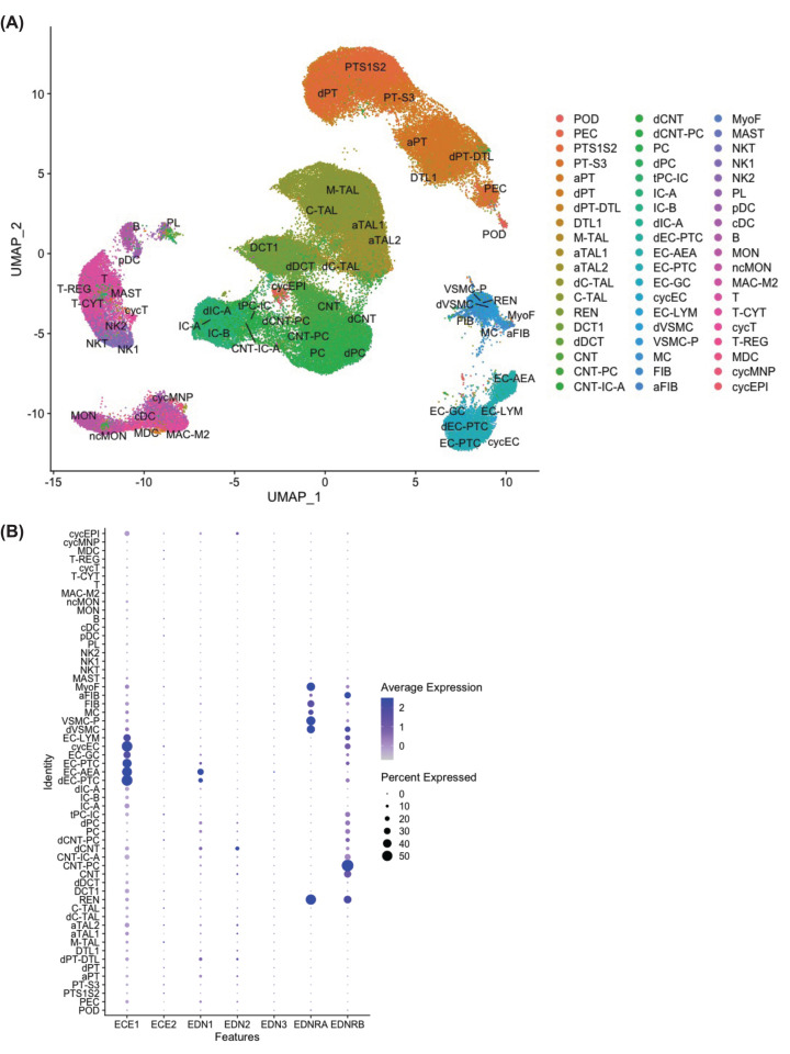
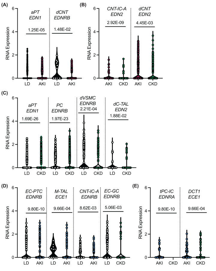
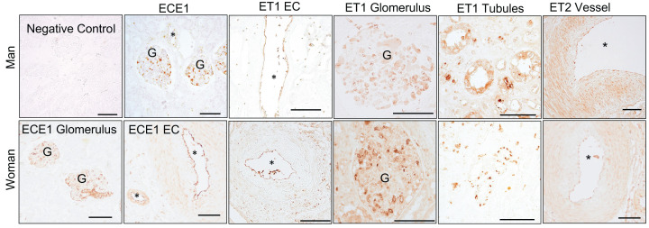
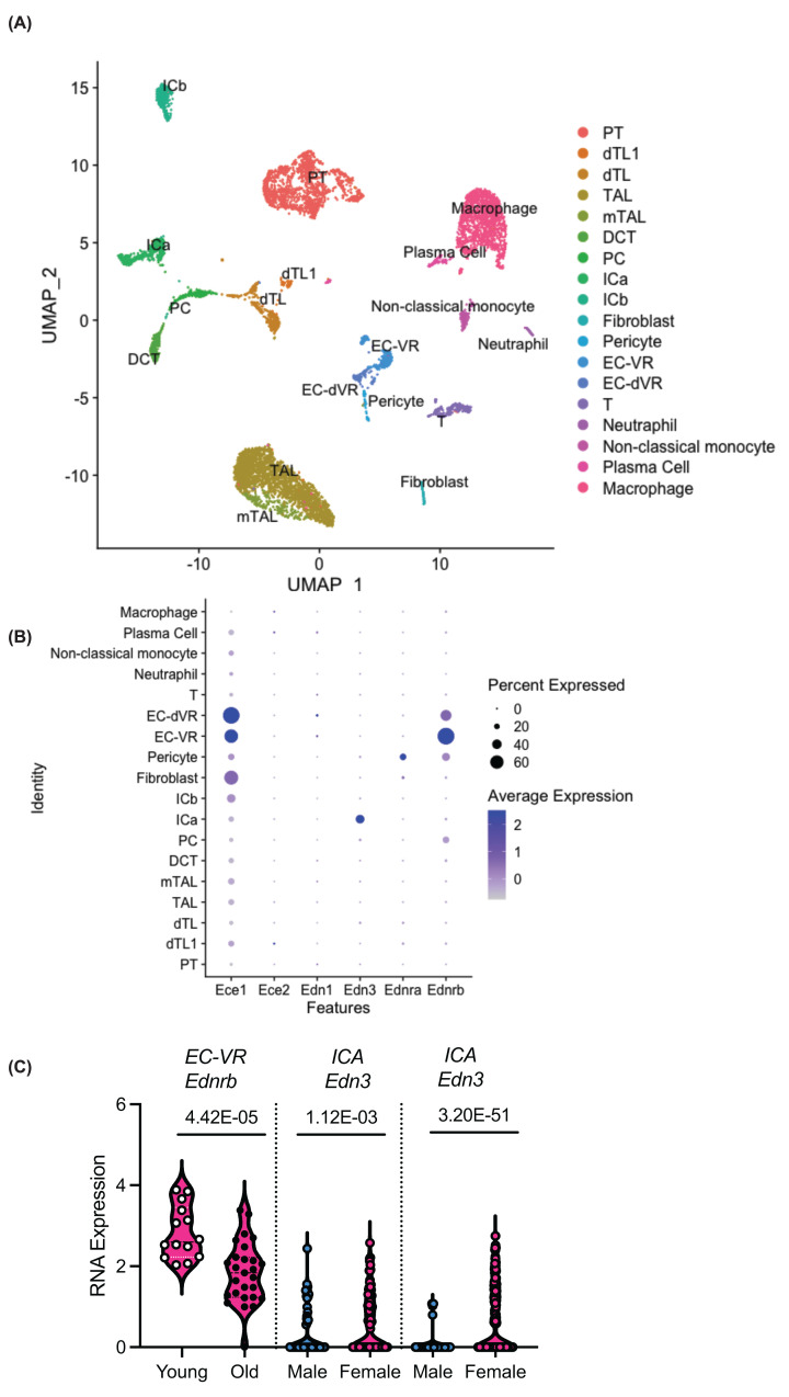
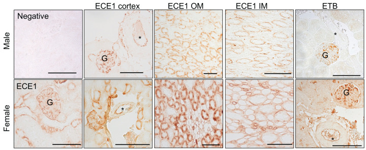
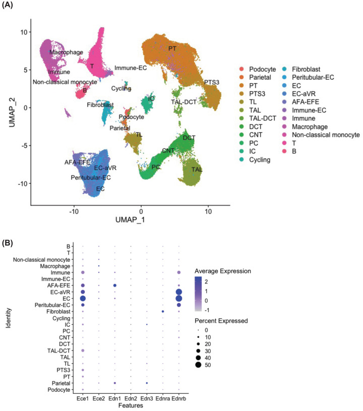
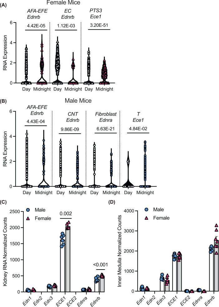
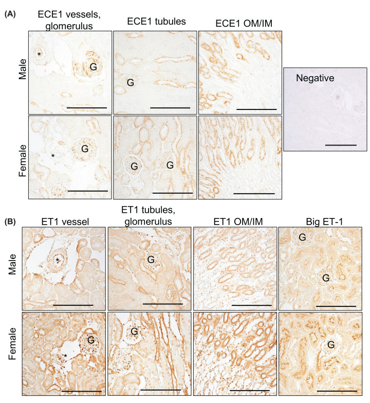
The intrarenal endothelin (ET) system is an established moderator of kidney physiology and mechanistic contributor to the pathophysiology and progression of chronic kidney disease in humans and rodents. The aim of the present study was to characterize ET system by combining single cell RNA sequencing (scRNA-seq) data with immunolocalization in human and rodent kidneys of both sexes. Using publicly available scRNA-seq data, we assessed sex and kidney disease status (human), age and sex (rats), and diurnal expression (mice) on the kidney ET system expression. In normal human biopsies of both sexes and in rodent kidney samples, the endothelin-converting enzyme-1 (ECE1) and ET-1 were prominent in the glomeruli and endothelium. These data agreed with the scRNA-seq data from these three species, with ECE1/Ece1 mRNA enriched in the endothelium. However, the EDN1/Edn1 gene (encodes ET-1) was rarely detected, even though it was immunolocalized within the kidneys, and plasma and urinary ET-1 excretion are easily measured. Within each species, there were some sex-specific differences. For example, in kidney biopsies from living donors, men had a greater glomerular endothelial cell endothelin receptor B (Ednrb) compared with women. In mice, females had greater kidney endothelial cell Ednrb than male mice. As commercially available antibodies did not work in all species, and RNA expression did not always correlate with protein levels, multiple approaches should be considered to maintain required rigor and reproducibility of the pre- and clinical studies evaluating the intrarenal ET system.

Keywords: Endothelin; RNA; chronic kidney disease; kidney; protein; sex.


Conflict of interest statement

The authors declare that there are no competing interests associated with the manuscript.

Figures

Figure 1
Figure 1. Human kidney single cell analyses from the Kidney Precision Medicine Program [36]
(A) Unsupervised clustering of the Kidney Precision Medicine Program human kidney single cell RNA-sequencing. A total of 11,0346 cells from living donor controls (n=18), chronic kidney disease (n =15), and acute kidney injury (n=12) subjects are included. (B) Dotplot of the RNA expression of the endothelin related genes across the clusters. Abbreviations: aFIB, Fibroblast (adaptive/maladaptive/repairing); aPT, Proximal Tubule Epithelial Cell (adaptive/maladaptive/repairing); aTAL1, Thick Ascending Limb Cell Cluster 1 (adaptive/maladaptive/repairing); aTAL2, Thick Ascending Limb Cell Cluster 2 (adaptive/maladaptive/repairing); B, B cell; C-TAL, Cortical Thick Ascending Limb Cell; cDC, Classical Dendritic Cell; CNT, Connecting Tubule Cell; CNT-IC-A, Connecting Tubule Intercalated Cell Type A; CNT-PC, Connecting Tubule Principal Cell; cycEC, Endothelial Cell (cycling); cycEPI, Epithelial cell (cycling); cycMNP, Mononuclear Phagocyte (cycling); cycT, T cell (cycling); dC-TAL, Cortical Thick Ascending Limb Cell (degenerative); dCNT, Connecting Tubule Cell (degenerative); dCNT-PC, Connecting Tubule cell-Principal cell (degenerative); DCT1, Distal Convoluted Tubule Cell Type 1; dDCT, Distal Convoluted Tubule Cell (degenerative); dEC-PTC, Peritubular Capillary Endothelial Cell (degenerative); dIC-A, Intercalated Cell Type A (degenerative); dPC, Principal cell (degenerative); dPT, Proximal Tubule Epithelial Cell (degenerative); dPT-DTL, Proximal Tubule Epithelial Cell/Descending Thin Limb Cell (degenerative); DTL1, Descending Thin Limb Cell Type 1; dVSMC, Vascular Smooth Muscle Cell (degenerative); EC-AEA, Afferent/Efferent Arteriole Endothelial Cell; EC-GC, Glomerular Capillary Endothelial Cell; EC-LYM, Lymphatic Endothelial Cell; EC-PTC, Peritubular Capillary Endothelial Cell; FIB, Fibroblast; IC-A, Intercalated Cell Type A; IC-B, Intercalated Cell Type B; M-TAL, Medullary Thick Ascending Limb Cell; MAC-M2, M2-Macrophage; MAST, Mast cell; MC, Mesangial Cell; MDC, Monocyte-derived Cell; MON, monocyte; MyoF, Myofibroblast; ncMON, Non-classical Monocyte; NK1, Natural killer 1; NK2, Natural killer 2; NKT, Natural Killer T Cell; PC, Principal cell; pDC, Plasmacytoid Dendritic Cell; PEC, Parietal Epithelial Cell; PL, Plasma Cell; POD, Podocyte; PT-S3, Proximal Tubule Epithelial Cell Segment 3; PTS1S2, Proximal Tubule Epithelial Cell Segment 1&2; REN, Renin-positive Juxtaglomerular Granular Cell; T, T Cell; T-CYT, T cytotoxic; T-REG, T regs; tPC-IC, Principal-Intercalated Cell (transitional); VSMC-P, Vascular Smooth Muscle Cell/Pericyte.
Figure 2
Figure 2. Violin plots with individual kidney cells from human biopsies of the differentially expressed genes related to the endothelin pathway
(A) Living donor (LD) compared with acute kidney injury (AKI) samples from women, (B) AKI compared with chronic kidney disease (CKD) samples from women, (C) LD compared with CKD samples from women. (D) LD compared with AKI or CKD kidney samples from men, (E) AKI compared with CKD samples from men. P-values adjusted for multiple comparisons reported. aPT, adaptive proximal tubules; CNT-ICA, connecting tubule intercalated cell alpha; CNT-PC, connecting tubule principal cell; dCNT, degenerative connecting tubule; dCNT-PC, degenerative connecting tubule principal cell; DCT1, distal convoluted tubule-1; dC-TAL, degenerative cortical thick ascending limb; dM-TAL, degenerative medullary thick ascending limb; dVSMC, degenerative vascular smooth muscle cell; EC-GC, glomerular endothelial capillary; EC-PTC, peritubular capillary endothelial cell; M-TAL, medullary thick ascending limb; PC, principal cell; tPC-IC, transitional principal-intercalated cell.
Figure 3
Figure 3. Representative images from human kidney biopsies used for immunolocalization of the ET system
The negative control lacked the primary antibody. Endothelin converting enzyme -1 (ECE1), endothelin-1 (ET-1), endothelin-2 (ET2) were abundant in the endothelium (EC) of large kidney blood vessels, and glomeruli (G). ET-1 was also found in some collecting ducts. Asterisks (*) marks blood vessels. Scale bar represents 100 micrometers.
Figure 4
Figure 4. Unsupervised clustering of kidney single cells from male and female rats published by [37]
A total of 8051 cells were RNA sequenced from a young female (5 months old), and aged female (27 months old), a young male (5 months) and aged male (27 months). (B) Dotplot of the RNA expression of the endothelin related genes across the clusters. (C) The only differentially expressed genes related to the endothelin system in this data set. P-values in the figure are adjusted for multiple comparisons. PT, proximal tubule; dTL1, descending thin limb-1; dTL, descending thin limb; TAL, thick ascending limb; mTAL, medullary thick ascending limb; DCT, distal convoluted tubule; PC, principal cell; ICA, intercalated cells alpha; ICB, intercalated cell β; EC-VR, endothelial cells-vasa recta; EC-dVR, endothelial cell descending vasa recta; T, T cell.
Figure 5
Figure 5. Representative images of rat kidneys use in immunolocalization of the ET system
The negative control lacked the primary antibody. The ECE1 was expressed in the glomeruli (G), endothelium of blood vessels (*), thick ascending limbs, vasa recta and inner medullary (IM) collecting ducts. The ETB was abundant in the glomerulus and endothelium of the large kidney blood vessels of female rats. The scale bar represents 100 microns.
Figure 6
Figure 6. Unsupervised clustering of kidney single cells and nuclei from male and female mice
(A) Unsupervised clustering of 99588 kidney single cells and single nuclei RNA sequenced from C57bl/6J male and female mice from [30,38,40–46]. (B) Dotplot of the RNA expression of the endothelin related genes across the clusters.
Figure 7
Figure 7. Violin plots of the normalized RNA expression of genes
Violin plots of the normalized RNA expression of genes at the individual cells/nuclei level comparing day and midnight samples, for (A) female mice and (B) male mice. P values are reported above the plots for each pair and are adjusted P values for multiple comparisons. (C,D) Normalized counts of RNA from (C) whole mouse kidney or (D) mouse inner medulla. P values reported are adjusted for multiple comparisons. Individual animals are plotted. Blue circles represent male mice and pink triangles female mice. AFA-EFE, afferent-efferent arteriole; EC, endothelial cell; PTS3, proximal tubule segment 3; CNT, connecting tubule; T, T cell.
Figure 8
Figure 8. Representative images of mouse kidneys
Representative images of mouse kidneys collected during the day time and used to immunolocalize the endothelin converting enzyme (ECE1), endothelin-1 (ET1), and big ET-1 of the ET system. (A) ECE1 was abundant in the glomerulus, and thick ascending limbs. Outer medulla, OM; Inner medulla, IM. Negative control lacked primary antibody. Asterisk (*) marks blood vessels. (B) ET-1 was abundant in the kidney blood vessels, glomerulus, and thick ascending limbs. There was also ET-1 found in interstitial cells. Big ET-1 was highly expressed in the S1 and S2 segments of the proximal tubules. Scale bar represents 100 microns.

Similar articles

Cited by

References

    1. Hyndman K.A. and Evans D.H. (2007) Endothelin and endothelin converting enzyme-1 in the fish gill: evolutionary and physiological perspectives. J. Exp. Biol. 210, 4286–4297, 210/24/4286 [pii] 10.1242/jeb.009969 - DOI - PubMed
    1. Hyndman K.A., Miyamoto M.M. and Evans D.H. (2009) Phylogeny, taxonomy, and evolution of the endothelin receptor gene family. Mol. Phylogenet. Evol. 52, 677–687, S1055-7903(09)00145-6 [pii] 10.1016/j.ympev.2009.04.015 - DOI - PubMed
    1. Davenport A.P., Hyndman K.A., Dhaun N., Southan C., Kohan D.E., Pollock J.S.et al. . (2016) Endothelin. Pharmacol. Rev. 68, 357–418 10.1124/pr.115.011833 - DOI - PMC - PubMed
    1. Stuart D., Rees S., Woodward S.K., Koesters R., Strait K.A. and Kohan D.E. (2012) Disruption of the endothelin A receptor in the nephron causes mild fluid volume expansion. BMC Nephrol. 13, 166 10.1186/1471-2369-13-166 - DOI - PMC - PubMed
    1. Bugaj V., Mironova E., Kohan D.E. and Stockand J.D. (2012) Collecting duct-specific endothelin B receptor knockout increases ENaC activity. Am. J. Physiol. Cell Physiol. 302, C188–C194, ajpcell.00301.2011 [pii] 10.1152/ajpcell.00301.2011 - DOI - PMC - PubMed

Publication types

MeSH terms

Grants and funding