. 2024 Jun 21;316(7):428.
doi: 10.1007/s00403-024-03131-9.

Potential of cannabidiol as acne and acne scar treatment: novel insights into molecular pathways of pathophysiological factors

Affiliations

Potential of cannabidiol as acne and acne scar treatment: novel insights into molecular pathways of pathophysiological factors

Jun Hyo Lee et al. Arch Dermatol Res. .

Abstract

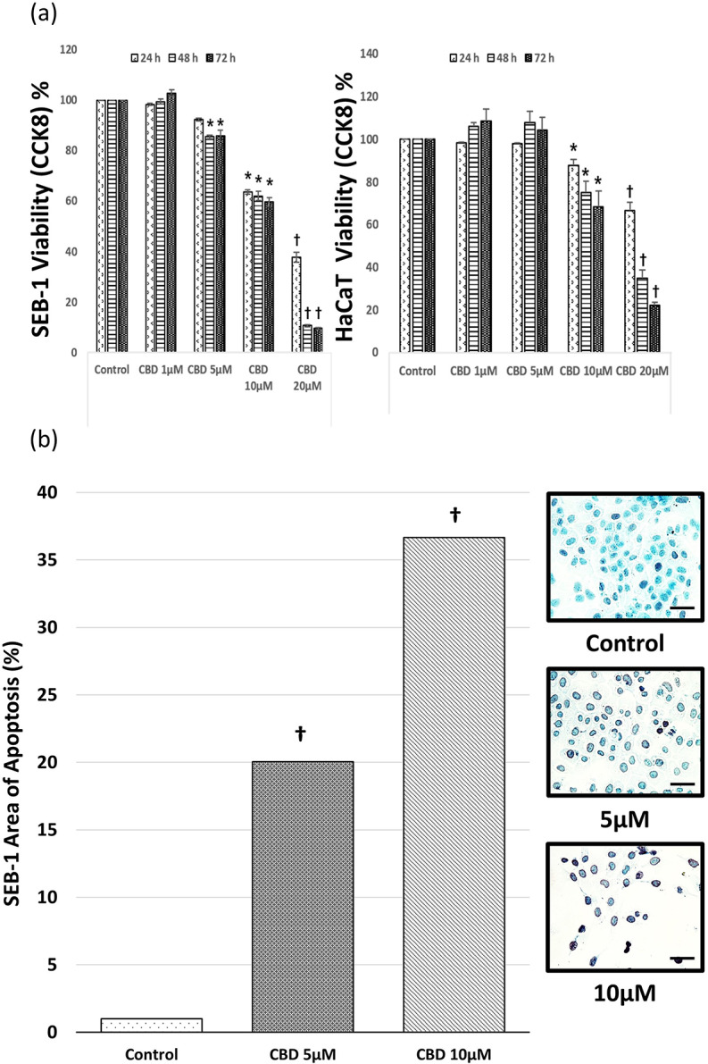
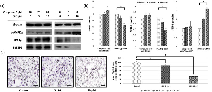
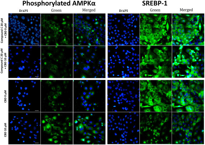
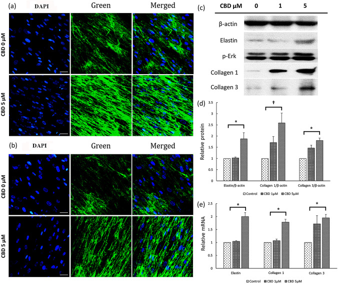
Cannabidiol (CBD), which is derived from hemp, is gaining recognition because of its anti-inflammatory and lipid-modulating properties that could be utilized to treat acne. We conducted experiments to quantitatively assess the effects of CBD on acne-related cellular pathways. SEB-1 sebocytes and HaCaT keratinocytes were exposed to various CBD concentrations. CBD exhibited a concentration-dependent impact on cell viability and notably reduced SEB-1 viability; furthermore, it induced apoptosis and a significant increase in the apoptotic area at higher concentrations. Additionally, CBD remarkably reduced pro-inflammatory cytokines, including CXCL8, IL-1α, and IL-1β. Additionally, it inhibited lipid synthesis by modulating the AMPK-SREBP-1 pathway and effectively reduced hyperkeratinization-related protein keratin 16. Simultaneously, CBD stimulated the synthesis of elastin, collagen 1, and collagen 3. These findings emphasize the potential of CBD for the management of acne because of its anti-inflammatory, apoptotic, and lipid-inhibitory effects. Notably, the modulation of the Akt/AMPK-SREBP-1 pathway revealed a novel and promising mechanism that could address the pathogenesis of acne.

Keywords: Acne; Cannabidiol; Inflammation; Keratinocytes; Lipid modulation; Sebocytes.


Conflict of interest statement

Jun Hyo Lee, Dong Hyo Kim, Ji Young Yoon and Dae Hun Suh declare no conflict of interest. The funders had no role in the design of the study; in the collection, analyses, or interpretation of data; in the writing of the manuscript; or in the decision to publish the results. Yoon Gyung Kwon, Geun-Hyeong Kim, and Byoung Jun Park are employees of Kolmar Korea, but they did not influence the interpretation of the experimental data.

Figures

Fig. 1
Fig. 1
Chemical structure of CBD
Fig. 2
Fig. 2
CBD induces apoptosis in SEB-1 cells and HaCaT cells. (a) SEB-1 cells and HaCaT cells were exposed to CBD at concentrations of 1, 5, 10 or 20 µM for 48 h, followed by confirmation of apoptosis using the TUNEL assay method. (b) Quantitative analysis of apoptosis staining was conducted and the results were graphed. Representative micrograph images of SEB-1 cells treated with CBD showing the induction of apoptosis (400X magnification). Data are presented from a sample size of n = 9 independent experiments. All are at the same magnification (bar = 50 μm). CBD, cannabidiol; TUNEL, terminal deoxynucleotidyl transferase dUTP nick end labeling. *, P < 0.05, †, P < 0.001
Fig. 3
Fig. 3
CBD modulates inflammatory cytokine expression in SEB-1 cells exposed to C. acnes. SEB-1 cells were treated with CBD at concentrations of 0 µM, 5 µM, and 10 µM in the presence of C. acnes to assess changes in inflammatory cytokines. (a) Quantitative real-time PCR validated reduced mRNA expression levels of CXCL8, IL-1α, IL-1β, and TNFα post-CBD treatment compared to the untreated control. (b) Western blot analysis quantified protein levels of CXCL8 and TNFα. Quantified data were graphically represented, showcasing decreased protein levels in response to CBD treatment. CBD, cannabidiol; C. acnes, Cutibacterium acnes. *, P < 0.05
Fig. 4
Fig. 4
Effect of CBD on lipogenesis pathway in SEB-1 cells. CBD reduces SEB-1 lipogenesis by inhibiting the expression of sterol regulatory element binding protein (SREBP)-1 through the AMP-activated protein kinase (AMPK) pathway. (a) SEB-1 cells were pretreated by CBD with or without 20 µM of compound C for 24 h before western blot. Levels of phospho-/total AMPK, PPARγ, and SREBP1 were measured. (b) Western blot bands of SREBP-1/β-actin, PPARγ/β-actin, and phospho-/total AMPKα were quantified and presented in a graph. (c) Oil red staining analysis was performed and graphed (400X). All are at the same magnification (bar = 50 μm). CBD, cannabidiol; SREBP, sterol regulatory element binding protein; AMPK, AMP activated protein kinase; PPARγ, peroxisome proliferator-activated receptor gamma. *, P < 0.05; †, P < 0.001
Fig. 5
Fig. 5
Representative DAPI staining images of CBD treated SEB-1 cells. Representative DAPI staining images of CBD treated SEB-1 cells. SEB-1 cells were treated with CBD at concentrations of 0 µM and 10 µM for a duration of 48 h. DAPI staining was performed to assess the impact of CBD treatment. Phosphorylated AMPKα expression and SREBP-1 expression were evaluated using DAPI staining (400X). All are at the same magnification (bar = 50 μm). CBD, cannabidiol; SREBP, sterol regulatory element binding protein; AMPK, AMP activated protein kinase; DAPI, 4’,6-diamidino-2-phenylindole
Fig. 6
Fig. 6
Inhibition of hyperkeratinization by CBD treatment in HaCaT cells. HaCaT cells were subjected to CBD treatment at concentrations of 0 µM, 5 µM, and 10 µM for a duration of 24 h. (a) Western blot analysis was employed to validate the reduction in keratin 16 levels. (b) Alterations in the mRNA expression of keratin 16 were verified through real-time PCR. CBD, cannabidiol. *, P < 0.05
Fig. 7
Fig. 7
Changes in the expression of proteins related to scar formation induced by CBD. (a, b) Fibroblasts were treated with CBD at concentrations of 0 µM and 5 µM for a duration of 48 h. DAPI staining was performed to assess the impact of CBD treatment. Collagen 1 (a) and collagen 3 (b) expression were evaluated using DAPI staining (400×). All are at the same magnification (bar = 50 μm). (c) Western blot analysis confirmed changes in the expression of elastin, collagen 1, collagen 3, and p-Erk at CBD concentration of 1 µM or 5 µM. (d) Quantification of western blot bands for elastin/β-actin, collagen 1/β-actin, and collagen 3/β-actin was performed. (e) Quantitative real-time analysis confirmed the mRNA expression levels of elastin, collagen 1, and collagen 3. CBD, cannabidiol; DAPI, 4’,6-diamidino-2-phenylindole; p-ERK, phosphorylated ERK. *, P < 0.05; †, P < 0.00

References

    1. Lynn DD, Umari T, Dunnick CA, Dellavalle RP (2016) The epidemiology of acne vulgaris in late adolescence. Adolesc Health Med Ther 7:13–25 - PMC - PubMed
    1. Hay RJ, Johns NE, Williams HC, Bolliger IW, Dellavalle RP, Margolis DJ et al (2014) The global burden of skin disease in 2010: an analysis of the prevalence and impact of skin conditions. J Invest Dermatol 134:1527–1534 10.1038/jid.2013.446 - DOI - PubMed
    1. Dreno B (2017) What is new in the pathophysiology of acne, an overview. J Eur Acad Dermatol Venereol 31:8–12 10.1111/jdv.14374 - DOI - PubMed
    1. Fox L, Csongradi C, Aucamp M, Du Plessis J, Gerber M (2016) Treatment modalities for acne. Molecules 21:1063 - PMC - PubMed
    1. Van Beugen S, Schut C, Kupfer J, Bewley AP, Finlay AY, Gieler U et al (2023) Perceived stigmatization among dermatological outpatients compared with controls: an observational Multicentre Study in 17 European countries. Acta Dermato-Venereologica 103:adv6485 - PMC - PubMed

MeSH terms

LinkOut - more resources