. 2024 Aug;21(6):867-879.
doi: 10.1007/s13770-024-00648-y. Epub 2024 Jun 21.

Transplantation of Neural Progenitor Cells Derived from Stem Cells from Apical Papilla Through Small-Molecule Induction in a Rat Model of Sciatic Nerve Injury

Affiliations

Transplantation of Neural Progenitor Cells Derived from Stem Cells from Apical Papilla Through Small-Molecule Induction in a Rat Model of Sciatic Nerve Injury

Junhao Koh et al. Tissue Eng Regen Med. 2024 Aug.

Abstract

Background: Stem cell-based transplantation therapy holds promise for peripheral nerve injury treatment, but adult availability is limited. A cell culture protocol utilizing a small-molecule cocktail effectively reprogrammed stem cells from apical papilla (SCAPs) into neural progenitor cells, subsequently differentiating into neuron-like cells. This study aims to evaluate neural-induced SCAPs, with and without small-molecule cocktail, for sciatic nerve repair potential.

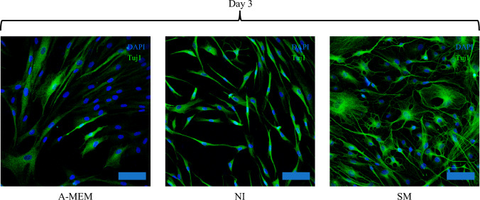
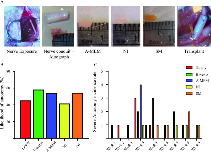
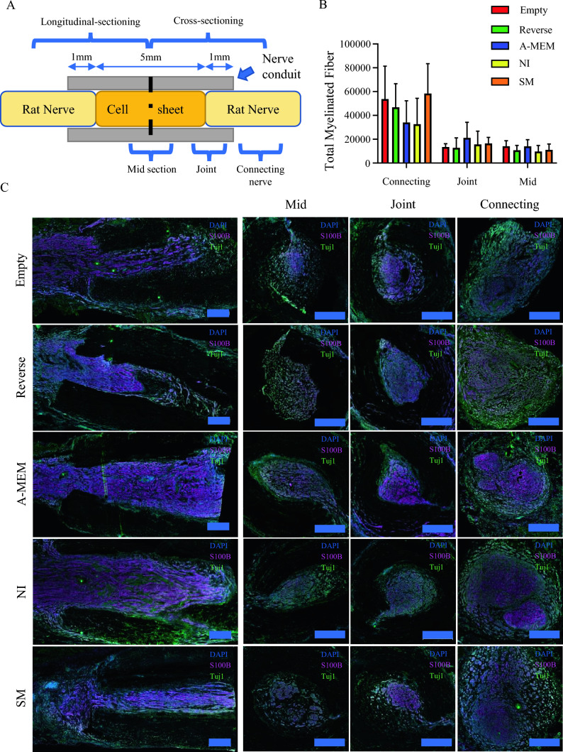
Methods: A scaffold-free cell sheet technique was used to construct a three-dimensional cell sheet. Subsequently, this cell sheet was carefully rolled into a tube and seamlessly inserted into a collagen conduit, which was then transplanted into a 5 mm sciatic nerve injury rat model. Functional sciatic nerve regeneration was evaluated via toe spread test, walking track analysis and gastrocnemius muscle weight. Additionally, degree of sciatic nerve regeneration was determined based on total amount of myelinated fibers.

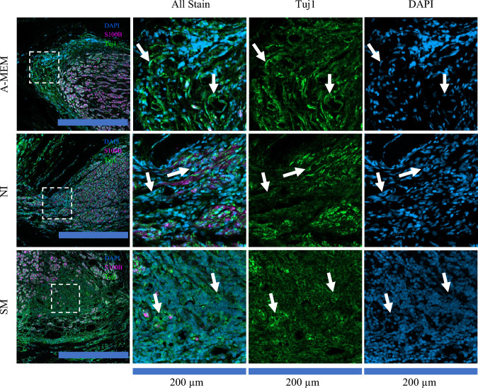
Results: Small-molecule cocktail induced SCAPs enhanced motor function recovery, evident in improved sciatic function index and gastrocnemius muscle retention. We also observed better host myelinated fiber retention than undifferentiated SCAPs or neural-induced SCAPs without small-molecule cocktail. However, clusters of neuron-like cell bodies (surrounded by sparse myelinated fibers) were found in all cell sheet-implanted groups in the implantation region. This suggests that while the implanted cells likely survived transplantation, integration was poor and would likely hinder long-term recovery by occupying the space needed for host nerve fibers to project through.

Conclusion: Neural-induced SCAPs with small-molecule cocktail demonstrated promising benefits for nerve repair; further research is needed to improve its integration and optimize its potential for long-term recovery.

Keywords: Cell sheet engineering; Nerve conduit; Peripheral nerve regeneration; SCAPs; Stem cells.


Conflict of interest statement

The authors declare that there are no commercial or financial conflict of interest in conducting this research.

Figures

Fig. 1
Fig. 1
Expression of early neuronal marker of SCAP after 3 days of induction. Immunocytofluorescence for detection of tuj1 (green) expression in SCAP cultured in A-MEM, NI, and SM at day 3. Nuclei were counterstained with DAPI (blue). Scale bar = 100 µm. A-MEM, alpha modified Minimum Essential Medium; NI, Neural induction medium; SM, neural induction with small-molecule cocktail; Tuj1, β-Tubulin III
Fig. 2
Fig. 2
Delayed onset of autotomy in neural-induced SCAP-derived cell sheet groups. A Surgical procedure for inserting the cell sheet into a nerve conduit before transplanting into the 5-mm sciatic nerve gap. (From left to right: Exposure of left sciatic nerve; representative images of nerve conduit and excised nerve length; A-MEM cell sheet; NI cell sheet; SM cell sheet; the connected nerve in the nerve conduit.) Incidence of autotomy by group as shown by B percentage and C number of animals in each group by week. A-MEM, alpha modified Minimum Essential Medium; NI, Neural induction medium; SM, neural induction with small-molecule cocktail
Fig. 3
Fig. 3
Assessment of functional recovery after cell sheet implantation. A At week 10, the SFI showed that the SM group had significantly more improvement than the NI group (*p < 0.05). Additionally, at week 12, the SM group outperformed the Empty (*p < 0.05), Reverse (*p < 0.05), and A-MEM (**p < 0.01) groups. B A bar chart of toe spread scores at week 12 post-injury revealed functional motor recovery in some animals in the Reverse and SM groups, but this was not statistically significant. Only the SM group demonstrated recovery in walking track analysis, as indicated by the SFI score. C Bar chart of the severity of chronic flexion contracture in each group at week 12 post-injury. D Representative images of toe spreading in each group showing the state of functional motor recovery of the foot at week 12. Data are presented as mean ± SD. A-MEM, alpha modified Minimum Essential Medium; NI, Neural induction medium; SFI, sciatic function index; SM, neural induction with small-molecule cocktail
Fig. 4
Fig. 4
Gastrocnemius muscle retention following cell sheet implantation. A Representative left and right gastrocnemius muscle between groups and comparison of the wet gastrocnemius muscle ratio between left and right limb. B The bar chart shows that the Empty group retained significantly higher muscle mass than the A-MEM or NI groups (*p < 0.01). Data are presented as mean ± SD. A-MEM, alpha modified Minimum Essential Medium; NI, Neural induction medium; SM, neural induction with small-molecule cocktail
Fig. 5
Fig. 5
Effects of different cell sheet transplants on myelinated fiber regeneration. A Schematic diagram illustrating the sectional arrangement of the nerve for staining. B Bar chart showing total myelinated fibers. C Representative immunofluorescent staining of the operated sciatic nerve showing the longitudinal sectioning of the nerve and cross-sectioning of the mid-section of the conduits (Mid), the joint between the connecting nerve with the implanted section (Joint), and the connecting part of the nerve (Connecting) for each group. Nuclei were counterstained with DAPI (blue), Myelin-sheet were counterstained with S100b (purple) and neuronal fibers were counterstained with Tuj1(Green). Scale bar for all images = 500 µm. A-MEM, alpha modified Minimum Essential Medium; NI, Neural induction medium; S100b, S100 calcium-binding protein B; SM, neural induction with small-molecule cocktail; Tuj1, β-Tubulin III
Fig. 6
Fig. 6
Cross-section of the mid-section showing transplanted cells surviving at 12 weeks. Clusters of non-myelinated cell bodies expressing Tuj1 were observed in the cell sheet groups (A-MEM, NI, and SM) but not in the Empty or Reverse groups. White arrows highlight the cell clusters. Nuclei were counterstained with DAPI (blue), Myelin-sheet were counterstained with S100b (purple) and neuronal fibers were counterstained with Tuj1(Green). The leftmost images are enlarged images from 200 µm × 200 µm dashed squares; the scale bar for the leftmost images = 500 µm. A-MEM, alpha modified Minimum Essential Medium; NI, Neural induction medium; S100b, S100 calcium-binding protein B; SM, neural induction with small-molecule cocktail; Tuj1, β-Tubulin III

References

    1. Frattini F, Lopes FR, Almeida FM, Rodrigues RF, Boldrini LC, Tomaz MA, et al. Mesenchymal stem cells in a polycaprolactone conduit promote sciatic nerve regeneration and sensory neuron survival after nerve injury. Tissue Eng Part A. 2012;18:2030–9. 10.1089/ten.tea.2011.0496 - DOI - PubMed
    1. Zheng C, Zhu Q, Liu X, Huang X, He C, Jiang L, et al. Improved peripheral nerve regeneration using acellular nerve allografts loaded with platelet-rich plasma. Tissue Eng Part A. 2014;20:3228–40. 10.1089/ten.tea.2013.0729 - DOI - PMC - PubMed
    1. Huang GT, Gronthos S, Shi S. Mesenchymal stem cells derived from dental tissues vs. those from other sources: their biology and role in regenerative medicine. J Dent Res. 2009;88:792–806. 10.1177/0022034509340867 - DOI - PMC - PubMed
    1. Huang GT, Sonoyama W, Liu Y, Liu H, Wang S, Shi S. The hidden treasure in apical papilla: the potential role in pulp/dentin regeneration and bioroot engineering. J Endod. 2008;34:645–51. 10.1016/j.joen.2008.03.001 - DOI - PMC - PubMed
    1. Kolar MK, Itte VN, Kingham PJ, Novikov LN, Wiberg M, Kelk P. The neurotrophic effects of different human dental mesenchymal stem cells. Sci Rep. 2017;7:12605. 10.1038/s41598-017-12969-1 - DOI - PMC - PubMed

Publication types

LinkOut - more resources