. 2024;14(5):941-964.
doi: 10.3233/JPD-240080.

Serotonergic Regulation of Synaptic Dopamine Levels Mitigates L-DOPA-Induced Dyskinesia in a Mouse Model of Parkinson's Disease

Affiliations

Serotonergic Regulation of Synaptic Dopamine Levels Mitigates L-DOPA-Induced Dyskinesia in a Mouse Model of Parkinson's Disease

Yuan-Hao Chen et al. J Parkinsons Dis. 2024.

Abstract

Background: The serotonin (5-HT) system can manipulate the processing of exogenous L-DOPA in the DA-denervated striatum, resulting in the modulation of L-DOPA-induced dyskinesia (LID).

Objective: To characterize the effects of the serotonin precursor 5-hydroxy-tryptophan (5-HTP) or the serotonin transporter (SERT) inhibitor, Citalopram on L-DOPA-induced behavior, neurochemical signals, and underlying protein expressions in an animal model of Parkinson's disease.

Methods: MitoPark (MP) mice at 20 weeks of age, subjected to a 14-day administration of L-DOPA/Carbidopa, displayed dyskinesia, referred to as LID. Subsequent investigations explored the effects of 5-HT-modifying agents, such as 5-HTP and Citalopram, on abnormal involuntary movements (AIMs), locomotor activity, neurochemical signals, serotonin transporter activity, and protein expression in the DA-denervated striatum of LID MP mice.

Results: 5-HTP exhibited duration-dependent suppressive effects on developing and established LID, especially related to abnormal limb movements observed in L-DOPA-primed MP mice. However, Citalopram, predominantly suppressed abnormal axial movement induced by L-DOPA in LID MP mice. We demonstrated that 5-HTP could decrease L-DOPA-upregulation of DA turnover rates while concurrently upregulating 5-HT metabolism. Additionally, 5-HTP was shown to reduce the expressions of p-ERK and p-DARPP-32 in the striatum of LID MP mice. The effect of Citalopram in alleviating LID development may be attributed to downregulation of SERT activity in the dorsal striatum of LID MP mice.

Conclusions: While both single injection of 5-HTP and Citalopram effectively mitigated the development of LID, the difference in mitigation of AIM subtypes may be linked to the unique effects of these two serotonergic agents on L-DOPA-derived DA and 5-HT metabolism.

Keywords: 5-HTP; Citalopram; L-DOPA-induced dyskinesia; Parkinson’s disease; Serotonin system; dopamine.


Conflict of interest statement

The authors have no conflict of interest to report.

Figures

Fig. 1
Fig. 1
The experimental protocol for this study involves categorizing the experimental mice into four groups: normal animals (WT), PD mice (MP), MP mice with L-DOPA-induced dyskinesia (MP LID), and the MP LID treatment group. L-DOPA and Carbidopa were administered daily to the MP LID mice from Day 1 to Day 6, while WT and MP mice received only L-DOPA and Carbidopa from Day 3 to Day 6. The experiment commenced after administering L-DOPA and Carbidopa from Day 3 to Day 5. On Day 6, 10 min after administering L-DOPA and Carbidopa, the mice were euthanized by cervical dislocation, and their brains were then removed for subsequent experiments.
Fig. 2
Fig. 2
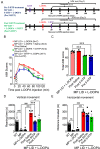
Administering 5-HTP (50 mg/kg) 30 min before L-DOPA (10 mg/kg) treatment was an effective method for mitigating the abnormal AIM scores induced by L-DOPA in L-DOPA-primed MP mice (MP LID mice). A) The influence of pre-treatment with 5-HTP at time intervals of 30, 60, 90, and 120 min before the administration of L-DOPA on the L-DOPA-induced dyskinesia in MP LID mice. B) MP LID mice were pre-administered 5-HTP, followed by the administration of L-DOPA at different time intervals. Following L-DOPA administration, AIM scores were assessed at 20-min intervals, with a total of seven evaluations. Two-way ANOVA followed by a Bonferroni post hoc test for multiple comparisons. At 30 min compared to baseline, *p < 0.05, ***p < 0.001; at 60 min compared to baseline, ##p < 0.01, ###p < 0.001. Observation of the seven scores separately across the time span of 20 to 140 min post-L-DOPA administration, (C) the maximum AIM score from the seven scores (maximum AIM score), and (D) the sum of the seven scores (total AIM score). The maximum AIM score and the total AIM score were both lower when L-DOPA was administered 30 min after pre-treatment with 5-HTP (represented by the blue bar/rectangular symbol; 30 min), as compared to the other pre-treatment 5-HTP groups and the group without 5-HTP pre-treatment (represented by the red bar/rectangular symbol; Baseline). In particular, the administration of L-DOPA 60 min after pre-treatment with 5-HTP (represented by the purple bar/triangle symbol; 60 min) resulted in an elevation in the total AIM score compared to the group without 5-HTP pre-treatment (Baseline vs. 60 min, p < 0.01). One-way ANOVA followed by a Bonferroni post hoc test for multiple comparisons. At 30 min compared to the other group, *p < 0.05, **p < 0.01, ***p < 0.001; at 60 min compared to Baseline, ##p < 0.01.
Fig. 3
Fig. 3
Either pre-treatment or post-treatment with 5-HTP can effectively alleviate L-DOPA-induced dyskinesia in MP LID mice. A) The protocol for administering 5-HTP in MP LID mice involved the following steps: The assessment of AIM scores began immediately after the injection of L-DOPA and continued at 20-min intervals for a total duration of 120 min. B) Although dyskinesia in MP LID mice was found following administration of L-DOPA, the subsequent treatment with 5-HTP alleviated the abnormal symptoms in these mice. Following a 20-min administration of L-DOPA, post-treatment with 5-HTP reduces the abnormal AIM scores at the 80th and 100th min (Two-way ANOVA followed by a Bonferroni post hoc test for multiple comparisons. MP LID + L-DOPA + Saline compared to MP LID + 5-HTP + L-DOPA, ***p < 0.001), (C) as well as reduces the cumulative AIM score from the 20th to the 120th min. One-way ANOVA followed by a Bonferroni post hoc test for multiple comparisons. MP LID + L-DOPA + Saline compared to MP LID + 5-HTP + L-DOPA, ***p < 0.001. D) Locomotor analysis showed significant increases in MP LID groups after L-DOPA injection. The increase in vertical movement observed in the MP LID group following L-DOPA injection was mitigated by pre- or post-treatment with 5-HTP. (MP LID + L-DOPA + saline vs. MP LID + 5-HTP + L-DOPA, p < 0.001; MP LID + L-DOPA + saline vs. MP LID + L-DOPA + 5-HTP, p < 0.05). One-way ANOVA followed by a Bonferroni post hoc test for multiple comparisons. *p < 0.05, ***p < 0.001, compared to MP LID + L-DOPA + Saline; MP + L-DOPA compared to MP LID + L-DOPA + Saline, ###p < 0.001; WT compared to MP, $$$p < 0.001. E) The administration of 5-HTP as a pre-treatment or post-treatment did not result in a reduction of the increased horizontal movement observed after L-DOPA injection. One-way ANOVA followed by a Bonferroni post hoc test for multiple comparisons. MP + L-DOPA compared to MP LID + L-DOPA + Saline, ###p < 0.001; WT compared to MP, $$p < 0.01.
Fig. 4
Fig. 4
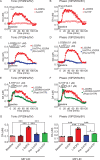
The administration of 5-HTP in the dorsal striatal slices from MP LID mice reduces dopamine release induced by supplementary L-DOPA, as measured using FSCV (fast-scan cyclic voltammetry). Dopamine release following L-DOPA infusion showed a significant increase in MP LID mice in both tonic release (induced by a single pulse at 25 Hz with 5 volts stimulation intensity) and phasic release (induced by 10 pulses at 25 Hz with 5 volts stimulation intensity), as illustrated in graphs (A) and (B) respectively. The increased release of dopamine caused by the infusion of L-DOPA could be suppressed by 5-HTP, whether administered beforehand (C, D) or afterward (E, F). G) With tonic stimulation, the co-administration of 5-HTP effectively reduced L-DOPA-induced dopamine release in the MP LID groups. One-way ANOVA followed by a Bonferroni post hoc test for multiple comparisons. ***p < 0.001, compared to MP LID + L-DOPA (red bar). H) With phasic stimulation, pre-treatment (blue bar, MP LID + 5-HTP + L-DOPA) or post-treatment (green bar, MP LID + L-DOPA + 5-HTP) of 5-HTP exhibited a significant suppression of L-DOPA-induced dopamine release in striatal slices. One-way ANOVA followed by a Bonferroni post hoc test for multiple comparisons. ***p < 0.001, compared to MP LID + L-DOPA; MP LID + 5-HTP + L-DOPA compared to MP LID + L-DOPA + 5-HTP, #p < 0.05.
Fig. 5
Fig. 5
Pharmacological modulation of the serotonin system can potentially affect the release of dopamine that is implicated in the development and manifestation of LID in MP mice. In addition to 5-HTP, Citalopram, a selective serotonin reuptake inhibitor (SERT inhibitor), was co-administered with L-DOPA. Subsequently, the AIM score was monitored using the experimental protocol depicted in (A). B) The AIM scores of each group were documented every 20 min following the injection of L-DOPA, over a duration of 2 h. C) The administration of 5-HTP (red bar: MP LID + Saline + L-DOPA vs. blue bar: MP LID + 5-HTP + L-DOPA, p < 0.01) or Citalopram (red bar: MP LID + Saline + L-DOPA vs. purple bar: MP LID + Citalopram + L-DOPA, p < 0.01) resulted in a significant decrease in the total AIM scores in L-DOPA-primed MP. D) The administration of Citalopram (p < 0.05) suppresses the occurrence of abnormal axial movements in the MP LID mice. The abnormal increase in (E) limb movements and (F) standing behavior observed in MP LID mice can be mitigated by 5-HTP (p < 0.05). G) The administration of either 5-HTP or Citalopram ameliorates the abnormal increase in vertical activity in MP LID mice. One-way ANOVA followed by a Bonferroni post hoc test for multiple comparisons. *p < 0.05, **p < 0.01, ***p < 0.001, compared to MP LID + Saline + L-DOPA.
Fig. 6
Fig. 6
DA release in striatal slices detected by FSCV. Both L-DOPA-induced increments in tonic (A) and phasic (B) release were reduced after Citalopram (0.1μM) infusion in brain slices from MP LID groups. Overall, the administration of 5-HTP or Citalopram, effectively suppressed both tonic (C) and phasic (D) dopamine release induced by L-DOPA infusion in the MP LID group. One-way ANOVA followed by a Bonferroni post hoc test for multiple comparisons. *p < 0.05, **p < 0.01, ***p < 0.001, compared to MP LID + L-DOPA.
Fig. 7
Fig. 7
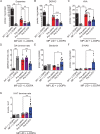
The concentrations of DA, serotonin, and their metabolites in the striatum were measured using high-performance liquid chromatography (HPLC) in the presence or absence of L-DOPA treatment. A) The DA concentration in the striatum of MP mice was lower than that of WT mice. The DA concentration in the striatum of MP LID mice receiving L-DOPA was higher than that in MP mice. The significant increase in DA concentration induced by L-DOPA injection in the MP LID group can be effectively suppressed by pre-treatment with 5-HTP or Citalopram. Only the administration of 5-HTP was able to suppress the increase in dopamine metabolites (red bar: MP LID + Saline + L-DOPA vs. blue bar: MP LID + 5-HTP + L-DOPA, p < 0.01), including DOPAC (p < 0.001) (B) and HVA (p < 0.001) (C), in the MP LID group following L-DOPA administration. The administration of Citalopram reduces only the increase in tissue concentration of HVA induced by L-DOPA in the MP LID group. D) The DA turnover rate increased significantly in MP and MP LID groups, which was suppressed by the administration of 5-HTP (p < 0.01). The concentration of serotonin (p < 0.05) (E), 5-HIAA (p < 0.01) (F), and the 5-HT turnover rate (p < 0.01) (G) exhibited significant increases in the striatum in MP LID mice given 5-HTP and L-DOPA, compared to MP LID mice given saline and L-DOPA. However, the administration of Citalopram did not have any effect on these measures. One-way ANOVA followed by a Bonferroni post hoc test for multiple comparisons. *p < 0.05, **p < 0.01, ***p < 0.001, compared to MP LID + Saline + L-DOPA; MP compared to MP LID + Saline + L-DOPA, #p < 0.05, ###p < 0.001; WT compared to MP, $$$p < 0.001.
Fig. 8
Fig. 8
The expression of proteins involved in dopaminergic and serotonergic transmission was analyzed in the striatum of WT, MP, and MP LID mice following co-treatment of L-DOPA with 5-HTP or Citalopram. A) Dopaminergic and serotonergic transmission-related protein expression in the striatum were illustrated. B) There was no significant change in D1 receptor protein expression among these groups. However, the administration of 5-HTP effectively suppressed the increased phosphorylation of signaling molecules in the downstream D1 pathway, specifically ERK (red bar: MP LID + Saline + L-DOPA vs. blue bar: MP LID + 5-HTP + L-DOPA, p < 0.01) (C) and DARPP32 (p < 0.01) (D), in the MP LID mice. In contrast, the administration of Citalopram did not have a significant effect on the phosphorylation levels of these molecules. There were no significant changes in the expressions of 5-HT1A receptor (E) and 5-HT1B receptor (F) proteins among these groups. One-way ANOVA followed by a Bonferroni post hoc test for multiple comparisons. **p < 0.01, compared to MP LID + Saline + L-DOPA; MP compared to MP LID + Saline + L-DOPA, ##p < 0.01.
Fig. 9
Fig. 9
PET scans of serotonin transporter (SERT) activity in WT, MP and MP LID mice. A) The bioactivity or availability of the serotonin transporter (SERT) was assessed using [18F]FE-ADAM radioactivity (SUR) measured in PET scans. Experimental animals were given L-DOPA and carbidopa for 20 min before undergoing PET scans. A lower activity was seen in MP mice compared to WT mice (WT + L-DOPA vs. MP + L-DOPA, p < 0.01), while an increase in activity was observed in MP LID mice. B) The SUR values in each group, plotted on a panel, revealed that the availability of SERT in MP LID mice was higher compared to MP mice (MP + L-DOPA vs. MP LID + Saline + L-DOPA, p < 0.001). Citalopram treatment significantly attenuated the increased SERT availability in MP LID mice (MP LID + Saline + L-DOPA vs. MP LID + Citalopram + L-DOPA, p < 0.05). However, the administration of 5-HTP did not result in any significant effect on the availability of SERT in MP LID animals. One-way ANOVA followed by a Bonferroni post hoc test for multiple comparisons. MP LID + Saline + L-DOPA compared to MP LID + Citalopram + L-DOPA, *p < 0.05; MP + L-DOPA compared to MP LID + Saline + L-DOPA, ###p < 0.001; WT + L-DOPA compared to MP + L-DOPA, $$p < 0.01.
Fig. 10
Fig. 10
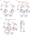
Graphical illustration delineates the effects of 5-HTP and Citalopram on DA release induced by the administration of L-DOPA in MP LID mice. A) The administration of L-DOPA induces aberrant release of DA in the synaptic cleft. The release of L-DOPA-derived DA may be regulated by the 5-HT neurons. B) Through the administration of 5-HTP, which competes with L-DOPA for AADC, the amount of DA in synaptic vesicles is reduced, thereby partially normalizing the abnormal DA levels in the synaptic cleft. Moreover, 5-HTP-derived 5-HT could activate 5-HT1 receptors, subsequently downregulating serotonin neuron-derived DA release in the DA-denervated striatum. C) Citalopram, by inhibiting SERT activity, may impede the reuptake of DA into presynaptic serotonin neurons, which attenuates the L-DOPA-induced aberrant DA release in the DA-denervated striatum. In addition, Citalopram administration increases serotonin concentration in the synaptic cleft by inhibiting SERT reuptake. Subsequently, excess 5-HT activates 5-HT1 receptors, subsequently inhibiting 5-HT neuron activity and reducing serotonin neuron-derived DA release after L-DOPA administration.

References

    1. Calabresi P, Ghiglieri V, Mazzocchetti P, Corbelli I, Picconi B (2015) Levodopa-induced plasticity: A double-edged sword in Parkinson’s disease? Proc R Soc Lond B Biol Sci 370, 20140184. - PMC - PubMed
    1. Bogetofte H, Alamyar A, Blaabjerg M, Meyer M (2020) Levodopa therapy for Parkinson’s disease: History, current status and perspectives. CNS Neurol Disord 19, 572–583. - PubMed
    1. Santini E (2009) Molecular basis of L-DOPA-induced dyskinesia: Studies on striatal signaling, Karolinska Institutet, Sweden.
    1. Bastide MF, Meissner WG, Picconi B, Fasano S, Fernagut P-O, Feyder M, Francardo V, Alcacer C, Ding Y, Brambilla R (2015) Pathophysiology of L-dopa-induced motor and non-motor complications in Parkinson’s disease. Prog Neurobiol 132, 96–168. - PubMed
    1. de la Fuente-Fernández R, Sossi V, Huang Z, Furtado S, Lu J-Q, Calne DB, Ruth TJ, Stoessl AJ (2004) Levodopa-induced changes in synaptic dopamine levels increase with progression of Parkinson’s disease: implications for dyskinesias. Brain 127, 2747–2754. - PubMed

Publication types

MeSH terms

LinkOut - more resources