. 2024 Jul 23;43(7):114382.
doi: 10.1016/j.celrep.2024.114382. Epub 2024 Jun 19.

Retrograde adenosine/A2A receptor signaling facilitates excitatory synaptic transmission and seizures

Affiliations

Retrograde adenosine/A2A receptor signaling facilitates excitatory synaptic transmission and seizures

Kaoutsar Nasrallah et al. Cell Rep. .

Abstract

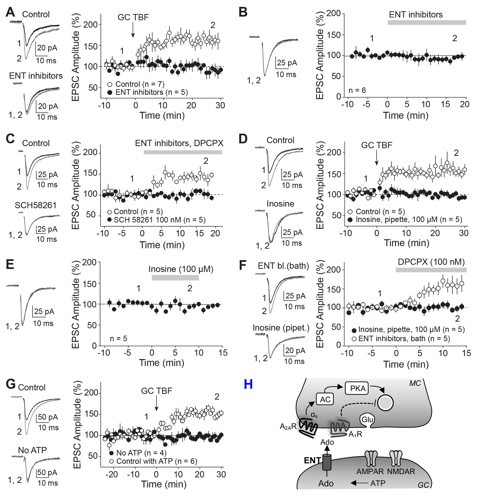
Retrograde signaling at the synapse is a fundamental way by which neurons communicate and neuronal circuit function is fine-tuned upon activity. While long-term changes in neurotransmitter release commonly rely on retrograde signaling, the mechanisms remain poorly understood. Here, we identified adenosine/A2A receptor (A2AR) as a retrograde signaling pathway underlying presynaptic long-term potentiation (LTP) at a hippocampal excitatory circuit critically involved in memory and epilepsy. Transient burst activity of a single dentate granule cell induced LTP of mossy cell synaptic inputs, a BDNF/TrkB-dependent form of plasticity that facilitates seizures. Postsynaptic TrkB activation released adenosine from granule cells, uncovering a non-conventional BDNF/TrkB signaling mechanism. Moreover, presynaptic A2ARs were necessary and sufficient for LTP. Lastly, seizure induction released adenosine in a TrkB-dependent manner, while removing A2ARs or TrkB from the dentate gyrus had anti-convulsant effects. By mediating presynaptic LTP, adenosine/A2AR retrograde signaling may modulate dentate gyrus-dependent learning and promote epileptic activity.

Keywords: BDNF; CP: Neuroscience; LTP; PKA; TrkB; dentate gyrus; epilepsy; hippocampus; mossy cell; presynaptic; retrograde signaling.


Conflict of interest statement

Declaration of interests The authors declare no competing interests.

Figures

Figure 1:
Figure 1:. Theta-burst firing of a single GC induces presynaptic LTP at the MC-GC synapse
(A) Left, Diagram illustrating the recording configuration. MC and MPP EPSCs were recorded from the same GC and evoked with stimulation electrodes placed in the inner and middle molecular layer, respectively. Right, Current clamp recording showing GC theta-burst firing (GC TBF). LTP induction protocol (GC TBF) was composed of 10 bursts at 5 Hz of 5 action potentials at 50 Hz, repeated 4 times every 5 s. (B) Left, Representative traces before (1) and after (2) GC TBF delivery. Right, Time-course plot showing that GC TBF induced LTP at MC-GC but not at MPP-GC synapses. (C) GC TBF-induced LTP was associated with significant reduction in PPR and CV (n = 13 cells). ** p < 0.01, *** p < 0.001. (D) LTP was abolished when TrkB was conditionally knocked out from from GCs (Post TrkB cKO, TrkBfl/fl mice injected in the dorsal blade with AAV5.CaMKII.Cre.GFP). LTP was unaffected in control animals (Control, TrkBfl/fl mice injected in the dorsal blade with AAV5.CaMKII.eGFP). (E) LTP was normally induced when loading PKI6-22 (2.5 μM) in GCs via the recording pipette but completely blocked when the cell-permeable PKA inhibitor PKI14-22 myristoylated (1 μM) was bath applied. (F) Summary bar graph showing the magnitude of GC TBF-induced LTP in the presence of DGC-IV (1 μM), when TrkB was conditionally knocked out from MCs (Pre TrkB cKO), when loading the PKI6-22 (2.5 μM) in GCs, and in the presence of D-APV (50 μM). LTP was abolished in the presence of the TrkB antagonist ANA-12 (15 μM), when Botox (0.5 μM) was loaded postsynaptically, in postsynaptic BDNF and TrkB cKO mice, and during bath application of the PKA inhibitors H89 (10 μM) or myristoylated PKI14-22 μM). Time-course summary plots are shown in Figure S1. ** p < 0.01, *** p < 0.001. (G) Scheme illustrating the emerging model for the mechanism underlying GC TBF-LTP. GC TBF triggers postsynaptic BDNF release and subsequent TrkB activation in GCs (1). Presynaptic PKA is then engaged downstream of postsynaptic BDNF/TrkB signaling (2), suggesting the requirement of a retrograde signal. Lastly, presynaptic PKA activation resulted in a long-lasting increase in glutamate release (3). Numbers in parentheses indicate the number of cells. Data are presented as mean ± SEM.
Figure 2:
Figure 2:. MC-GC LTP requires activation of presynaptic A2ARs
(A) Bath application of the A2AR selective antagonists SCH 58261 (100 nM) and ZM241385 (50 nM) blocked GC TBF-induced LTP. (B) SCH 58261 (100 nM) did not change basal EPSC amplitude. (C) SCH 58261 (100 nM) abolished LTP induced by BDNF puffs (8 nM, 2 puffs of 3 s in the IML), as compared with interleaved controls. (D) Left, loading GDPβS in the recording pipette did not affect TBF-induced LTP. Right, interleaved, positive control showing that replacing GTP by 1 mM GDPβS in the internal solution efficiently abolished the GABAB receptor agonist (baclofen, 10 μM)-induced increase in holding current. (E) Left, A mix of AAV5.CamKII. Cre-mCherry and AAVDJ.hSyn.Flex.ChIEF.Tdtomato was injected unilaterally into the DG of Adora2afl/fl (cKO) or WT (control) mice. Right, Infrared/differential interference contrast (IR/DIC, top) and fluorescence (bottom) images show that ChIEF-TdTomato was selectively expressed in putative MC axons of contralateral IML. (F) Light-evoked MC EPSCs were recorded in contralateral DG. MC-GC LTP was abolished in presynaptic Adora2a conditional knockout mice as compared with controls. (G) Basal PPR was similar in control and presynaptic Adora2a cKO animals, n.s. p > 0.05., Numbers in parentheses represent number of cells. Data are presented as mean ± SEM.
Figure 3:
Figure 3:. A2AR activation is sufficient to trigger PKA-dependent LTP at MC-GC but not at MPP-GC synapses.
(A) Representative traces (top) and time-course summary plot (bottom) showing that bath application of the A2AR selective agonist CGS21680 (50 nM) potentiated MC-GC but not MPP-GC synaptic transmission. (B) CGS21680-induced potentiation at MC-GC synapse was associated with a significant reduction of both PPR and CV. ** p < 0.01; n = 9 cells. (C) AAV1-EF1a-DIO-hChR2(H134R)-eYFP was injected into the SuM of VGluT2-Cre mice. Light-evoked SuM EPSCs and electrically-triggered MC EPSCs were monitored in GCs. CGS21680 (50 nM) potentiated MC-GC but not SuM-GC synaptic transmission. (D) CGS21680-induced potentiation was abolished in presynaptic Adora2a cKO mice. A mix of AAV5.CamKII.Cre-mCherry and AAVDJ.hSyn.Flex.ChIEF.TdTomato was injected unilaterally into the DG of Adora2afl/fl. Light-evoked MC EPSCs were recorded in contralateral DG. (E) CGS21680 induced long-lasting potentiation even when the A2AR selective antagonist SCH 58261 (100 nM)was included during CGS21680 washout. CGS21680-induced LTP was completely abolished in continuous presence of SCH 58261. (F) Loading the selective PKA blocker PKI6-22 (2.5 μM) in GCs via the recording pipette did not impair CGS21680-induced LTP while bath application of the cell-permeable PKA inhibitor PKI14-22 myristoylated (1 μM) completely blocked LTP. (G) Light-evoked MC EPSCs showing that bath application of the adenylyl cyclase activator forskolin (50 μM) induced LTP in presynaptic Adora2a cKO mice. (H) Bath application of the TrkB antagonist ANA-12 (15 μM) did not impair CGS21680-induced LTP. (I) Positive control in interleaved experiments showing that ANA-12 (15 μM) efficiently blocked GC TBF-induced LTP. (J, K) Bath application of CGS21680 (50 nM, 15 min) occluded LTP induced with both BDNF (8 nM, 2 puffs of 3 s in the IML, J) and GC TBF (K). (L) Cartoon illustrating how activation of presynaptic A2ARs induces PKA-dependent long-lasting increase in glutamate (Glu) release. The presynaptic A2AR/PKA pathway is engaged downstream of postsynaptic BDNF/TrkB signaling during LTP induction. Numbers in parentheses represent number of cells. Data are presented as mean ± SEM.
Figure 4:
Figure 4:. Subcellular localization of A1 and A2A receptors in the molecular layer of the dentate gyrus.
(A-C, E-G) Electron micrographs of the molecular layer of the dentate gyrus showing immunoreactivity for A1Rs and A2ARs revealed by pre-embedding immunogold methods. (A-C) Both at MC-GC (A, B) and PP-GC (C) putative synapses, immunoparticles for A1R were mainly observed on the presynaptic plasma membrane (arrowheads) of axon terminals (at), with very low frequency in postsynaptic sites (arrows) of spines (s) or dendritic shafts (Den). (D) Quantitative analysis of the relative number of immunoparticles found in the presynaptic membrane for A1R at MC-GC and MPP-GC putative synapses. From the total number of immunoparticles detected (n = 490 for MC-GC; n = 419 for MPP-GC synapses, N = 3 mice), 468 (95.5%) and 402 (95.9%) were present in presynaptic sites of MC-GC and MPP-GC putative synapses, respectively. (E-H) Electron micrographs (E-G) and quantitative analysis (H) showing that, at putative MC-GC synapses (E, F and H), immunoparticles for A2AR were mainly detected presynaptically (91.0%) whereas they were mainly found on the postsynaptic plasma membrane (92.0%) of putative PP-GC synapses (G, H). Total number of immunoparticles: n = 668 for MC-GC; n = 690 for MPP-GC synapses, N = 3 mice. Scale bars: 200 nm. Data are presented as mean ± SEM.
Figure 5:
Figure 5:. MC-GC LTP requires passive release of adenosine from GCs, via ENTs.
(A) Representative traces (left) and time-course summary plot (right) showing that GC TBF failed to induce LTP in presence of the ENT blockers (20 μM of dipyridamole and 10 μM of NBMPR) as compared with interleaved controls. (B) Bath application of the ENT blockers (20 μM of dipyridamole and 10 μM of NBMPR) did not change basal MC-GC EPSC amplitude. (C) Co-application of the ENT blockers (20 μM of dipyridamole and 10 μM of NBMPR) and the A1R antagonist DPCPX (100 nM) increased EPSC amplitude in the control condition but not in presence of the A2AR antagonist SCH 58261 (100 nM). (D) Intracellular loading of inosine (100 μM) via the patch pipette abolished GC TBF-induced LTP. (E) Time course summary plot (right) and representative traces (left) showing that bath application of inosine (100 μM) did not affect basal EPSC amplitude. (F) Bath application of DPCPX (100 nM) increased EPSC amplitude when the ENT blockers (20 μM of dipyridamole and 10 μM of NBMPR) were included in the bath but not when inosine (100 μM) was loaded in the postsynaptic neuron via the patch pipette. (G) GC TBF failed to induce LTP when ATP was removed from the recording solution as compared to controls. To allow for complete intracellular dialysis, LTP induction protocol was applied 25-35 min after break-in. (H) Scheme summarizing the findings. Adenosine is passively released from GCs via ENTs. Adenosine then activates presynaptic A1Rs and A2ARs. A2AR activation induces a long-lasting PKA-dependent increase in glutamate (Glu) release, whereas A1R activation dampens synaptic transmission and LTP. Numbers in parentheses represent number of cells. Data are represented as mean ± SEM.
Figure 6:
Figure 6:. Induction of MC-GC LTP triggers a transient TrkB-dependent increase in extracellular adenosine level
(A) AAV9.hSyn.GRAB.Ado1.0m (GRABAdo) was injected unilaterally in the DG of WT mice (top). Two-photon image (bottom) showing GRABAdo was selectively expressed in putative MC axons in contralateral IML. (B, C) Two-photon images of the IML (B) showing GRABAdo fluorescence intensity increased during burst electrical stimulation of MC axon terminals (MC BS) in normal ACSF (control) but not in continuous presence of the A2A receptor antagonist SCH 58261 (100 nM). Time-course summary plot of the average fractional fluorescence changes (ΔF/F0) with time are shown in C. (D, E) Two-photon images of the IML (D) and time-course summary plot (E) showing how MC BS failed to increase GRABAdo fluorescence intensity in the continuous presence of the TrkB antagonist ANA-12 (15 μM) and when TrkB (Post TrkB cKO) or BDNF (Post Bdnf cKO) was conditionally knocked out from GCs. (F) Quantification of the averaged responses during burst stimulation of MCs (15-25 s) * p < 0.05, ** p < 0.01, *** p < 0.001; one-way ANOVA. Number of slices are shown between parentheses. Data are represented as mean ± SEM.
Figure 7:
Figure 7:. In vivo release of adenosine during acute seizures plays a pro-convulsant role by activating A2ARs
(A) Experimental timeline. A mix of AAV5.CamKII.Cre-mCherry and AAVDJ.hSyn.Flex.ChIEF.TdTomato was injected unilaterally into the DG of Adora2afl/fl (Adora2a cKO) or WT (control) mice. Seizures were induced using a single KA i.p. (20 mg/kg) injection and acute hippocampal slices were prepared 25 min after KA injection. Whole-cell recordings were performed from GC in the contralateral DG and MC-GC light-evoked synaptic responses were monitored. (B) KA-induced seizures decreased PPR in control but not in presynaptic Adora2a cKO conditions. ** p < 0.01. (C) AAV5-CaMKII-Cre-mCherry was injected bilaterally into ventral and dorsal DG of WT (control) and Adora2afl/fl (cKO) mice. Mouse behavior was assessed for 120 min following KA (20 mg/kg i.p.) administration. (D) Confocal images showing the viral expression in the DG of WT (control) and Adora2afl/fl (Adora2a cKO) mice. (E, F) Deletion of Adora2a from DG excitatory neurons (Adora2af/fl mice injected with AAV5-CaMKII-Cre-mCherry) significantly increased latency to convulsive seizures (E, F), but did not affect seizure severity as compared to control animals. Note the shift in seizure score time course toward the right (E). ** p < 0.01, n.s. p > 0.05. (G, H) AAV-CaMKII-eGFP (control) or AAV-CaMKII-Cre-GFP (cKO) was injected bilaterally into ventral and dorsal DG of TrkBfl/fl mice. Behavioral seizures were monitored and scored for 120 min (G). Deletion of TrkB from hippocampal excitatory neurons induced significant increase in latency to convulsive seizures (E) and a decrease in sum score (H) and as compared with controls. * p < 0.05. (I) Experimental timeline. AAV9.hSyn.GRAB.Ado1.0m (GRABAdo) was injected unilaterally in the DG of TrkBfl/fl mice. In the contralateral DG, both control (AAV-CaMKII-mCherry) or Cre-expressing AAV (AAV-CaMKII-Cre-mCherry, TrkB cKO) was injected and an optic fiber was implanted above the contralateral IML. Fiber photometry was performed 3-5 weeks later to assess GRABAdo fluorescence intensity before and after acute seizure induction with kainic acid (KA, 30 mg/kg i.p.), in control and TrkB cKO mice. (J) Confocal image showing fiber tract and GRABAdo expression in the contralateral IML. (K, L) Time-course of a control and a TrkB cKO representative experiments (K) and summary histogram (L) showing how KA (30 mg/kg i.p.) administration increased the average fractional fluorescence (ΔF/F0) of GRABAdo in control mice, an effect that was significantly reduced in TrkB cKO animals. ** p < 0.01, n.s. p > 0.05. Numbers in parentheses represent number of mice. Data are presented as mean ± SEM.

Comment in

  • Adenosine Makes a Scene.
    Scharfman HE. Scharfman HE. Epilepsy Curr. 2025 Apr 16;25(3):198-200. doi: 10.1177/15357597251323126. eCollection 2025 May-Jun. Epilepsy Curr. 2025. PMID: 40256115 Free PMC article. No abstract available.

References

    1. Regehr WG, Carey MR, and Best AR (2009). Activity-dependent regulation of synapses by retrograde messengers. Neuron 63, 154–170. 10.1016/j.neuron.2009.06.021. - DOI - PMC - PubMed
    1. Fitzsimonds RM, and Poo MM (1998). Retrograde signaling in the development and modification of synapses. Physiol Rev 78, 143–170. 10.1152/physrev.1998.78.1.143. - DOI - PubMed
    1. Castillo PE (2012). Presynaptic LTP and LTD of excitatory and inhibitory synapses. Cold Spring Harb Perspect Biol 4. 10.1101/cshperspect.a005728. - DOI - PMC - PubMed
    1. Monday HR, Younts TJ, and Castillo PE (2018). Long-Term Plasticity of Neurotransmitter Release: Emerging Mechanisms and Contributions to Brain Function and Disease. Annu Rev Neurosci 41, 299–322. 10.1146/annurev-neuro-080317-062155. - DOI - PMC - PubMed
    1. Davis GW, and Muller M (2015). Homeostatic control of presynaptic neurotransmitter release. Annu Rev Physiol 77, 251–270. 10.1146/annurev-physiol-021014-071740. - DOI - PubMed

Publication types

LinkOut - more resources