. 2024 Dec;39(1):2366236.
doi: 10.1080/14756366.2024.2366236. Epub 2024 Jun 18.

Development of a multi-targeted chemotherapeutic approach based on G-quadruplex stabilisation and carbonic anhydrase inhibition

Affiliations

Development of a multi-targeted chemotherapeutic approach based on G-quadruplex stabilisation and carbonic anhydrase inhibition

Alessio Nocentini et al. J Enzyme Inhib Med Chem. 2024 Dec.

Abstract

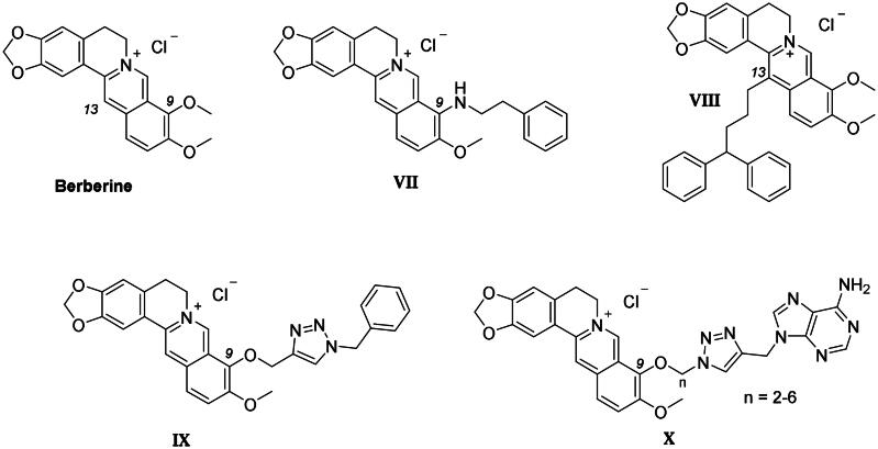
A novel class of compounds designed to hit two anti-tumour targets, G-quadruplex structures and human carbonic anhydrases (hCAs) IX and XII is proposed. The induction/stabilisation of G-quadruplex structures by small molecules has emerged as an anticancer strategy, disrupting telomere maintenance and reducing oncogene expression. hCAs IX and XII are well-established anti-tumour targets, upregulated in many hypoxic tumours and contributing to metastasis. The ligands reported feature a berberine G-quadruplex stabiliser scaffold connected to a moiety inhibiting hCAs IX and XII. In vitro experiments showed that our compounds selectively stabilise G-quadruplex structures and inhibit hCAs IX and XII. The crystal structure of a telomeric G-quadruplex in complex with one of these ligands was obtained, shedding light on the ligand/target interaction mode. The most promising ligands showed significant cytotoxicity against CA IX-positive HeLa cancer cells in hypoxia, and the ability to stabilise G-quadruplexes within tumour cells.

Keywords: G-Quadruplex; carbonic anhydrase; multitarget-directed ligands; tumours.


Conflict of interest statement

All authors except CTS report no conflict of interest. CT Supuran is Editor-in-Chief of the Journal of Enzyme Inhibition and Medicinal Chemistry. He was not involved in the assessment, peer review, or decision-making process of this paper. The authors have no relevant affiliations of financial involvement with any organisation or entity with a financial interest in or financial conflict with the subject matter or materials discussed in the manuscript. This includes employment, consultancies, honoraria, stock ownership or options, expert testimony, grants or patents received or pending, or royalties.

Figures

None
Graphical abstract
Figure 1.
Figure 1.
Rationale for the development of GQ ligand–CAI derivatives as innovative chemotherapeutics to disrupt tumour cell growth at multiple levels.
Figure 2.
Figure 2.
Chemical structure of the carbonic anhydrase inhibitors discussed here.
Figure 3.
Figure 3.
Drug design and general structure of the berberine-CAI derivatives proposed here.
Figure 4.
Figure 4.
Chemical structure of the berberine derivatives as GQ ligands discussed here.
Scheme 1.
Scheme 1.
Synthetic path to yield berberrubine 2 from berberine hydrochloride 1.
Scheme 2.
Scheme 2.
Synthetic pathway to yield the berberine azide (3–6) and alkyne (7, 8) intermediates.
Scheme 3.
Scheme 3.
Synthesis of benzenesulfonamide azide (11, 12) and alkyne (14, 16) intermediates.
Scheme 4.
Scheme 4.
Synthesis of coumarin azide (18–20 and 25) and alkyne (21–23) derivatives.
Scheme 5.
Scheme 5.
Synthesis of derivatives 26–39 of type 1 which include the triazole N1 berberine oriented. CuAAC: (i) CuSO4 and sodium ascorbate or (ii) Cu nanosized and tetramethylammonium chloride in tBuOH/H2O at 40 °C.
Scheme 6.
Scheme 6.
Synthesis of hybrid derivatives 40–45 of type 2 which include the triazole N1 CAI oriented. CuAAC: (i) CuSO4 and sodium ascorbate or Cu nanosized and tetramethylammonium chloride in tBuOH/H2O at 40 °C.
Scheme 7.
Scheme 7.
Synthesis of berberine derivative 49 bearing an aliphatic primary sulphonamide moiety.
Figure 5.
Figure 5.
(A) Monomolecular parallel GQs arranged in columns growing along the c axis. (B) OMIT electron density map contoured at 2σ level of S-3,2 (29) and skeleton of the ligand in the two disordered orientations—gray (Y orientation) and black (Z orientation); (C) and (D) interaction detail with GQ: (C) Y orientation; (D) Z orientation (T11 and T12 nucleic base atoms not shown). Final coordinates and structure factors (Table S3, Supporting Information) have been deposited with the Protein Data Bank (PDB accession number 7PNL).
Figure 6.
Figure 6.
(A) Representative fields showing G4 foci formation detected by immunofluorescence in HeLa cells treated for 24 h with 35.0 µM S-4,0, 40.0 µM C-4,2, and 37.5 µM C-5,2 or an equivalent amount of vehicle (0.1% DMSO). As a positive control, cells were treated with 2 µM RHPS4 (a well-established GQ ligand) for 24 h. Scale bar: 10 µm. Upper panels: the merged channels of BG4-stained GQ structures (magenta) and Hoechst-counterstained nuclei (grey) are reported. Lower panels: enlargements from the pictures in the upper panels. (B) Quantitative analysis of the fluorescence intensity of the total number of GQ foci. An average of 50 cells were screened for each condition and the results are expressed as a fold change of fluorescence intensity (anti-GQ signal) over the negative control (DMSO). Histograms show the mean ± SD of three independent experiments. The statistical significance was calculated using a one-way ANOVA test on GraphPad Prism 8.0.2 (ns: not significant, **: p < 0.01).
Figure 7.
Figure 7.
(A) Representative fields showing G4 foci formation detected by immunofluorescence in HeLa cells pre-treated with 50 μM cobalt (II) chloride hexahydrate for 60 h to induce hypoxia and then exposed to 1 µM C-4,2 or vehicle (0.1% DMSO) for the following 12 h, still in the presence of CoCl2. DMSO-treated normoxic HeLa cells are also shown. Scale bar: 10 µm. Upper panels: the merged channels of BG4-stained GQ structures (magenta) and Hoechst-counterstained nuclei (grey) are reported. Lower panels: enlargements from the pictures in the upper panels. (B) Quantification of the cellular BG4 foci in DMSO-treated normoxic HeLa cells, DMSO-treated hypoxic HeLa cells, and C-4,2-treated hypoxic HeLa cells. An average of 50 cells were screened for each condition. The number of GQ foci has been normalised to the mean of the corresponding control sample (DMSO). Histograms show the mean ± SD of three independent experiments. The statistical significance was calculated using a one-way ANOVA test on GraphPad Prism 8.0.2 (**: p < 0.01, ****: p < 0.0001).

References

    1. Bochman ML, Paeschke K, Zakian VA.. DNA secondary structures: stability and function of G-quadruplex structures. Nat Rev Genet. 2012;13(11):770–780. - PMC - PubMed
    1. Spiegel J, Adhikari S, Balasubramanian S.. The structure and function of DNA G-Quadruplexes. Trends Chem. 2020;2(2):123–136. - PMC - PubMed
    1. Teng FY, Jiang ZZ, Guo M, Tan XZ, Chen F, Xi XG, Xu Y, Quadruplex G.. DNA: a novel target for drug design. Cell Mol. Life Sci. 2021; 78:6557–6583. - PMC - PubMed
    1. Marsico G, Chambers VS, Sahakyan AB, McCauley P, Boutell JM, Antonio MD, Balasubramanian S.. Whole genome experimental maps of DNA G-quadruplexes in multiple species. Nucleic Acids Res. 2019;47(8):3862–3874. - PMC - PubMed
    1. Verma A, Yadav VK, Basundra R, Kumar A, Chowdhury S.. Evidence of genome-wide G4 DNA-mediated gene expression in human cancer cells. Nucleic Acids Res. 2009;37(13):4194–4204. - PMC - PubMed

MeSH terms