. 2024 Jun 20;187(13):3194-3219.
doi: 10.1016/j.cell.2024.05.027.

Toward developing human organs via embryo models and chimeras

Affiliations

Toward developing human organs via embryo models and chimeras

Jun Wu et al. Cell. .

Abstract

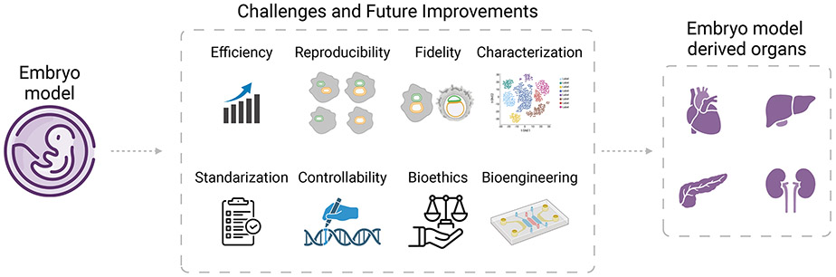
Developing functional organs from stem cells remains a challenging goal in regenerative medicine. Existing methodologies, such as tissue engineering, bioprinting, and organoids, only offer partial solutions. This perspective focuses on two promising approaches emerging for engineering human organs from stem cells: stem cell-based embryo models and interspecies organogenesis. Both approaches exploit the premise of guiding stem cells to mimic natural development. We begin by summarizing what is known about early human development as a blueprint for recapitulating organogenesis in both embryo models and interspecies chimeras. The latest advances in both fields are discussed before highlighting the technological and knowledge gaps to be addressed before the goal of developing human organs could be achieved using the two approaches. We conclude by discussing challenges facing embryo modeling and interspecies organogenesis and outlining future prospects for advancing both fields toward the generation of human tissues and organs for basic research and translational applications.

Keywords: blastocyst complementation; extraembryonic endoderm cells; hypoblast stem cells; interspecies chimeras; interspecies organogenesis; organ engineering; pluripotent stem cells; stem cell-based embryo models; trophoblast stem cells.


Conflict of interest statement

Declaration of interests The authors declare no competing interests.

Figures

Figure 1.
Figure 1.. A schematic overview of two innovative strategies for creating human organs from pluripotent stem cells (PSCs).
(A) In vitro generation of organ via stem cell-derived embryo models. Such models, mimicking the initial stages of embryonic development, could potentially be advanced through cultivation in bioreactors and other ex vivo methods to nurture the growth of organ primordia into sizable, functional organs. (B) In vivo generation of organ via interspecies chimeras. Chimera competent human PSCs can be injected into animal embryos that lack essential genes for organ formation. This process facilitates the production of human organs in animal within the animal host as it undergoes its natural developmental processes.
Figure 2.
Figure 2.. A summary of human stem cell derived embryo models and the developmental stages in vivo they represent.
(A) During early human development, the embryo develops from a zygote and proceeds through specific recognizable stages of (i) pre-implantation, (ii) peri-implantation, and (iii) organogenesis. During this process, cells in the human embryo differentiate and diversify while acting in a coordinated fashion to enact tissue morphogenesis and patterning programs to shape the body plan. (B) PSC-derived human embryo models are generated to mimic various in vivo developmental stages. (C) Chimera competent human PSCs are introduced into pre-implantation blastocysts or early post-implantation embryos of host animals. This process is designed to produce human-animal chimeras, along with tissues and organs enriched with humanb cells.
Figure 3.
Figure 3.. Challenges and future improvements in utilizing stem cell based embryo models for organ engineering.
Figure 4.
Figure 4.. Xenogeneic barriers.
(A) A notable competitive interaction was identified between primed PSCs from evolutionarily distant species (e.g., human-mouse, human-cow, human-rat) based on interspecies PSC co-culture experiments. The elimination of the “loser” cells (e.g., human PSCs when co-cultured with mouse epiblast stem cells [EpiSCs]) is governed by the NF-κB signaling pathway. Disabling the P65 gene (also known as RELA) or an upstream regulator (MYD88) of the NF-κB complex in human cells can overcome this competition, thus enhancing the survival and chimerism of human cells within early mouse embryos. In “winner” cells (e.g., mouse EpiSCs), the retinoic acid-inducible gene I (RIG-I)-like receptor (RLR) signaling pathway, an RNA sensor, appears to play an important role in determining the outcome of competitive interactions between co-cultured mouse and human PSCs. (B) Incompatibilities in cell adhesion, particularly among primed PSCs from different species, present a significant xenogeneic barrier. Employing 3D interspecies PSC co-cultures offers a valuable in vitro method to investigate this barrier. A notable approach to overcoming this issue involves engineering synthetic cell adhesion. This can potentially be achieved by leveraging membrane-anchored nanobody-antigen interactions to facilitate cell adhesion compatibility between PSCs from different species. (C) Heterochrony represents another xenogeneic barrier. Matching developmental timing of the donor PSCs with host embryos is an important consideration for the successful generation of intra- and inter-species chimeras. (D) Genomic evolution leading to mismatched ligand-receptor pairs poses another xenogeneic challenge.

Similar articles

Cited by

References

    1. Anand RP, Layer JV, Heja D, Hirose T, Lassiter G, Firl DJ, Paragas VB, Akkad A, Chhangawala S, Colvin RB, et al. (2023). Design and testing of a humanized porcine donor for xenotransplantation. Nature 622, 393–401. 10.1038/s41586-023-06594-4. - DOI - PMC - PubMed
    1. Rafat M, Jabbarvand M, Sharma N, Xeroudaki M, Tabe S, Omrani R, Thangavelu M, Mukwaya A, Fagerholm P, Lennikov A, et al. (2023). Bioengineered corneal tissue for minimally invasive vision restoration in advanced keratoconus in two clinical cohorts. Nat Biotechnol 41, 70–81. 10.1038/s41587-022-01408-w. - DOI - PMC - PubMed
    1. Hirsch T, Rothoeft T, Teig N, Bauer JW, Pellegrini G, De Rosa L, Scaglione D, Reichelt J, Klausegger A, Kneisz D, et al. (2017). Regeneration of the entire human epidermis using transgenic stem cells. Nature 551, 327–332. 10.1038/nature24487. - DOI - PMC - PubMed
    1. Ott HC, Matthiesen TS, Goh SK, Black LD, Kren SM, Netoff TI, and Taylor DA (2008). Perfusion-decellularized matrix: using nature's platform to engineer a bioartificial heart. Nat Med 14, 213–221. 10.1038/nm1684. - DOI - PubMed
    1. Lee A, Hudson AR, Shiwarski DJ, Tashman JW, Hinton TJ, Yerneni S, Bliley JM, Campbell PG, and Feinberg AW (2019). 3D bioprinting of collagen to rebuild components of the human heart. Science 365, 482–487. 10.1126/science.aav9051. - DOI - PubMed

LinkOut - more resources