. 2024 Jun 21;14(1):14361.
doi: 10.1038/s41598-024-65096-z.

MiR-503-5p alleviates peripheral neuropathy-induced neuropathic pain in T2DM mice by regulating SEPT9 to inhibit astrocyte activation

Affiliations

MiR-503-5p alleviates peripheral neuropathy-induced neuropathic pain in T2DM mice by regulating SEPT9 to inhibit astrocyte activation

Yuqing Guo et al. Sci Rep. .

Abstract

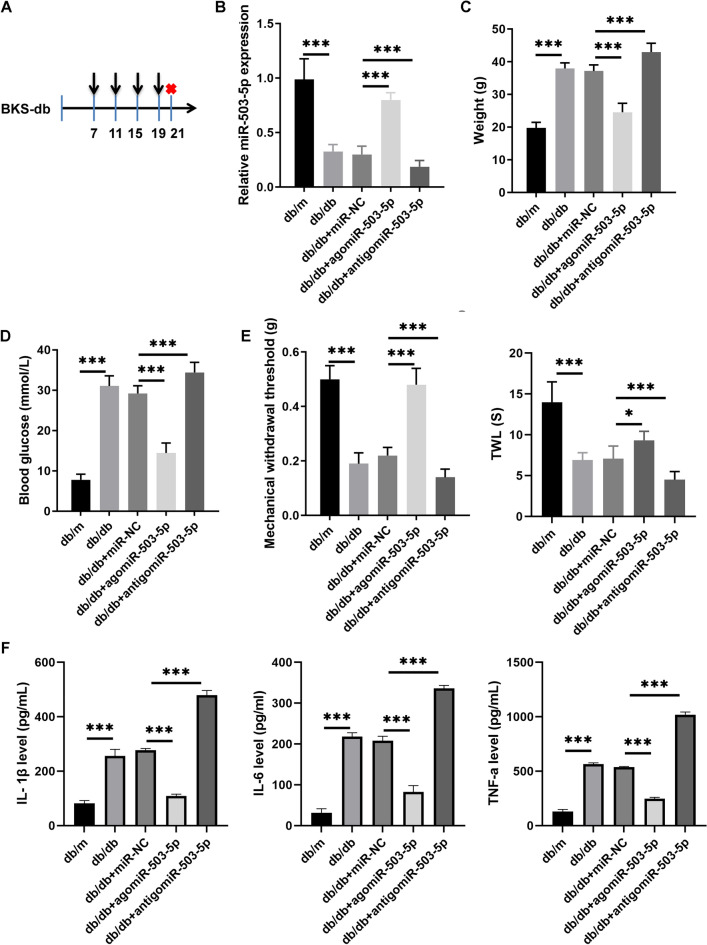
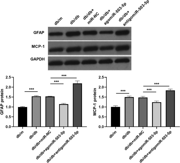
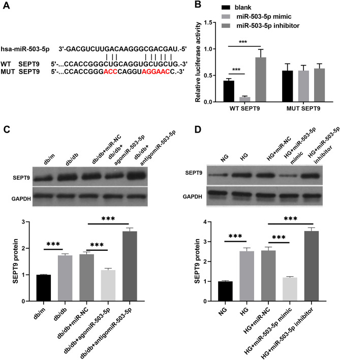
Diabetic peripheral neuropathy (DPN) is a common complication of type 2 diabetes mellitus (T2DM) that causes peripheral and autonomic nervous system dysfunction. Dysregulation of miRNAs plays a crucial role in DPN development. However, the role of miR-503-5p in DPN remains unknown. Herein, T2DM mice (db/db) were used as a DPN model in vivo, and astrocytes isolated from db/db mice were induced with high glucose levels as a DPN model in vitro. MiR-503-5p expression was analyzed using qRT-PCR. GFAP, MCP-1, and SEPT9 protein levels were analyzed using western blotting and immunofluorescence. Luciferase assays were performed to investigate the interaction between miR-503-5p and SEPT9. We found that miR-503-5p expression decreased in the spinal cord of DPN model mice and astrocytes treated with high glucose (HG). The db/db mice displayed higher body weight and blood glucose, lower mechanical withdrawal threshold and thermal withdrawal latency, and higher GFAP and MCP-1 protein levels than db/m mice. However, tail vein injection of agomiR-503-5p remarkably reversed these parameters, whereas antigomiR-503-5p enhanced them. HG markedly facilitated GFAP and MCP-1 protein expression in astrocytes, whereas miR-503-5p mimic or inhibitor transfection markedly blocked or elevated GFAP and MCP-1 protein expression, respectively, in astrocytes with HG. SEPT9 was a target of miR-503-5p. In addition, SEPT9 protein levels were found to be elevated in db/db mice and astrocytes treated with HG. Treatment with agomiR-503-5p and miR-503-5p mimic was able to reduce SEPT9 protein levels, whereas treatment with antigomiR-503-5p and miR-503-5p inhibitor led to inhibition of the protein. Furthermore, SEPT9 overexpression suppressed the depressing effect of miR-503-5p overexpression in astrocytes subjected to HG doses. In conclusion, miR-503-5p was found to alleviate peripheral neuropathy-induced neuropathic pain in T2DM mice by regulating SEPT9 expression.

Keywords: Astrocytes; Diabetic peripheral neuropathy; SEPT9; T2DM mice; miR-503-5p.


Conflict of interest statement

The authors declare no competing interests.

Figures

Figure 1
Figure 1
miR-503-5p overexpression reduces the peripheral neuropathy in mice in the Diabetic peripheral neuropathy (DPN) model in vivo. (A) Flowchart of the mouse experiment: Mice are adaptively fed for one week before being injected with miRNA, once every four days for a total of four times. Mice are euthanized two days after the final injection. (B) The miR-503-5p expression in the spinal cord ofdb/db mice injected with agomiR-503-5p, antigomiR-503-5p, or miR-NC (miRNA negative control) was detected with qRT-PCR. (C and D) The body weight with blood glucose levels in db/db mice was assessed after injections with agomiR-503-5p, antigomiR-503-5p, or miR-NC. (E) db/db mice injected with agomiR-503-5p, antigomiR-503-5p, or miR-NC were assessed by mechanical withdrawal threshold (MWT) and thermal withdrawal latency (TWL). (F) The IL-1β, IL-6, and TNF-α levels in serum were measured by ELISA. (*P < 0.05 and ***P < 0.001).
Figure 2
Figure 2
The levels of GFAP and MCP-1 proteins in the spinal cord were regulated by miR-503-5p. The GFAP and MCP-1 protein levels in the spinal cord of db/db mice injected with agomiR-503-5p, antigomiR-503-5p, or miR-NC (negative control) were measured using western blot. Full-length blots are presented in Supplementary Figure 1A (*** P < 0.001).
Figure 3
Figure 3
Overexpression and silencing of miR-503-5p influenced the activation of astrocytes treated with high glucose (HG). (A) The efficiency of transfection with miR-503-5p mimic or inhibitor in astrocytes exposed to HG was verified using qRT-PCR. (B) The levels of IL-1β, IL-6, and TNF-α in the culture supernatant were quantified by ELISA. (C) The expression of GFAP and MCP-1 proteins in astrocytes treated with HG following transfection with either miR-503-5p mimic or miR-503-5p inhibitor was evaluated using western blot. Full-length blots are presented in Supplementary Figure 1B. (D) The expression of GFAP and MCP-1 proteins in astrocytes treated with HG following transfection with miR-503-5p mimic or miR-503-5p inhibitor was assessed using immunofluorescence (***P < 0.001). For the HG treatment, an additional 30 mM glucose was added to the normal culture medium. NG: Astrocytes were cultured at normal glucose concentrations (5.5 mM).
Figure 4
Figure 4
SEPT9 was a target of miR-503-5p. (A)The predicted binding site between miR-503-5p and SEPT9. (B) The targeted relationship between miR-503-5p and SEPT9 was validated using a dual-luciferase reporter assay. (C and D) The expression of SEPT9 protein in the spinal cord of db/db mice treated with agomiR-503-5p/antigomiR-503-5p or astrocytes with HG and transfected with miR-503-5p mimic/miR-503-5p inhibitor, was measured using western blot. Full-length blots are presented in Supplementary Figure 1C and D. (***P < 0.001).WT: Wild type; MUT: Mutant type 3.
Figure 5
Figure 5
Overexpression of SEPT9 was able to reverse the suppressive effects of miR-503-5p overexpression in astrocytes treated with HG. (A) The expression of SEPT9, GFAP, and MCP-1 proteins in astrocytes with HG, following co-transfection with the miR-503-5p mimic and ov-SEPT9, was determined using western blot analysis. Full-length blots are displayed in Supplementary Fig.1E. (B) The expression of GFAP and MCP-1 proteins in astrocytes with HG, after co-transfection with the miR-503-5p mimic and ov-SEPT9, was evaluated using immunofluorescence. (C) The levels of IL-1β, IL-6, and TNF-α in the culture supernatant were quantified using ELISA after co-transfection with the miR-503-5p mimic and ov-SEPT9. (***P < 0.001).

References

    1. Cloete L. Diabetes mellitus: an overview of the types, symptoms, complications and management. Nurs. Stand. 2022;37(1):61–66. doi: 10.7748/ns.2021.e11709. - DOI - PubMed
    1. Yang K, Wang Y, Li YW, Chen YG, Xing N, Lin HB, Zhou P, Yu XP. Progress in the treatment of diabetic peripheral neuropathy. Biomed. Pharmacother. 2022;148:112717. doi: 10.1016/j.biopha.2022.112717. - DOI - PubMed
    1. Jeyam A, McGurnaghan SJ, Blackbourn LAK, McKnight JM, Green F, Collier A, McKeigue PM, Colhoun HM. Diabetic neuropathy is a substantial burden in people with type 1 diabetes and is strongly associated with socioeconomic disadvantage: a population-representative study from Scotland. Diabetes Care. 2020;43(4):734–742. doi: 10.2337/dc19-1582. - DOI - PubMed
    1. Li GZ, Hu YH, Lu YN, Yang QY, Fu D, Chen F, Li YM. CaMKII and Ca(V)3.2 T-type calcium channel mediate Connexin-43-dependent inflammation by activating astrocytes in vincristine-induced neuropathic pain. Cell Biol. Toxicol. 2023;39(3):679–702. doi: 10.1007/s10565-021-09631-y. - DOI - PubMed
    1. Kasimu A, Apizi X, Talifujiang D, Ma X, Fang L, Zhou X. miR-125a-5p in astrocytes attenuates peripheral neuropathy in type 2 diabetic mice through targeting TRAF6. Endocrinol. Diabetes Nutr. (Engl Ed) 2022;69(1):43–51. - PubMed