. 2024 Sep 3;65(8):1285-1297.
doi: 10.1093/pcp/pcae062.

A Comprehensive Study of Light Quality Acclimation in Synechocystis Sp. PCC 6803

Affiliations

A Comprehensive Study of Light Quality Acclimation in Synechocystis Sp. PCC 6803

Tomáš Zavřel et al. Plant Cell Physiol. .

Erratum in

Abstract

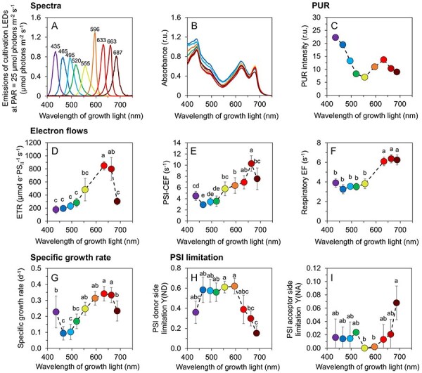
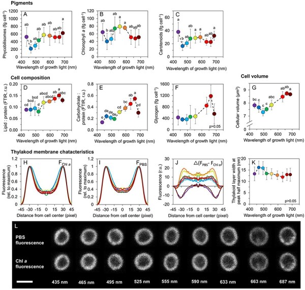
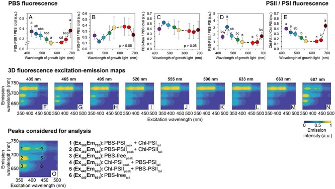
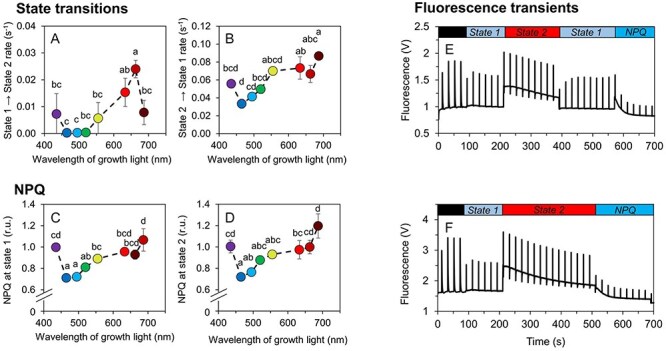
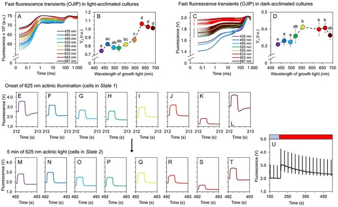
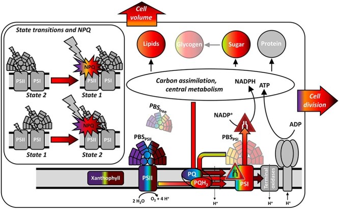
Cyanobacteria play a key role in primary production in both oceans and fresh waters and hold great potential for sustainable production of a large number of commodities. During their life, cyanobacteria cells need to acclimate to a multitude of challenges, including shifts in intensity and quality of incident light. Despite our increasing understanding of metabolic regulation under various light regimes, detailed insight into fitness advantages and limitations under shifting light quality remains underexplored. Here, we study photo-physiological acclimation in the cyanobacterium Synechocystis sp. PCC 6803 throughout the photosynthetically active radiation (PAR) range. Using light emitting diodes (LEDs) with qualitatively different narrow spectra, we describe wavelength dependence of light capture, electron transport and energy transduction to main cellular pools. In addition, we describe processes that fine-tune light capture, such as state transitions, or the efficiency of energy transfer from phycobilisomes to photosystems (PS). We show that growth was the most limited under blue light due to inefficient light harvesting, and that many cellular processes are tightly linked to the redox state of the plastoquinone (PQ) pool, which was the most reduced under red light. The PSI-to-PSII ratio was low under blue photons, however, it was not the main growth-limiting factor, since it was even more reduced under violet and near far-red lights, where Synechocystis grew faster compared to blue light. Our results provide insight into the spectral dependence of phototrophic growth and can provide the foundation for future studies of molecular mechanisms underlying light acclimation in cyanobacteria, leading to light optimization in controlled cultivations.

Keywords: Cyanobacteria; Light harvesting; Light quality; Photomorphogenesis; Photosynthesis; State transitions.


Figures

Fig. 1
Fig. 1
Growth rate and electron flows in Synechocystis cells grown under narrow-band cultivation lights. Emission spectra of cultivation LEDs at PAR of 25 µmol photons m−2 s−1 (A), baseline-corrected absorption spectra of Synechocystis cultures (B), photosynthesis-usable radiation, PUR (C; see Eq. (8) for details), electron transport rate through PSII (ETR, D), cyclic electron flow around PSI (PSI-CEF, E), respiratory electron flow (REF, F), specific growth rate (G) and donor side [Y(ND), H] and acceptor side [Y(NA), I] limitation of PSI in Synechocystis cells as cultivated under narrow-band LEDs. Both REF and PSI-CEF were determined from P700+ re-reduction kinetics, as measured after AL saturation pulse (635 nm, 100 ms). Both REF and PSI-CEF are expressed as 1/τ; the time constant τ was obtained by fitting the exponentially decaying absorption signal at 830 nm. The values in panels B and D–I represent mean ± SD (n = 3–6). Values in panel B (n = 3) are shown without error bars for clarity. The letters above the symbols in panels D–I indicate statistically significant differences within each parameter (P < 0.05).
Fig. 2
Fig. 2
Macromolecular composition and morphology of Synechocystis cells grown under narrow-band cultivation lights. Content of phycobilisomes (A), chlorophyll a (B) and carotenoids (C) in Synechocystis cells, lipid (D) and carbohydrate (E) relative to protein, and cellular content of glycogen (F). Cell volume (G). Parameters derived from confocal microscopy imaging: autofluorescence profiles of chlorophyll a (FChl a, H) and phycobilisomes (FPBS) I) across Synechocystis cells, difference between FPBS and FChl a (J) and thickness of the thylakoid membrane, determined from FChl a (H) as half maximum of the fluorescence peaks (K). All values represent mean ± SD [n = 3–5 (AG)/58–113 (HK)]. Error bars in panels HJ are omitted for clarity. Confocal microscopy images of representative Synechocystis cells as cultivated under narrow-spectrum lights are shown in panel L (scale bar: 2 µm). The letters above the symbols in panels AG and K indicate statistically significant differences within each parameter (P < 0.05).
Fig. 3
Fig. 3
77 K fluorescence excitation–emission maps and derived parameters under narrow-band cultivation lights. Fluorescence of phycobilisomes functionally attached to PSII (PBS–PSII, A), to PSI (PBS–PSI, B) and of phycobilisomes functionally uncoupled from both photosystems (PBS–free, C). PBS–PSII/PBS–PSI (D) and PSII/PSI ratio (E); excitation–emission maps of Synechocystis cultures cultivated under narrow-band cultivation LEDs (FN). The maps represent standardized averages (n = 3); error intervals are not shown for clarity. Representative fluorescence map with wavelengths used for analysis (O). For further details on the 77 K spectra processing see ‘Materials and Methods’ section (Eq. 9–16). The values in panels AE represent mean ± SD (n = 4–7). The letters above the symbols in panels AE indicate statistically significant differences within each parameter.
Fig. 4
Fig. 4
State transitions and NPQ in Synechocystis under narrow-band cultivation lights. Assessment of State 1 → State 2 (A) and State 2 → State 1 (B) transition rates as induced by weak blue (480 nm, 80 µmol photons m−2 s−1) and weak red (625 nm, 50 µmol photons m−2 s−1) AL, respectively. Qualitative assessment of NPQ induced by strong blue AL (1 800 µmol photons m−2 s−1) in State 1 (C) and State 2 (D). Example PAM protocol used for the estimation of State 2 → State 1 rate and NPQ in State 1 is shown in panel E, example protocol for the estimation of State 1 → State 2 rate and NPQ in State 2 is shown in panel F. Prior to each measurement, Synechocystis cells were dark-acclimated for 15 min. Fluorescence recordings from all cultivation lights are summarized in Supplementary Figures S9–S10. The values in panels AB and CD represent mean ± SD (n = 3), the letters above the symbols indicate statistically significant differences within each parameter (P < 0.05). We note that the amplitude of Fm’ under 480 nm AL was underestimated in MC-PAM (see Supplementary Figure S13 for details).
Fig. 5
Fig. 5
Redox state of the PQ pool and transient fluorescence drops after saturation pulses. Redox state of the PQ-pool was determined based on the relative fluorescence level at J point, VJ (B, D) derived from fast fluorescence kinetics (OJIP curves) in light-acclimated (A) and dark-acclimated Synechocystis cultures (C). Fluorescence traces during saturation pulses at the onset of 625 nm AL and after 5 min of 625 nm AL are summarized in panels EL and MT, respectively; an example pattern of a slow fluorescence kinetic trace is shown in panel U. All values represent means (n = 3), error bars in AD represent standard deviations. Error bars in panels ET are not shown for clarity. Fluorescence recordings from all cultivation lights are summarized in Supplementary Figures S9–S10. The letters above the symbols in panels B and D indicate statistically significant differences within each parameter (P < 0.05).
Fig. 6
Fig. 6
Summary of wavelength-dependent shifts in Synechocystis as found in this study. Individual processes and components are color-coded according to the narrow-band irradiance under which the corresponding processes/components were upregulated. Grey regions represent cellular components that were not determined (terminal oxidases, ATPase) or that are depicted only for illustrative purpose (insert showing state transition). Components/processes dependent on the cultivation wavelength are marked without transparency; glycogen, PBS–PSI and PBS–free are marked partially transparent as these componenets were identified independent of light quality. Colored arrows represent measured electron flows, black arrows represent pathways of which only the final sinks were determined.

Similar articles

Cited by

References

    1. Ajlani G. and Vernotte C. (1998) Construction and characterization of a phycobiliprotein-less mutant of Synechocystis sp. PCC 6803. Plant Mol. Biol. 37: 577–580. - PubMed
    1. Bečková M., Sobotka R. and Komenda J. (2022) Photosystem II antenna modules CP43 and CP47 do not form a stable ‘no reaction centre complex’ in the cyanobacterium Synechocystis sp. PCC 6803. Photosynth. Res. 152: 363–371. - PMC - PubMed
    1. Bernát G., Steinbach G., Kaňa R., Govindjee, Misra A.N., Prašil O. and Prašil O. (2018) On the origin of the slow M–T chlorophyll a fluorescence decline in cyanobacteria: interplay of short-term light-responses. Photosynth. Res. 136: 183–198. - PubMed
    1. Bernát G., Zavřel T., Kotabová E., Kovács L., Steinbach G., Vörös L., et al. (2021) Photomorphogenesis in the picocyanobacterium Cyanobium gracile includes increased phycobilisome abundance under blue light, phycobilisome decoupling under near far-red light, and wavelength-specific photoprotective strategies. Front Plant Sci. 12: 1–16. - PMC - PubMed
    1. Calzadilla P.I. and Kirilovsky D. (2020) Revisiting cyanobacterial state transitions. Photochem. Photobiol. Sci. 19: 585–603. - PubMed

MeSH terms

Substances

LinkOut - more resources