. 2024 Jul 2;18(26):16674-16683.
doi: 10.1021/acsnano.4c01640. Epub 2024 Jun 22.

Enhanced Prostate-specific Membrane Antigen Targeting by Precision Control of DNA Scaffolded Nanoparticle Ligand Presentation

Affiliations

Enhanced Prostate-specific Membrane Antigen Targeting by Precision Control of DNA Scaffolded Nanoparticle Ligand Presentation

Deblin Jana et al. ACS Nano. .

Abstract

Targeted nanoparticles have been extensively explored for their ability to deliver their payload to a selective cell population while reducing off-target side effects. The design of actively targeted nanoparticles requires the grafting of a ligand that specifically binds to a highly expressed receptor on the surface of the targeted cell population. Optimizing the interactions between the targeting ligand and the receptor can maximize the cellular uptake of the nanoparticles and subsequently improve their activity. Here, we evaluated how the density and presentation of the targeting ligands dictate the cellular uptake of nanoparticles. To do so, we used a DNA-scaffolded PLGA nanoparticle system to achieve efficient and tunable ligand conjugation. A prostate-specific membrane antigen (PSMA) expressing a prostate cancer cell line was used as a model. The density and presentation of PSMA targeting ligand ACUPA were precisely tuned on the DNA-scaffolded nanoparticle surface, and their impact on cellular uptake was evaluated. It was found that matching the ligand density with the cell receptor density achieved the maximum cellular uptake and specificity. Furthermore, DNA hybridization-mediated targeting chain rigidity of the DNA-scaffolded nanoparticle offered ∼3 times higher cellular uptake compared to the ACUPA-terminated PLGA nanoparticle. Our findings also indicated a ∼ 3.7-fold reduction in the cellular uptake for the DNA hybridization of the non-targeting chain. We showed that nanoparticle uptake is energy-dependent and follows a clathrin-mediated pathway. Finally, we validated the preferential tumor targeting of the nanoparticles in a bilateral tumor xenograft model. Our results provide a rational guideline for designing actively targeted nanoparticles and highlight the application of DNA-scaffolded nanoparticles as an efficient active targeting platform.

Keywords: DNA; cell uptake; ligand density; nanoparticles; surface chemistry.


Conflict of interest statement

The authors declare no competing financial interest.

Figures

Figure 1
Figure 1
Fabrication and characterization of ACUPA-functionalized DNA-scaffolded PLGA nanoparticle. (a) Schematic diagram of the factors associated with the receptor-binding efficiency of DNA-scaffolded PLGA nanoparticles. (b) Fabrication of PLGA nanoparticles with surface DNA scaffolds. (c) Absorption spectra of nanoparticle concentration as measured by OD at 550 nm (linear range of 0.1–1 OD). (d) Single-particle tracking (SPT) of the nanoparticle at 0.0001 OD. The particle size is 190 nm, and the concentration is 2.8 × 10¹⁰ particles per mL for 1 OD. (e) Functionalization of DNA-scaffolded nanoparticles with ACUPA. ACUPA-azide was conjugated to the complementary oligo and hybridized onto the particle surface. (f) Surface-loading quantity of ACUPA. The grafting ratio is based on the maximal loading capacity of the scaffolded DNA strand (Supplementary Table 3). ACUPA/nanoparticle numbers are calculated using the SPT and absorption data.
Figure 2
Figure 2
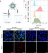
Ligand presentation-dependent nanoparticle uptake. (a) Schematic illustration of cellular internalization. The PSMA (+) PC3-pip cells were incubated with ACUPA-functionalized DNA-scaffolded nanoparticles at a final concentration of 0.1 OD for 2 h. (b) CLSM images of nanoparticle uptake in PC3-pip cells (scale bar = 20 μm). Red: AF647. Green: PE-anti-PSMA. Hoechst 33342. (c) Schematic illustration of receptor density and ligand density relationship on nanoparticle uptake in accordance with the confocal data. (d) Maximal intensity projection of nanoparticles. Red: AF647. Green: PE-anti-PSMA. Hoechst 33342. (c) Schematic illustration of receptor density and ligand density relationship on nanoparticle uptake in accordance with the confocal data. (d) Maximal intensity projection of nanoparticles. Red: AF647. Green: PE-anti-PSMA. Hoechst 33342. (e) Schematic illustration of the ligand presentation difference between ACUPA-functionalized DNA-scaffolded nanoparticles and ACUPA-functionalized PLGA–PEG nanoparticles. (f) 3D CLSM images of nanoparticle uptake in PC3-pip cells. Red: AF647. Green: PE-anti-PSMA. Hoechst 33342.
Figure 3
Figure 3
Non-targeting ligand-dependent nanoparticle uptake. (a) Schematic illustration of mixed DNA-scaffolded nanoparticle preparation with ratiometric control. The nanoparticles were functionalized with ACUPA with G-DNA, and the non-targeting B-DNA were either hybridized or non-hybridized. (b) DLS analysis of G1-B9. (c) Flow cytometry analysis of the nanoparticles G1-A-B9 and G1-A-B9-compB. (d) CLSM images of nanoparticle uptake in PC3-pip cells (scale bar: 20 μm). Red: AF647. Green: PE-anti-PSMA. Blue: Hoechst 33342. (e) Schematic illustration showing hybridization of non-targeting DNA chains can impede cellular internalization.
Figure 4
Figure 4
Selectivity of the G1-A-B9 nanoparticle. (a) Schematic illustration of nanoparticle cellular internalization in PSMA(+) PC3-pip and PSMA(−) PC3-flu cell lines. (b) Flow cytometry analysis of G1-A-B9 nanoparticle uptake in PC3-pip and PC3-flu cells. (c) CLSM images of G1-A-B9 nanoparticle uptake in PC3-pip and PC3-flu cells (scale bar = 20 μm). Red: AF647. Green: PE-anti-PSMA. Blue: Hoechst 33342.
Figure 5
Figure 5
Intracellular uptake pathway. (a) Flow cytometry images of G1-A-B9 nanoparticle uptake at 37 and 4 °C. (b) Flow cytometry analysis of nanoparticle uptake in the presence or absence of endocytosis inhibitors. (c) CLSM images of nanoparticle uptake in the presence or absence of endocytosis inhibitors (scale bar = 20 μm). Red: AF647. Green: PE-anti-PSMA. Blue: Hoechst 33342. (d) Flow cytometry analysis of time-dependent nanoparticle uptake. (e) Schematic of the nanoparticle intracellular uptake through an energy-dependent and clathrin-mediated endocytosis.
Figure 6
Figure 6
In vivo tumor accumulation. (a) Schematic representation of the experimental design for in vivo evaluation of the DiR-tagged nanoparticles in mice bearing dual xenografts of PSMA(+) PC3-pip (left flank) and PSMA(–) PC3-flu (right flank). (b) Ex vivo fluorescence imaging of tumor tissues at 48 h postinjection. Average radiant efficiency of tumors of two groups: (c) DNA-scaffolded nanoparticles and (d) PLGA–PEG nanoparticles. Data are represented as mean ± s.e.m. (n = 3 mice).

References

    1. Bhatia S. N.; Chen X.; Dobrovolskaia M. A.; Lammers T. Cancer nanomedicine. Nat. Rev. Cancer 2022, 22, 550–556. 10.1038/s41568-022-00496-9. - DOI - PMC - PubMed
    1. van der Meel R.; Sulheim E.; Shi Y.; Kiessling F.; Mulder W. J. M.; Lammers T. Smart cancer nanomedicine. Nat. Nanotechnol. 2019, 14, 1007–1017. 10.1038/s41565-019-0567-y. - DOI - PMC - PubMed
    1. Veiga N.; Diesendruck Y.; Peer D. Targeted nanomedicine: Lessons learned and future directions. J. Controlled Release 2023, 355, 446–457. 10.1016/j.jconrel.2023.02.010. - DOI - PubMed
    1. Li J.; Kataoka K. Chemo-physical Strategies to Advance the in Vivo Functionality of Targeted Nanomedicine: The Next Generation. J. Am. Chem. Soc. 2021, 143, 538–559. 10.1021/jacs.0c09029. - DOI - PubMed
    1. Csizmar C. M.; Petersburg J. R.; Perry T. J.; Rozumalski L.; Hackel B. J.; Wagner C. R. Multivalent Ligand Binding to Cell Membrane Antigens: Defining the Interplay of Affinity, Valency, and Expression Density. J. Am. Chem. Soc. 2019, 141, 251–261. 10.1021/jacs.8b09198. - DOI - PMC - PubMed

Publication types