Review
. 2024 Sep:75:103211.
doi: 10.1016/j.redox.2024.103211. Epub 2024 May 30.

Ferroptosis in health and disease

Carsten Berndt  1 Hamed Alborzinia  2 Vera Skafar Amen  3 Scott Ayton  4 Uladzimir Barayeu  5 Alexander Bartelt  6 Hülya Bayir  7 Christina M Bebber  8 Kivanc Birsoy  9 Jan P Böttcher  10 Simone Brabletz  11 Thomas Brabletz  11 Ashley R Brown  12 Bernhard Brüne  13 Giorgia Bulli  14 Alix Bruneau  15 Quan Chen  16 Gina M DeNicola  17 Tobias P Dick  18 Ayelén Distéfano  19 Scott J Dixon  20 Jan B Engler  21 Julia Esser-von Bieren  22 Maria Fedorova  23 José Pedro Friedmann Angeli  3 Manuel A Friese  21 Dominic C Fuhrmann  13 Ana J García-Sáez  24 Karolina Garbowicz  25 Magdalena Götz  26 Wei Gu  27 Linda Hammerich  15 Behrouz Hassannia  28 Xuejun Jiang  29 Aicha Jeridi  30 Yun Pyo Kang  31 Valerian E Kagan  32 David B Konrad  33 Stefan Kotschi  34 Peng Lei  35 Marlène Le Tertre  36 Sima Lev  37 Deguang Liang  29 Andreas Linkermann  38 Carolin Lohr  39 Svenja Lorenz  40 Tom Luedde  39 Axel Methner  41 Bernhard Michalke  42 Anna V Milton  33 Junxia Min  43 Eikan Mishima  40 Sebastian Müller  44 Hozumi Motohashi  45 Martina U Muckenthaler  36 Shohei Murakami  45 James A Olzmann  46 Gabriela Pagnussat  19 Zijan Pan  47 Thales Papagiannakopoulos  48 Lohans Pedrera Puentes  49 Derek A Pratt  50 Bettina Proneth  40 Lukas Ramsauer  10 Raphael Rodriguez  44 Yoshiro Saito  51 Felix Schmidt  41 Carina Schmitt  33 Almut Schulze  52 Annemarie Schwab  11 Anna Schwantes  13 Mariluz Soula  9 Benedikt Spitzlberger  53 Brent R Stockwell  54 Leonie Thewes  1 Oliver Thorn-Seshold  33 Shinya Toyokuni  55 Wulf Tonnus  56 Andreas Trumpp  57 Peter Vandenabeele  58 Tom Vanden Berghe  59 Vivek Venkataramani  60 Felix C E Vogel  52 Silvia von Karstedt  61 Fudi Wang  43 Frank Westermann  25 Chantal Wientjens  62 Christoph Wilhelm  62 Michele Wölk  23 Katherine Wu  48 Xin Yang  63 Fan Yu  16 Yilong Zou  64 Marcus Conrad  65
Affiliations
Review

Ferroptosis in health and disease

Carsten Berndt et al. Redox Biol. 2024 Sep.

Abstract

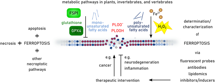
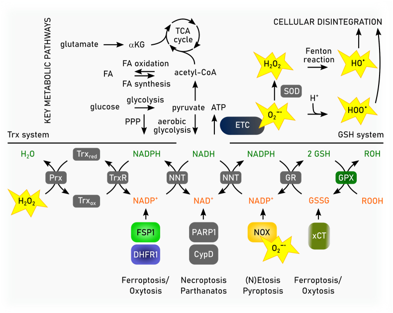
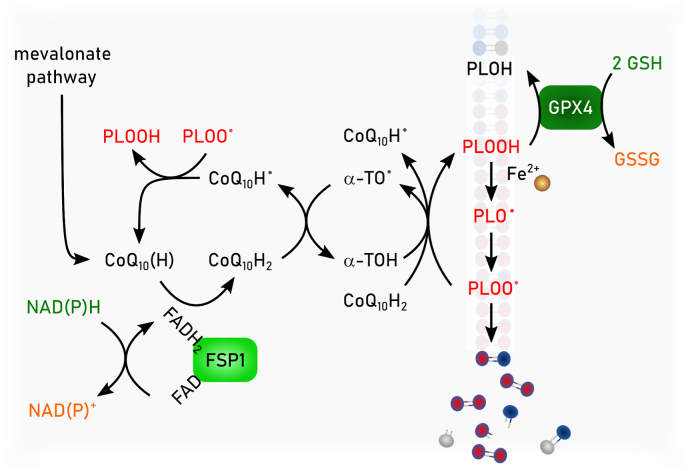
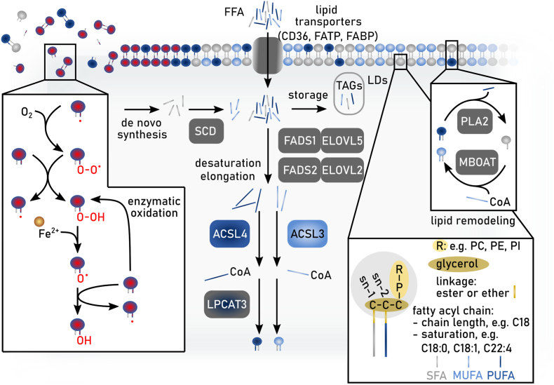
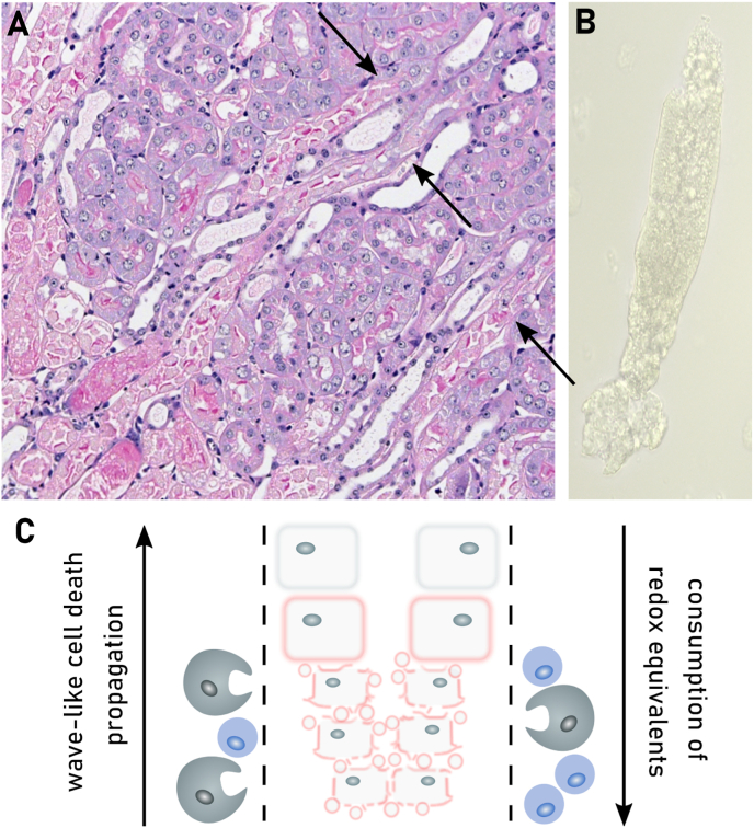
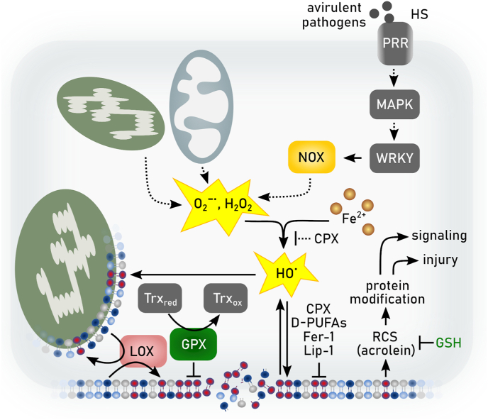
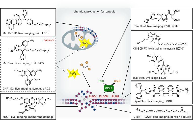
Ferroptosis is a pervasive non-apoptotic form of cell death highly relevant in various degenerative diseases and malignancies. The hallmark of ferroptosis is uncontrolled and overwhelming peroxidation of polyunsaturated fatty acids contained in membrane phospholipids, which eventually leads to rupture of the plasma membrane. Ferroptosis is unique in that it is essentially a spontaneous, uncatalyzed chemical process based on perturbed iron and redox homeostasis contributing to the cell death process, but that it is nonetheless modulated by many metabolic nodes that impinge on the cells' susceptibility to ferroptosis. Among the various nodes affecting ferroptosis sensitivity, several have emerged as promising candidates for pharmacological intervention, rendering ferroptosis-related proteins attractive targets for the treatment of numerous currently incurable diseases. Herein, the current members of a Germany-wide research consortium focusing on ferroptosis research, as well as key external experts in ferroptosis who have made seminal contributions to this rapidly growing and exciting field of research, have gathered to provide a comprehensive, state-of-the-art review on ferroptosis. Specific topics include: basic mechanisms, in vivo relevance, specialized methodologies, chemical and pharmacological tools, and the potential contribution of ferroptosis to disease etiopathology and progression. We hope that this article will not only provide established scientists and newcomers to the field with an overview of the multiple facets of ferroptosis, but also encourage additional efforts to characterize further molecular pathways modulating ferroptosis, with the ultimate goal to develop novel pharmacotherapies to tackle the various diseases associated with - or caused by - ferroptosis.

Keywords: Cancer; Cell death; Iron; Ischemia/reperfusion; Lipid peroxidation; Neurodegeneration.


Conflict of interest statement

Declaration of competing interest The authors declare the following financial interests/personal relationships which may be considered as potential competing interests: SJD is a co-founder of Prothegen, Inc. and holds patents related to ferroptosis. XJ is an inventor on patents related to autophagy and cell death and holds equity of and consults for Exarta Therapeutics and Lime Therapeutics. JAO is a member of the scientific advisory board for Vicinitas Therapeutics and has patent applications related to ferroptosis. TP reports grants from Dracen Pharmaceuticals, Kymera Therapeutics, Bristol-Myers Squibb, Agios Pharmaceuticals, personal fees from Vividion Therapeutics, Tohoku University, and personal fees from Faeth Therapeutics outside the submitted work; in addition, TP has a patent for US-20210361603-A1 pending and a patent for US-20210085763-A1 pending. BRS is an inventor on patents and patent applications involving ferroptosis, holds equity in and serves as a consultant to Exarta Therapeutics, and ProJenX Inc, holds equity in Sonata Therapeutics, and serves as a consultant to Weatherwax Biotechnologies Corporation and Akin Gump Strauss Hauer & Feld LLP. TVB holds patents related to ferroptosis inhibitors. CWil is a consultant for Odyssey Therapeutics and Orphagen Pharmaceuticals. YZ is a consultant for Keen Therapeutics. BP and MC hold patents for some of the compounds described herein, and is co-founder and shareholder of ROSCUE Therapeutics.

Figures

Image 1
Graphical abstract
Fig. 1
Fig. 1
Timeline with milestones in ferroptosis research. The presented timeline summarizes milestones in selenium and (seleno-)cyteine research as prerequisites for the understanding of ferroptosis and more recent research directly connected to ferroptosis. ACSL4: acyl-CoA synthetase long chain family member 4, FSP1: ferroptosis suppressor protein 1, GCH1: GTP cyclohydrolase 1, GPX4: glutathione peroxidase 4, RTA: radical trapping antioxidant, SLC7A11: solute carrier family 7 member 11, 7-DHC: 7-dehydrocholesterin.
Fig. 2
Fig. 2
Deregulated redox metabolome drives regulated necrosis. The two key thiol-dependent antioxidant systems in mammalian cells, the thioredoxin (Trx) and the glutathione (GSH) redox cycles are dependent on the presence of NAD(P)H cofactor that is restored by nicotinamide nucleotide transhydrogenase (NNT) within the mitochondria. Trx reductase (TrxR) utilizes NADPH to reduce Trx and other low molecular weight compounds. Peroxiredoxins (Prxs), on the other hand, accept electrons from Trx and subsequently scavenges hydrogen peroxide (H2O2) and other organic hydroperoxides. Like TrxR glutathione reductase (GSR) uses electrons from NADPH to restore reduced GSH from its oxidized state (GSSG). GSH in turn is the main substrate of glutathione peroxidases (GPXs). Members of the GPX protein family convert hydrogen peroxide and lipid hydroperoxides into water and alcohols, respectively. Different redox couples are affected by different types of regulated necrosis. Ac-CoA: Acetyl-CoA, αKG: α-ketoglutarate, DHFR1: dihydrofolate reductase 1, FA: fatty aci, NAD: nicotinamide adenine dinucleotide, NADPH: nicotinamide adenine dinucleotide phosphate, Prx: peroxiredoxin, SOD: superoxide dismutase.
Fig. 3
Fig. 3
Enzymatic reactivities of GPX4 and FSP1. Glutathione peroxidase 4 (GPX4) reduces in a glutathione (GSH)-dependent reaction phospholipid hydroperoxides (PLOOH) to phospholipid alcohols (PLOH), whereas ferroptosis suppressor protein 1 (FSP1) convert phospholipid peroxyl radicals (PLOO) to PLOOH. NAD(P)H reduces the flavin adenine dinucleotide (FAD) bound to FSP1. Via this reduced FAD (FADH2) FSP1 reduces quinone (CoQ10(H)) to hydroquinone (CoQ10H2). CoQ10H2 promotes the reaction from PLOO to PLOOH either by its own reaction with PLOO• or by restoring α-tocopherol (α-TO) facilitating the same reaction. Semihydroquinone (CoQ10H) reacts with another PLOO to form again CoQ10(H).
Fig. 4
Fig. 4
Lipid metabolism and ferroptosis. Schematic overview of the main pathways involved in fatty acid biosynthesis and uptake, lipid synthesis and lipid remodeling that regulate ferroptosis sensitivity in cells. In general, two fatty acyl chains are linked to a glycerol backbone via an ester or ether bond. The fatty acids at the sn-1 position are generally saturated fatty acids (SFAs) or mono-unsaturated fatty acids (MUFAs), whereas the fatty acids at the sn-2 position can be SFAs, MUFAs or poly-unsaturated (PUFAs). Lipids attached to the glycerol can be e.g. phosphatidylcholine (PC), phosphatidylethanolamine (PE), or phosphatidylinositol (PI). Free fatty acids (FFA) from the microenvironment can be taken up via CD36, fatty acid transport proteins (FATP) or fatty acid-binding proteins (FABP). The intracellular pool of fatty acids is comprised of SFA as product of de novo fatty acid synthesis or uptake, together with either MUFAs or PUFAs as products of fatty acid desaturation and elongation. Noteworthy, certain PUFAs are essential and thus have to be taken up by aforementioned extracellular space. Depending on substrate specificity, fatty acids are desaturated by stearoyl-CoA desaturase (SCD) and fatty acid desaturases (FADS1 or FADS2). Further, different elongases e.g. ELOVL2 or ELOVL5 are synthesizing fatty acids (SFA, MUFA or PUFA) with longer carbon chains. Members of the acyl-CoA synthetase long chain family of proteins (e.g. ACSL3 or ACSL4) convert free long-chain fatty acids into fatty acyl-CoA esters that then can be utilized to become incorporated into membrane phospholipids (PL) mediated by e.g. lysophosphatidylcholine acyltransferase LPCAT3. Additionally, intracellular fatty acids can be stored into lipid droplets (LDs) as they are converted into triacylglyceride. Existing membrane phospholipids can be remodeled via the Lands Cycle. Here, hydrolysis of the fatty acid (e.g. PUFA) at the sn-2 position by PLA2 produces a lyso-PL and a following re-esterification of another fatty acid-CoA (e.g. MUFA) mediated by MBOAT1 leads to the remodeled membrane PL.
Fig. 5
Fig. 5
Iron metabolism and ferroptosis. Iron metabolism is regulated at the systemic and the cellular level. Iron efflux from duodenal enterocytes and macrophages is controlled by hepcidin, a peptide hormone produced by the liver which binds to and induces degradation of the iron exporter ferroportin. In iron overload diseases (e.g. haemochromatosis), the hepcidin/ferroportin axis is disturbed and iron is released in excess into the bloodstream. The capacity of transferrin to bind iron is exceeded and a potentially toxic form of iron appears, called non-transferrin bound iron (NTBI). TBI (transferrin-bound iron) is imported into cells in a controlled manner via the transferrin receptor Tfr, whereas NTBI is taken up in an uncontrolled manner via NTBI importers such as ZIP14 and CD44. Iron uptake enlarges the labile iron pool (LIP). Through the generation of hydroxyl (HO) or hydroperoxyl radicals (HOO) by the Fenton reaction, free and redox-active iron can trigger lipid peroxidation and ferroptosis. Iron storage in ferritin (Fer) or iron export via ferroportin reduce the LIP and therefore protect cells against ferroptosis. The release of free iron from Fer by NCOA4 mediated ferritinophagy or from heme by heme oxygenase (HO-1) increases the LIP and potentially sensitizes the cells to ferroptosis. A growing number of studies suggests that ferroptosis plays a critical role in the iron-mediated hepatic (cirrhosis, hepatocellular carcinoma), pancreatic (insulin-dependent diabetes) or cardiac (cardiac arrhythmia, cardiac failure) complications observed in iron overload disorders.
Fig. 6
Fig. 6
Ferroptosis protection by the GCH1/BH4/DHFR axis. Tetrahydrobiopterin (BH4) is synthesized through a series of steps catalyzed by GTP cyclohydrolase 1 (GCH1, the rate-limiting enzyme), 6-pyruvoyl-tetrahydropterin synthase (PTS), and sepiapterin reductase (SPR). BH4 acts as a radical trapping antioxidant in lipid membranes, directly quenching lipid radicals. BH4 can regenerate oxidized a-tocopherol (vitamin E), an endogenous lipophilic antioxidant. Oxidized BH4 (BH2) is recycled back to BH4 by DHFR1. Additionally, BH4 may contribute the synthesis of CoQ10, promoting the protective effects of the NAD(P)H/FSP1/CoQ10 system.
Fig. 7
Fig. 7
Cysteine metabolism and ferroptosis. Schematic overview of the main pathways involved in cysteine uptake, de novo cysteine synthesis, and cysteine utilization that regulate ferroptosis sensitivity in cells. Cysteine can be taken up in its oxidized form cystine via the xCT transporter, or synthesized de novo from methionine and serine via the transulfuration enzymes cystathionine β-synthase (CBS) and cystathionine γ-lyase (CSE). Production of glutathione (GSH) from cysteine supports the activity of glutathione peroxidase 4 (GPX4) to detoxify phospholipid hydroperoxides (PLOOH) and mitigate the propogation of the lipid peroxidation cycle. Moreover, cysteine-derived sulfane sulfur species (hydropersulfides, RSSH) act as radical trapping agents that can suppress the initiation and propagation of the radical chain reaction driving lipid peroxidation.
Fig. 8
Fig. 8
Selenium metabolism and selenoprotein synthesis. Selenocysteine (Sec) is a unique amino acid synthesized on transfer RNA (tRNA) by incorporating inorganic selenium. Initially, Sec is converted by the enzyme Sec lyase (SCLY) to selenide and then further delivered to selenophosphate by SEPHS2. Selenite and other inorganic forms of selenium can also enter this pathway through cellular reduction processes. Concurrently, Serine (Ser) is bound to tRNA[Ser]Sec, which harbors the UGA anticodon, by seryl-tRNA synthetase (SerS), followed by phosphorylation facilitated by phosphoseryl-tRNA kinase (PTSK). Sec is then produced on tRNA by Sec synthase (SecS/SEPSECS). During translation, Sec is incorporated into selenoproteins via the Sec-insertion sequence (SECIS), a stable loop structure located in the 3′-UTR of selenoprotein mRNA. The complex formed by Sec-tRNA[Ser]Sec and associated factors, such as eukaryotic elongation factor for Sec translation (eEFSEC) and SECIS binding protein 2 (SBP2/SECISBP2), facilitates the translation process. At the UGA codon, Sec-tRNA[Ser]Sec is supplied from the SECIS complex to ensure accurate incorporation of Sec into the growing peptide chain and preventing premature termination.
Fig. 9
Fig. 9
Calcium signaling and energy metabolism in ferroptosis. Increased cytosolic calcium is one of the hallmarks of ferroptosis. It remains unclear, however, whether calcium ions enter the cell through membrane nanopores, through endogenous channels at the plasma membrane, or whether the ER or mitochondria are responsible for calcium transport, or a combination of those. Cytosolic calcium contributes to ferroptosis and to the subsequent release of intracellular components. Yet, calcium signaling also connects plasma membrane damage with the activation of membrane repair mechanisms that counterbalance cell death. Energetic metabolism centered around mitochondrial activity is the major source for the generation of free radicals and reactive oxygen species in the cell. Therefore, energetic metabolism plays a central role in ferroptosis induction. Related processes, such as glutamine anaplerosis, TCA cycle and eletron transfer chain (ETC) activity, have all been demonstrated to be involved in ferroptosis. AMPK: AMP-activated protein kinase, ESCRTIII: endosomal sorting complex required for transport III, MICU1: Mitochondrial calcium uptake 1, ORAI: calcium channel protein, RyR: ryanodine receptor, SOCE: store-operated calcium entry, VGCC: voltage-gated calcium channels.
Fig. 10
Fig. 10
NRF2 target genes involved in the ferroptosis regulation. NRF2 is bound by KEAP1 and degraded in proteasome in unstressed conditions. Exposure to oxidative stress, such as hydrogen peroxide and 4-hydroxynonenal, leads to KEAP1 inactivation and NRF2 stabilization. NRF2 suppresses ferroptosis by promoting NADPH production, glutathione synthesis, and cystine uptake and by decreasing labile iron. The increased cystine uptake leads to increased sulfane sulfur production. GPX4 and FSP1 appear to be context dependent NRF2 target genes. GCLC and GCLM: catalytic and regulatory subunits of gamma-glutamylcysteine ligase, G6PD: glucose-6-phosphate dehydrogenase, IDH1: isocitrate dehydrogenase 1, ME1: malic enzyme 1, PGD: phosphogluconate dehydrogenase, RSSH: hydropersulfide, xCT: representing cystine/glutamate antiporter.
Fig. 11
Fig. 11
Regulation of ferroptosis-related proteins via posttranslational modifications. The activities or levels of several pro- and anti-ferroptotic proteins are regulated by (linear) ubiquitination, SUMOylation, glutathionylation, phosphorylation, lipidation, or acetylation ultimately leading to either increased (orange) or decreased (green) susceptibility towards ferroptosis. ACSL4: acyl-CoA synthetase long chain family member 4, FSP1: ferroptosis suppressor protein 1, GPX4: glutathione peroxidase 4, OTUB1: OTU deubiquitinase ubiquitin aldehyde binding 1, STAT3: signal transducer and activator of transcription 3, xCT: representing cystine/glutamate antiporter.
Fig. 12
Fig. 12
Ferroptosis and immunity. Ferroptosis is induced in diseases such as rheumatoid arthritis (RA) via transient receptor potential cation channel, subfamily M, member 7 (TRPM7) and ferroptosis suppressor protein (FSP1), inflammatory bowel disease (IBD), ulcerative colitis (UC), Crohn's disease (CD) via decreasing glutathione peroxidase 4 (GPX4), and psoriasis via fatty acid-CoA ligase 4 (ACSL4), prostaglandin-endoperoxide synthase 2 (Ptgs2) and transferrin receptor (TfRc). Thereby, ferroptosis leads to exposure of programmed cell death ligand-1 (PD-L1) and calreticulin (CALR) as well as release of ATP and high mobility group box 1 (HMGB1) which facilitate immune cell recruitment. As potential therapeutic inducers of ferroptosis IKE in combination with TNF antagonists are discussed. In addition, interferon (INF) γ released by natural killer NK cells and T cells, which relay on GPX4 during development, contributes to ferroptosis induction. Ferroptosis is mediated in dendritic cells by peroxisome proliferator-activated receptor (PPAR) γ. During systemic lupus erythematosus (SLE) ferroptosis is facilitated by autoantibodies and TNFα which induce cAMP-responsive element modulator (CREM) α, a transcriptional repressor for GPX4, in neutrophils. In macrophages interleukin (IL) −4 and −13 contributes to ferroptosis by inducing lipoxygenases (LOX) and blocking GPX4 and inducible nitric oxide synthase (iNOS). Nevertheless, it inhibits ferroptosis via l-amino acid oxidase IL-4 induced gene-1 (IL4iL). Further, nuclear factor erythroid-2-related factor 2 (NRF2) and ferritin (Fer) protect macrophages from ferroptosis.
Fig. 13
Fig. 13
Chemical structures of ferroptosis inducers.
Fig. 14
Fig. 14
Mode of action and structures of ferroptosis inhibitors.
Fig. 15
Fig. 15
Schematic overview of the complexity of different pathways modulating ferroptosis in cancer cells. p53 and BAP1 suppress SLC7A11 to restrain the uptake of cystine. NRF2 and MYC/MYCN inhibit ferroptosis in both GPX4-or FSP1-dependent manner. KRAS G12D and ARF positively or negatively modulate NRF2 activities, respectively. NF2-YAP1 and RB-E2F axis regulate cell ferroptosis vulnerability by modulating ACSL4 expression and PUFA-PL content. Conversely, AR-MBOAT2, ER-MBOAT1, or PI3K-AKT-mTORC1-SERBP1-SCD pathways decrease cell sensitivity to ferroptosis by modulating MUFA-PL content. ACSL: acyl-CoA synthetase long-chain family member, AKT1: AKT serine/threonine kinase 1, ALOX: lipoxygenase, AR: androgen receptor, ARF: Tumor suppressor ARF, BAP1: BRCA1 associated protein 1, BACH1: BTB domain and CNC homolog 1, DECR1: 2,4-dienoyl-CoA reductase 1, E2F: E2F transcription factor 1, ER: estrogen receptor, FSP1: ferroptosis suppressor protein 1, GPX4: glutathione peroxidase 4, 2-HG: 2-hydroxyglutarate, KRAS: KRAS proto-oncogene, GTPase, MBOAT: O-acyltransferase domain containing, mTORC1: mammalian target of rapamycin complex 1, MYC: MYC proto-oncogene, MYCN: MYCN proto-oncogene, NF2: neurofibromin 2, NRF2: nuclear factor erythroid 2-related factor 2; PI3K: phosphatidylinositol 3-kinase; PTEN: phosphatase and tensin homolog, RB: RB transcriptional corepressor 1, SCD: stearoyl-CoA desaturase, SREBP1: sterol regulatory element-binding protein 1, xCT: SLC7A11 - solute carrier family 7 member 11, YAP1: Yes1 associated transcriptional regulator.
Fig. 16
Fig. 16
Schematic representation of ferroptosis impairing the diseased brain, and its potential targeting as a novel approach for neuronal replacement. Astrocytes: dark grey, neurons: bright grey.
Fig. 17
Fig. 17
Wave-like cell death propagation along renal tubules. A) Following murine renal ischemia/reperfusion injury, typical tubular necroses can be observed. Arrows demark transition zones where ferroptotic cell death is just ongoing. B) A typical granular cast as observed in a patients urine sediment considered as an equivalent of acute tubular necrosis. C) Schematic depiction of acute tubular necrosis, which follows the consumption of redox equivalents. Note the innate immune cells, which are specifically recruited to necrotic areas.
Fig. 18
Fig. 18
Proposed model for plant ferroptosis. Ferroptosis can be induced by avirulent pathogens or by heat stress (HS) via pattern recognition receptors (PRR). After an attempted infection, a MAPK cascade is activated, phosphorylating WRKY transcription factors which in turn induce NOX expression, leading to ROS accumulation. The pathway involves GSH depletion, ROS accumulation and iron-dependent lipid peroxidation. Cytoplasmic ROS accumulation might occur through NADPH oxidase activity (NOX) and by the generation of toxic lipid‐peroxides. Those lipid peroxides could originate from enzymatic or non‐enzymatic processes on membrane poly-unsaturated fatty acids (PUFA-PLs) from chloroplasts and cytoplasmic membrane, involving either lipoxygenases (LOXs) activity or Fenton chemistry. Lipid ROS can also be degraded to reactive carbonyl species (RCS), such as acrolein, which are also related to cell death. Glutathione peroxidases (GPXs) are proposed to act as negative regulators, detoxifying the cell from lipid peroxides. In plants, GPXs uses thioredoxin (Trx) as a main reductant agent, but they might also utilize GSH. Ciclopirox olamine (CPX), D-PUFAs, ferroststin 1 (Fer-1), and liproxstatin-1 (Lip-1) inhibit ferroptosis. RSL3 induces ferroptosis. Dashed lines indicate indirect evidence.
Fig. 19
Fig. 19
Chemical probes for detecting ferroptosis. These probes can report on phospholipid peroxidation (C11-BODIPY, H4BPMHC, LiperFluo; including in mitochondria via MitoPeDPP); loss of plasma membrane integrity (MDG1); and depletion of antioxidant defenses e.g. GSH levels (RealThiol). Fluorescent “dihydro" probes claimed to report ROS levels (MitoSox, DHR-123) should only be used with caution.

Similar articles

Cited by

References

    1. Wiernicki B., Dubois H., Tyurina Y.Y., Hassannia B., Bayir H., Kagan V.E., Vandenabeele P., Wullaert A., Vanden Berghe T. Excessive phospholipid peroxidation distinguishes ferroptosis from other cell death modes including pyroptosis. Cell Death Dis. 2020;11:922. doi: 10.1038/s41419-020-03118-0. - DOI - PMC - PubMed
    1. Conrad M., Pratt D.A. The chemical basis of ferroptosis. Nat. Chem. Biol. 2019;15:1137–1147. doi: 10.1038/s41589-019-0408-1. - DOI - PubMed
    1. Hirata Y., Cai R., Volchuk A., Steinberg B.E., Saito Y., Matsuzawa A., Grinstein S., Freeman S.A. Lipid peroxidation increases membrane tension, Piezo1 gating, and cation permeability to execute ferroptosis. Curr. Biol. 2023;33:1282–1294 e1285. doi: 10.1016/j.cub.2023.02.060. - DOI - PubMed
    1. Zilka O., Shah R., Li B., Friedmann Angeli J.P., Griesser M., Conrad M., Pratt D.A. On the mechanism of cytoprotection by ferrostatin-1 and liproxstatin-1 and the role of lipid peroxidation in ferroptotic cell death. ACS Cent. Sci. 2017;3:232–243. doi: 10.1021/acscentsci.7b00028. - DOI - PMC - PubMed
    1. Dixon S.J., Lemberg K.M., Lamprecht M.R., Skouta R., Zaitsev E.M., Gleason C.E., Patel D.N., Bauer A.J., Cantley A.M., Yang W.S., Morrison B., 3rd, Stockwell B.R. Ferroptosis: an iron-dependent form of nonapoptotic cell death. Cell. 2012;149:1060–1072. doi: 10.1016/j.cell.2012.03.042. - DOI - PMC - PubMed