. 2024 Jul 11;111(7):1383-1404.
doi: 10.1016/j.ajhg.2024.05.023. Epub 2024 Jun 21.

Truncated variants of MAGEL2 are involved in the etiologies of the Schaaf-Yang and Prader-Willi syndromes

Affiliations

Truncated variants of MAGEL2 are involved in the etiologies of the Schaaf-Yang and Prader-Willi syndromes

David Heimdörfer et al. Am J Hum Genet. .

Erratum in

Abstract

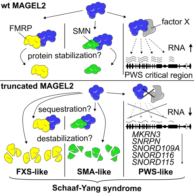
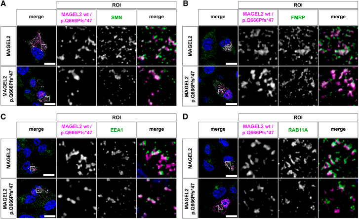
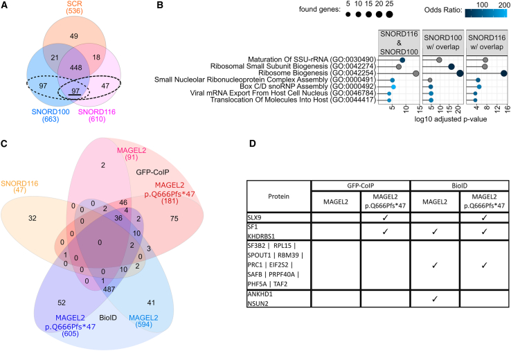
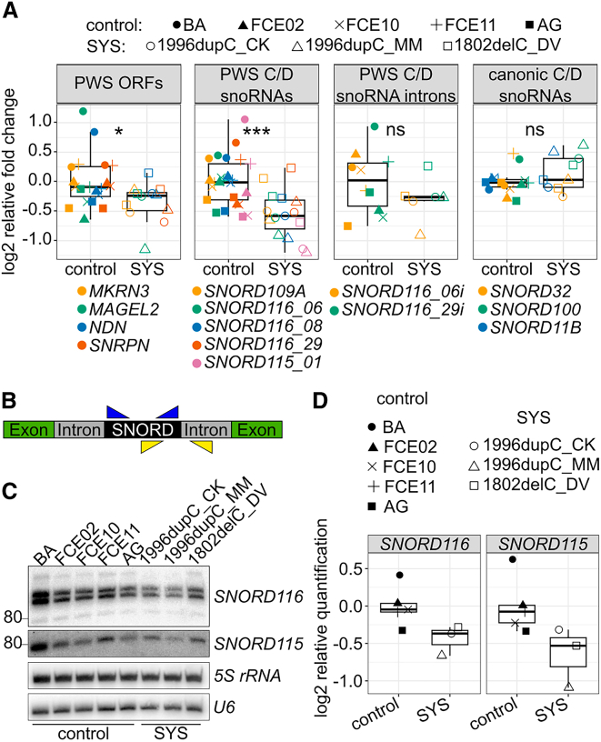
The neurodevelopmental disorders Prader-Willi syndrome (PWS) and Schaaf-Yang syndrome (SYS) both arise from genomic alterations within human chromosome 15q11-q13. A deletion of the SNORD116 cluster, encoding small nucleolar RNAs, or frameshift mutations within MAGEL2 result in closely related phenotypes in individuals with PWS or SYS, respectively. By investigation of their subcellular localization, we observed that in contrast to a predominant cytoplasmic localization of wild-type (WT) MAGEL2, a truncated MAGEL2 mutant was evenly distributed between the cytoplasm and the nucleus. To elucidate regulatory pathways that may underlie both diseases, we identified protein interaction partners for WT or mutant MAGEL2, in particular the survival motor neuron protein (SMN), involved in spinal muscular atrophy, and the fragile-X-messenger ribonucleoprotein (FMRP), involved in autism spectrum disorders. The interactome of the non-coding RNA SNORD116 was also investigated by RNA-CoIP. We show that WT and truncated MAGEL2 were both involved in RNA metabolism, while regulation of transcription was mainly observed for WT MAGEL2. Hence, we investigated the influence of MAGEL2 mutations on the expression of genes from the PWS locus, including the SNORD116 cluster. Thereby, we provide evidence for MAGEL2 mutants decreasing the expression of SNORD116, SNORD115, and SNORD109A, as well as protein-coding genes MKRN3 and SNRPN, thus bridging the gap between PWS and SYS.

Keywords: Schaaf-Yang syndrome, Prader-Willi syndrome, MAGEL2, SNORD116, SMN, FMRP.


Conflict of interest statement

Declaration of interests The authors declare no competing interests.

Figures

None
Graphical abstract
Figure 1
Figure 1
Subcellular localization of MAGEL2 WT and p.Gln666Profs47 (A and B) MAGEL2/p.Gln666Profs47 fusion proteins employed for investigation of their subcellular localization, tagged with either an N-terminal mCherry (A) or GFPspark (B). (C and D) Confocal immunofluorescence microscopy of recombinant fusion proteins WT mCherry-MAGEL2 or mCherry-p.Gln666Profs47 (magenta) in transiently transfected HEK293T cells, 24 h after transfection. Nuclei were counterstained with DAPI (blue). The scale bar is 10 μm. White arrows indicate sites of spatial proximity between WT MAGEL2 and p.Gln666Profs47 with endogenous early endosomes (C) and recycling endosomes (D), which were visualized with anti-EEA1 and anti-RAB11A antibodies (green), respectively. At least 5 cells were imaged in three independent replicates and representative cells are depicted. (E) MAGEL2 WT and truncated p.Gln666Profs47 with computationally predicted putative NLS (black boxes) and NES (gray boxes) motifs. (F and G) HeLa cells were transfected with the GFP-tagged wild-type MAGEL2 or MAGEL2 c.1996dupC mutant. 48 h after transfection cells were treated with leptomycin B (20 nM) or left untreated for 20 h. Nuclei were counterstained with DAPI (blue). (F) HeLa cells with and without leptomycin B treatment. Cells from three independent transfection experiments were used for quantification. The scale bar is 40 μm. (G) Quantification of the nuclear/cytosolic ratio of individual cells from three independent experiments. Wilcoxon tests were calculated employing rstatix.
Figure 2
Figure 2
MAGEL2 WT and p.Gln666Profs47 show high affinity for proteins involved in RNA metabolism and chromosome condensation (A) Fusion-protein constructs for BioID. (B–D) Analyses were carried out for proteins identified in at least two of three replicates per experiment. (B) Combined Venn diagram interaction partners of MAGEL2 WT and p.Gln666Profs47 identified by BioID and GFP-CoIP. (C) Protein-protein association networks (gene names are depicted) of the 36 common interaction partners identified in (B), colored by the top 5 biological processes in (D). (D) Top 5 hits by combined score of gene enrichment analysis employing enrichR for biological processes (2023) in either BioID or GFP-CoIP. Only GO terms with an adjusted p value <0.05 were included.
Figure 3
Figure 3
MAGEL2 WT and p.Gln666Profs47 interact with proteins involved in RNA metabolism and gene expression as well as disease related SMN (spinal muscular atrophy) and FMRP (fragile-X syndrome) (A) Venn diagrams of protein interaction partners for MAGEL2 WT and p.Gln666Profs47 identified by BioID or GFP-CoIP. (B) Gene enrichment analysis for Biological Processes (2023) in either BioID or GFP-CoIP employing enrichR. Shades of blue depict top 5 hits by combined score. Gray circles indicate that the respective GO term is among the top hits in another condition. GO terms with an adjusted p value > 0.05 were excluded. (C) Heatmap for enriched proteins (gene names are depicted) in MAGEL2 WT and p.Gln666Profs47 by BioID and GFP-CoIP. GO term enrichment analyses for Biological Process and KEGG Pathways from Figure S1 were grouped to broader functional terms as shown in Table S9 are written in the respective colors (Gene Ontology). Gene ontology “others” include novel interaction partners of interest which could not be assigned to the remaining annotated terms. (D) Western blots of proteins which co-immunoprecipitated with WT GFP-MAGEL2 or GFP-p.Gln666Profs47 (n = 3). 5% of the lysate were used in input fractions.
Figure 4
Figure 4
MAGEL2 WT and p.Gln666Profs47 co-localize with EEA1, RAB11, SMN and FMRP in mouse Neuro-2a cells Mouse Neuro-2a cells were transiently transfected with WT mCherry-MAGEL2 or mCherry-p.Gln666Profs47 (magenta) fusion proteins and subcellular localization was analyzed employing confocal immunofluorescence microscopy 24 h after transfection. Nuclei were counterstained with DAPI (blue). The scale bar is 10 μm. SMN (A), FMRP (B), endogenous early endosomes (C), and recycling endosomes (D) were visualized with anti-SMN, anti-FMRP, anti-EEA1, and anti-RAB11A antibodies (green), respectively. A white signal in the overlap images indicated signal overlap or co-localization. At least 3 transfected cells were imaged in three independent replicates and representative cells are depicted.
Figure 5
Figure 5
SNORD116 does not directly interact with WT MAGEL2 or p.Gln666Profs47 RNA-coIP was performed with biotinylated SNORD116, SNORD100, or a SCR control with HEK293T IP lysates. Analyses were carried out with proteins identified in at least 2 of 3 replicates per condition. (A) Venn diagrams showing overlapping proteins identified via RNA-coIP. The 97, 194, and 144 proteins found in RNA-coIPs for SNORD116 and SNORD100 (underlined), SNORD100 with overlap (dashed circles), and SNORD116 with overlap (dashed circles) were employed for the analysis in (B). (B) GO term enrichment analysis employing enrichR for Biological Processes (2023) for proteins identified in SNORD116 and SNORD100, but not in the SCR control. Top 5 hits by Combined Score are shown in shades of blue. Gray circles indicate that the respective GO term is among top hits in another condition. Only GO terms with an adjusted p value < 0.05 were included. (C) Venn diagram displaying the overlap between unique SNORD116 interacting proteins (A) and those identified in 2 of 3 replicates of the BioID or GFP-CoIP performed with WT MAGEL2 or p.Gln666Profs47. (D) Table showing overlapping proteins from (C).
Figure 6
Figure 6
Influence of MAGEL2 frameshift mutations on gene expression, assessed in SYS smNPCs compared to healthy controls (A) RT-qPCR analysis of genes of interest (GOI) within and outside chromosome 15q11–q13. Relative expression was calculated with primer efficiency normalized N0 values (linRegPCR50). N0 values were normalized to the geometric mean of housekeeping genes TBP, GAPDH, and HPRT and to the geometric mean of healthy control samples. Genes were combined into groups depending on type and genomic localization. Wilcoxon tests were applied employing rstatix. (B) Scheme of primer position to detect snoRNAs before (yellow) and after (blue) splicing and processing. (C) Northern blot analysis of 19 and 30 copies of SNORD116 and SNORD115, respectively. (D) Northern blot bands were quantified employing Fiji and normalized to geometric mean areas of 5S rRNA and U6 snRNA signals.

References

    1. Fountain M.D., Schaaf C.P. Prader-Willi Syndrome and Schaaf-Yang Syndrome: Neurodevelopmental Diseases Intersecting at the MAGEL2 Gene. Dis. Basel Switz. 2016;4 doi: 10.3390/diseases4010002. - DOI - PMC - PubMed
    1. Chamberlain S.J. RNAs of the human chromosome 15q11-q13 imprinted region. Wiley Interdiscip. Rev. RNA. 2013;4:155–166. doi: 10.1002/wrna.1150. - DOI - PMC - PubMed
    1. Höybye C., Tauber M. Approach to the Patient With Prader–Willi Syndrome. J. Clin. Endocrinol. Metab. 2022;107:1698–1705. doi: 10.1210/clinem/dgac082. - DOI - PMC - PubMed
    1. Cassidy S.B., Schwartz S., Miller J.L., Driscoll D.J. Prader-Willi syndrome. Genet. Med. 2012;14:10–26. doi: 10.1038/gim.0b013e31822bead0. - DOI - PubMed
    1. Szabadi S., Sila Z., Dewey J., Rowland D., Penugonda M., Ergun-Longmire B. A Review of Prader–Willi Syndrome. Endocrines. 2022;3:329–348. doi: 10.3390/endocrines3020027. - DOI

Publication types

LinkOut - more resources