. 2024 Jul 10;4(7):100590.
doi: 10.1016/j.xgen.2024.100590. Epub 2024 Jun 21.

Inverted triplications formed by iterative template switches generate structural variant diversity at genomic disorder loci

Affiliations

Inverted triplications formed by iterative template switches generate structural variant diversity at genomic disorder loci

Christopher M Grochowski et al. Cell Genom. .

Abstract

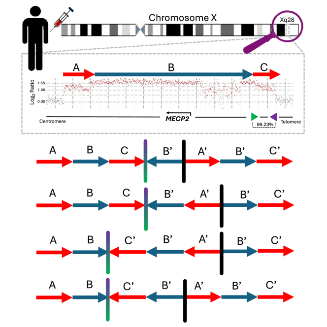
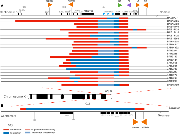
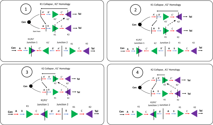
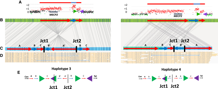
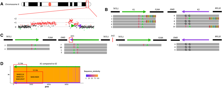
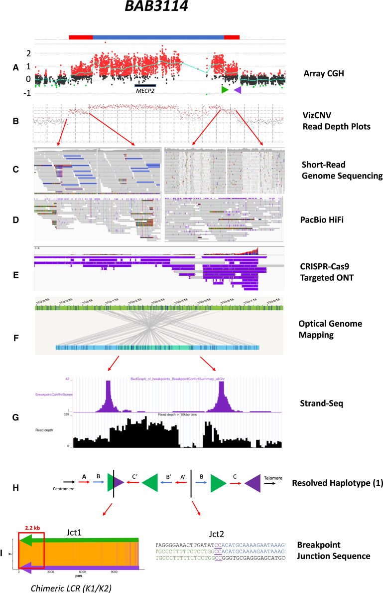
The duplication-triplication/inverted-duplication (DUP-TRP/INV-DUP) structure is a complex genomic rearrangement (CGR). Although it has been identified as an important pathogenic DNA mutation signature in genomic disorders and cancer genomes, its architecture remains unresolved. Here, we studied the genomic architecture of DUP-TRP/INV-DUP by investigating the DNA of 24 patients identified by array comparative genomic hybridization (aCGH) on whom we found evidence for the existence of 4 out of 4 predicted structural variant (SV) haplotypes. Using a combination of short-read genome sequencing (GS), long-read GS, optical genome mapping, and single-cell DNA template strand sequencing (strand-seq), the haplotype structure was resolved in 18 samples. The point of template switching in 4 samples was shown to be a segment of ∼2.2-5.5 kb of 100% nucleotide similarity within inverted repeat pairs. These data provide experimental evidence that inverted low-copy repeats act as recombinant substrates. This type of CGR can result in multiple conformers generating diverse SV haplotypes in susceptible dosage-sensitive loci.

Keywords: MECP2 duplication syndrome; MMBIR; Mendelian diseases; Xq28; break-induced replication; copy-number variant; inversions; recombination; segmental duplication; template switching.


Conflict of interest statement

Declaration of interests Baylor College of Medicine and Miraca Holdings have formed a joint venture with shared ownership and governance of BG, which performs clinical microarray analysis, clinical ES, and clinical biochemical studies. J.R.L. serves on the scientific advisory board of the BG. J.R.L. has stock ownership in 23andMe, is a paid consultant for Genomics International, and is a co-inventor on multiple US and European patents related to molecular diagnostics for inherited neuropathies, eye diseases, genomic disorders, and bacterial genomic fingerprinting. E.H. and S.J. are employees of ONT and shareholders and/or share option holders of ONT. D.P. provides consulting services for Ionis Pharmaceuticals. F.J.S. receives research support from Genetech, Illumina, Pacbio, and ONT.

Figures

None
Graphical abstract
Figure 1
Figure 1
Probands carrying DUP-TRP/INV-DUP genomic structure (A) Genomic region spanning Xq28, including the MECP2 critical region, is shown with the location of selected genes and inverted repeats mediating DUP-TRP/INV-DUP formation in this cohort (43202a/43202b; 43221a/43221b [K1/K2, green and purple arrows]; 43231a/43231b [L1/L2]). The relative genomic locations of the duplication (red) and triplication (blue) are shown. Uncertainty as to the precise location of the start/end of either the duplication or triplication due to lack of probes on aCGH or low mapping quality in short-read GS within a given interval are depicted in light red and light blue, respectively. (B) A single individual female (BAB12566) is shown with a DUP-TRP/INV-DUP within Xq21, along with the relative position of inverted LCR pairs (37696a/37696b). The naming scheme for 43221a/43221b (K1/K2) and 43231a/43231b (L1/L2) are derived from previous work detailing the DUP-TRP/INV-DUP structures at the MECP2 locus on the X chromosome.,, Genes included in this panel have an associated phenotype in OMIM.
Figure 2
Figure 2
Predictive model for DUP-TRP/INV-DUP formation At least 4 haplotype sub-structures can be derived from rearrangement involving a pair of inverted LCRs. This figure depicts the LCRs K1 and K2 (green and purple arrowheads) within the MECP2 locus used as substrates during an intrachromosomal event. The same model can be applied to other DUP-TRP/INV-DUPs formed through inverted LCRs pairs nearby dosage-sensitive genes. The formation of the DUP-TRP/INV-DUP event may start due to a replication fork stall and collapse at or nearby the LCR (K1), denoted as a green arrowhead. Homology drives strand invasion at the inverted LCR (K2′) on the opposite strand (denoted in purple), producing junction 1. DNA replication continues in the opposite direction until a second replication fork collapse and repair on the original strand through either MMBIR or NHEJ resolves the second junction. The 4 conformer possibilities shown here are determined by the replication fork collapsing and jumping (TS denoted by dashed black arrows) from either K1 to K2′ or K2 to K1′.
Figure 3
Figure 3
Haplotype resolution using OGM Structural haplotype determination and conformer configuration was established based upon single-molecule support through junction 1 and junction 2 in cis. Two samples in the cohort are highlighted, BAB14604 (left) and BAB15418 (right). (A) ArrayCGH plots for each sample show a similarly sized DUP-TRP-DUP event both mediated by inverted LCRs (shown as green and purple arrows) downstream of MECP2 (black rectangle). (B) OGM reference (green rectangle) shows in silico motifs throughout the MECP2 locus. The red arrows correspond to the duplicated segments, whereas the blue arrow corresponds to the triplicated segments. The length of the CNVs is proportional to the aCGH CNV. (C) OGM de novo assembly from proband samples are shown in blue rectangles. Sequence motifs aligned to the reference shown as connecting gray lines enable restriction fragment genome mapping and pattern recognition. Red and blue arrows are overlayed to represent the position and orientation of each amplified genomic fragment within the DUP-TRP-DUP structure. The connection points forming junctions 1 and 2 are shown as black vertical dashed lines/bars. (D) Single DNA molecules that span both junctions 1 and 2 are highlighted in blue, confirming that both junctions are present in cis. (E) Hypothesized resolved haplotypes based on CNV and in cis junction analysis. Although both samples show nearly identical aCGH patterns, BAB14604 has conformer haplotype 3 and BAB15418 shows conformer haplotype 4.
Figure 4
Figure 4
Refined position of TS within LCR K1/K2 Identification of PSVs through inverted LCRs allows for a determination of the relative position of the breakpoint junction within the inverted LCRs for BAB2727. (A) aCGH showing a DUP-TRP-DUP structure, with MECP2 locus highlighted and magnified. The inverted LCRs K1 and K2 (shown as green and purple arrows) are located flanking the terminal/3′ end duplication in the structure. (B) Positions of K1 and K2 are shown with representative HiFi data below, highlighting sequence reads that span the region. Ancestral reads denote HiFi reads are uniquely aligned with LCR (e.g., reads 2, 4, and 5). Breakpoint reads denote HiFi reads that begin in unique sequence and show soft clipping as they exit the LCR (e.g., reads 1 and 3). PSVs are visualized in LCR K2 with the green (A nucleotide) and red (T nucleotide) positions (ChrX:153,615,342 and ChrX:153,615,645, respectively) that are found breakpoint reads in K2 and are present within points of homology in K1. (C) Linearized structure showing the reads found within each position. The chimeric K1/K2 shows the positioning of PSVs used to refine the position of Jct1. (D) Percentage of uniquely aligned base in slide window of 20 bp (i.e., sequence similarities were shown as a heatmap). A “hot” color, orange, denotes a 100% match, while a “cold” color, purple, denotes reduced similarity. The position of the PSV can be used to estimate the distance the replication fork proceeds before the TS to K2 occurred. Samples in this cohort could be narrowed to a 2.2- or 5.5-kb region.
Figure 5
Figure 5
Multipronged approach resolving DUP-TRP/INV-DUP events Sample BAB3114, including the methodology used to fully resolve the SV haplotype and breakpoint junctions 1 and 2. (A) aCGH showing a DUP-TRP-DUP structure at Xq28, including the MECP2 gene and LCRs K1 (green arrow) and K2 (purple arrow). (B) Illumina short-read GS showing the read depth for the region as visualized in the VizCNV plotting program. (C) Red arrows denote the regions of copy-number change as seen in the short-read sequencing in the Integrative Genomics Viewer. Of note, soft clipping can be seen in the regions of unique sequence (left) versus the unmapped reads at the region with K1 and K2 due to sequence similarity of the region. (D) PacBio HiFi data show the reads that include the breakpoint region (shown as soft clipping) within both junction 1 and junction 2. (E) CRISPR-Cas9-targeted ONT facilitated ultra-long molecule (>500 kb) sequencing to capture the haplotype structure within a single DNA molecule. (F) Bionano OGM shows orientation and connection points of amplified genomic fragments forming junctions 1 and 2 within the structure. (G) Strand-seq data showing the points of breakpoint (purple peaks) with the inverted genomic sequence between. (H) Resolved haplotype structure 1 for BAB3114 shows the triplication and initial duplication in an inverted orientation. (I) Junction 1 shows a heatmap of K1/K2 similarity. The point of fork stall/collapse and strand invasion to the inverted LCR occurs within a 2.2-kb stretch of the LCR K1/K2 (as shown with the red arrow). Junction 2 can be determined to nucleotide-level resolution and shows a 2-bp microhomology.

References

    1. Carvalho C.M.B., Lupski J.R. Mechanisms underlying structural variant formation in genomic disorders. Nat. Rev. Genet. 2016;17:224–238. - PMC - PubMed
    1. Pellestor F., Anahory T., Lefort G., Puechberty J., Liehr T., Hédon B., Sarda P. Complex chromosomal rearrangements: origin and meiotic behavior. Hum. Reprod. Update. 2011;17:476–494. - PubMed
    1. Liu P., Carvalho C.M.B., Hastings P.J., Lupski J.R. Mechanisms for recurrent and complex human genomic rearrangements. Curr. Opin. Genet. Dev. 2012;22:211–220. - PMC - PubMed
    1. Schuy J., Grochowski C.M., Carvalho C.M.B., Lindstrand A. Complex genomic rearrangements: an underestimated cause of rare diseases. Trends Genet. 2022;38:1134–1146. - PMC - PubMed
    1. Carvalho C.M.B., Ramocki M.B., Pehlivan D., Franco L.M., Gonzaga-Jauregui C., Fang P., McCall A., Pivnick E.K., Hines-Dowell S., Seaver L.H., et al. Inverted genomic segments and complex triplication rearrangements are mediated by inverted repeats in the human genome. Nat. Genet. 2011;43:1074–1081. - PMC - PubMed

MeSH terms