Review
. 2024 Sep:123:105626.
doi: 10.1016/j.meegid.2024.105626. Epub 2024 Jun 20.

Strategies of rational and structure-driven vaccine design for Arenaviruses

Affiliations
Review

Strategies of rational and structure-driven vaccine design for Arenaviruses

Antonia Sophia Peter et al. Infect Genet Evol. 2024 Sep.

Abstract

The COVID-19 outbreak has highlighted the importance of pandemic preparedness for the prevention of future health crises. One virus family with high pandemic potential are Arenaviruses, which have been detected almost worldwide, particularly in Africa and the Americas. These viruses are highly understudied and many questions regarding their structure, replication and tropism remain unanswered, making the design of an efficacious and molecularly-defined vaccine challenging. We propose that structure-driven computational vaccine design will contribute to overcome these challenges. Computational methods for stabilization of viral glycoproteins or epitope focusing have made progress during the last decades and particularly during the COVID-19 pandemic, and have proven useful for rational vaccine design and the establishment of novel diagnostic tools. In this review, we summarize gaps in our understanding of Arenavirus molecular biology, highlight challenges in vaccine design and discuss how structure-driven and computationally informed strategies will aid in overcoming these obstacles.

Keywords: Arenavirus; Computational vaccine design; Lassa virus; Mammarenavirus; Vaccine.


Conflict of interest statement

Declaration of competing interest C.T.S. has received unrelated research funds from Navigo Protein GmbH, Halle (Saale), Germany. The authors declare that they have no known competing financial interests or personal relationships that could have appeared to influence the work reported in this article.

Figures

Fig. 1.
Fig. 1.
Arenavirus virion structure and Glycoprotein complex domains. Arenaviruses are enveloped viruses with a diameter of approximately 120 nm that contain two or three segments of single stranded RNA, a Large protein (L) that is an RNA-dependent RNA polymerase, a Matrix protein (Z) and the nucleoprotein (Np). Additionally, ribosomes that are of host cell origin are packaged into the virions (A). The only viral protein on the surface is the glycoprotein complex (GPC), comprised of the glycoprotein 1 (GP1) domain, the GP2 domain and the stable signal peptide. GPC is processed from a precursor protein through proteolytic cleavage, mediated by the signal peptidase (SPase) or subtilisin kexin isozyme-1 (SKI-1)/site-1 protease (S1P) (indicated by the triangles) (B). The GP1 domain contains the receptor binding motif whilst the GP2 domain contains a fusion peptide (FP), Internal fusion loop (IFL, two heptad repeat domains (HR1 and 2), a T-Loop domain that acts as a linker, a Membrane proximal external domain (MPER), a transmembrane domain (TM) and a C terminal domain (CT) that is located intracellularly. Not to scale (Garry, 2023; Pennington and Lee, 2022; Stott et al., 2020; Zhang et al., 2019).
Fig. 2.
Fig. 2.
Structure and sequence conservation of the hinge region of Arenaviruses. The structure of GPC (PDB 7UL7) of the OW Mammarenavirus LASV is shown from the side (A), with an image detail displaying the interactions between the GP1 hinge and the backing GP2 β-sheet. (B) Schematic representation of the proposed breathing mechanism enabled by the hinge region shown in magenta. (C) Sequence alignment of the GP1 hinge region and the GP2 backing β-sheet located of the indicated viruses. The degree of similarity was determined using a Blosum62 matrix. The Alignment is based on the following PDBs or Uniprot sequences: LASV (7UL7) (Buck et al., 2022), LCMV (8DMI) (Moon-Walker et al., 2023), C5ILC1 (LUJV), D2CFR7 (JUNV), Q6IUF7 (MACV), Q8AYW1 (GOTV), Q90037 (SABV), and Q911P0 (WWAV).
Fig. 3.
Fig. 3.
Putative attachment factors for OW and NW Arenaviruses. OW and NW Arenaviruses can use putative attachment factors for entering host cells. Shown are the respective factors as well as their usage by OW or NW viruses. Receptor dendritic cell specific intercellular adhesion molecule-3-(ICAM-3) grabbing non integrin (DC-SIGN), liver/lymph node specific ICAM-3-grabbing non-integrin (L-SIGN), Tyro3/Axl/Mer (TAM), T-cell immunoglobulin mucin I (TIM-1), voltage-gated calcium channels (VGCC) and Niemann-Pick C1 (NPC1).
Fig. 4.
Fig. 4.
Arenavirus GPC glycosylation overview. Shown are the GPC precursor proteins of the Old World (OW) Lassa Virus (LASV), Lymphocytic choriomenengitis virus (LCMV) and the New World (NW) Lujo Virus (LUJV), Junin Virus (JUNV), Machupo virus (MACV), Guanarito Virus (GOTV), Sabia Virus (SABV), White Water Arroyo Virus (WWAV) and Chapre Virus (CHAV) with their respective predicted glycosylation sites within the SSP (Stable Signal Peptide), GP1 (Glycoprotein 1) and GP2 (Glycoprotein 2). The black arrows indicate proteolytic cleavage sites. Glycosylation predictions were made based on the following sequences Uniprot LASV (P08669), LCMV (P09991), C5ILC1 (LUJV), D2CFR7 (JUNV), Q6IUF7 (MACV), Q8AYW1 (GOTV), Q90037 (SABV), Q911P0 (WWAV) and GeneBank EU260463.1 (CHAV) with the NetNGlyc - 1.0 tool that predicts N-Glycosylation sites by examining the sequence context of N-X-S/T sequons using artificial neural networks (Gupta and Brunak, 2002). Not to scale.
Fig. 5.
Fig. 5.
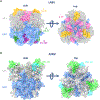
LASV residues contacted by 18.5C, 19.7E, 25.10C and 8.9F and JUNV residues contacted by CR1–28 and CR1–07. Shown are the structures of the LASV (PDB 7UL7) (A) and JUNV (B) from the side and the top. The residues contacted by the antibodies indicated are colored correspondingly. JUNV was modeled based on P26313 (Uniprot) with ColabFold (Mirdita et al., 2022). The GPCs were visualized using ChimeraX (Pettersen et al., 2004).
Fig. 6.
Fig. 6.
Strategies for a pan-Arenavirus vaccine generation. Several strategies can be employed for a pan-Arenavirus vaccine. Such as glycan masking of unwanted epitopes (A), the generation of a chimeric GPC, that is comprised of several different Arenavirus GPC domains, the generation of a consensus construct (C) or the sequential immunization with different Arenavirus GPCs (D). A vaccine strategically activating T cells might also be beneficial. A strategy for such a vaccine might be the generation of nanoparticles encompassing T cell activating peptides and presenting GPCs on their surface (E). Another approach would be to administer germline targeting immunogens that lead to the proliferation of naïve B cells that have specific B cell receptors after priming (F). Through so called shepherding immunizations and the final polishing immunization the desired B cells can be stimulated further.

References

    1. A Lassa Fever Vaccine Trial in Adults and Children Residing in West Africa, 2024. A Lassa Fever Vaccine Trial in Adults and Children Residing in West Africa [Internet]. [cited 2024 Apr 8]. Available from: https://clinicaltrials.gov/study/NCT05868733#more-information.
    1. Abraham J, Corbett KD, Farzan M, Choe H, Harrison SC, 2010. Apr 1. Structural basis for receptor recognition by New World hemorrhagic fever arenaviruses. Nat. Struct. Mol. Biol. 17 (4), 438–444. - PMC - PubMed
    1. Adolf-Bryfogle J, Kalyuzhniy O, Kubitz M, Weitzner BD, Hu X, Adachi Y, et al., 2018. Apr 27. RosettaAntibodyDesign (RAbD): a general framework for computational antibody design. PLoS Comput. Biol. 14 (4) e1006112. - PMC - PubMed
    1. Albariño César G., Bird Brian H., Chakrabarti Ayan K., Dodd Kimberly A., Mike Flint, Éric Bergeron, et al., 2011. Oct 1. The major determinant of attenuation in mice of the Candid1 vaccine for argentine hemorrhagic fever is located in the G2 glycoprotein transmembrane domain. J. Virol. 85 (19), 10404–10408. - PMC - PubMed
    1. Aliza Borenstein-Katz, Anastasiya Shulman, Hedva Hamawi, Orith Leitner, Ron Diskin, 2019. Apr 3. Differential antibody-based immune response against Isolated GP1 receptor-binding domains from Lassa and Junín viruses. J. Virol. 93 (8) 10.1128/jvi.00090-19. - DOI - PMC - PubMed

Publication types

LinkOut - more resources