Review
. 2024 Sep;22(9):2393-2403.
doi: 10.1016/j.jtha.2024.06.004. Epub 2024 Jun 21.

Venous thromboembolic disease genetics: from variants to function

Affiliations
Review

Venous thromboembolic disease genetics: from variants to function

Mary Underwood et al. J Thromb Haemost. 2024 Sep.

Abstract

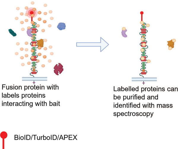
Venous thromboembolic disease (VTE) is a prevalent and potentially life-threatening vascular disease, including both deep vein thrombosis and pulmonary embolism. This review will focus on recent insights into the heritable factors that influence an individual's risk for VTE. Here, we will explore not only the discovery of new genetic risk variants but also the importance of functional characterization of these variants. These genome-wide studies should lead to a better understanding of the biological role of genes inside and outside of the canonical coagulation system in thrombus formation and lead to an improved ability to predict an individual's risk of VTE. Further understanding of the molecular mechanisms altered by genetic variation in VTE risk will be accelerated by further human genome sequencing efforts and the use of functional genetic screens.

Keywords: CRISPR; anticoagulants; genome-wide association study; secretory pathway; venous thrombosis.


Conflict of interest statement

Declaration of competing interests K.C.D., C.B., and M.U. have no conflicts of interest to report.

Figures

Figure 1.
Figure 1.. Gene Collapsing rare variant burden.
Next generation sequencing identifies rare variants in a hypothetical population of cases and controls where A) illustrates a gene with equal numbers of qualifying rare variants and B) displays a gene with higher frequency of qualifying rare variants in cases compared to controls. If the difference is significant after correction for multiple observations (number of genes examined) then the gene is a candidate risk factor. Down arrow indicates position of DNA variant in coding sequence.
Figure 2.
Figure 2.. CRISPR knock out screen.
A lentiviral library is prepared targeting all genes in genome or a restricted set of candidate genes. Each lentiviral particle encodes a single guide RNA and CRISPR/Cas9 proteins. The lentiviral library is titrated to transduce cells so that most cells are infected by a single particle and results in the loss of function of a single targeted gene. Transduced cells are selected and separated by flow cytometry based on abundance of target protein. Different bins of sorted cells are collected and DNA encoding the guide RNA are identified with next generation sequencing. Genes associated with altered target protein abundance are identified by the enrichment of complementary guide RNA in each bin. Figure created with BioRender.com
Figure 3.
Figure 3.. Deep Mutational Scan.
A library of cells is transfected with a pooled mammalian expression vector encoding all possible single missense variants in a protein or protein domain of interest. Often cells are transfected into a specific locus to normalize expression levels. Transfected cells are selected and sorted based on intracellular fluorescence generated by a fluorescent fusion protein or antibody binding. Alteration of intracellular signal is used as a proxy for the efficiency of secretion of the protein. Sorted cells are collected and DNA encoding the variants are identified with next generation sequencing. Figure created with BioRender.com
Figure 4.
Figure 4.. Massively parallel reporter assay.
A) Using putative cis regulatory domain sequence at a gene locus, tiled sequences are synthesized in a massive oligonucleotide array. B) These tiles of genomic DNA sequence are synthesized with a unique DNA sequence barcode and ultimately cloned into individual mammalian expression vectors where a minimal promoter separates the tile from its reporter and/or barcode. Cells are transfected with this library to identify which tiles transcription of RNA barcodes or translation of a reporter protein. RNA seq quantifies the expression level of each tile/barcode, identifying the area of the cis regulatory domain responsible for the enhanced expression. Altered translation of a reporter protein can be measured by flow cytometry and subsequent DNA sequencing. Figure created with BioRender.com
Figure 5.
Figure 5.. Proximity labelling scheme.
cDNA encoding a fusion protein is cloned and expressed in a cell line adding a proximity labeling protein (BioID or APEX) to a bait protein identified in GWAS or rare variant study. Additions of reagents induce biotinylation in vitro. Cell lysates are collected and biotinylated proteins are purified and identified by mass spectroscopy. Figure illustrates an endocytosed transmembrane protein with a terminal proximity labeling fusion indicated by the red lollipop. Figure created with BioRender.com

References

    1. Zoller B, Ohlsson H, Sundquist J, Sundquist K. A sibling based design to quantify genetic and shared environmental effects of venous thromboembolism in Sweden. Thromb Res. 2016. - PubMed
    1. Goswami C, Chattopadhyay A, Chuang EY. Rare variants: data types and analysis strategies. Ann Transl Med. 2021;9(12):961. - PMC - PubMed
    1. Torkamaneh D, Belzile F. Accurate Imputation of Untyped Variants from Deep Sequencing Data. Methods Mol Biol. 2021;2243:271–281. - PubMed
    1. Hindy G, Tyrrell DJ, Vasbinder A, et al. Increased soluble urokinase plasminogen activator levels modulate monocyte function to promote atherosclerosis. J Clin Invest. 2022;132(24). - PMC - PubMed
    1. Klarin D, Busenkell E, Judy R, et al. Genome-wide association analysis of venous thromboembolism identifies new risk loci and genetic overlap with arterial vascular disease. Nat Genet. 2019;51(11):1574–1579. - PMC - PubMed

LinkOut - more resources