. 2024 Jun 22;14(1):14419.
doi: 10.1038/s41598-024-64103-7.

Development of a single-chain variable antibody fragment against a conserved region of the SARS-CoV-2 spike protein

Affiliations

Development of a single-chain variable antibody fragment against a conserved region of the SARS-CoV-2 spike protein

Tingyu Gao et al. Sci Rep. .

Abstract

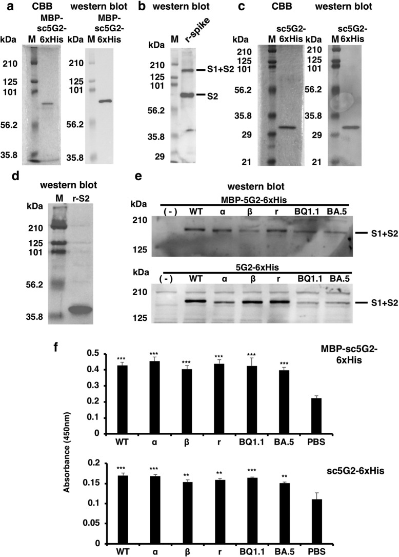
Severe acute respiratory syndrome coronavirus 2 (SARS-CoV-2) has prolonged the duration of the pandemic because of the continuous emergence of new variant strains. The emergence of these mutant strains makes it difficult to detect the virus with the existing antibodies; thus, the development of novel antibodies that can target both the variants as well as the original strain is necessary. In this study, we generated a high-affinity monoclonal antibody (5G2) against the highly conserved region of the SARS-CoV-2 spike protein to detect the protein variants. Moreover, we generated its single-chain variable antibody fragment (sc5G2). The sc5G2 expressed in mammalian and bacterial cells detected the spike protein of the original SARS-CoV-2 and variant strains. The resulting sc5G2 will be a useful tool to detect the original SARS-CoV-2 and variant strains.


Conflict of interest statement

The authors declare no competing interests.

Figures

Figure 1
Figure 1
The GTH peptide on the SARS-CoV-2 spike protein. (a) The position of the GTH peptide in the spike proteins of the α, β, γ and o BA.5 and BQ1.1 mutant strains. The vertical blue lines show the mutation sites in the spike proteins; the purple boxes show the position of the GTH peptide. SP: signal peptide. HR1 and HR2: heptad repeat 1 and 2, respectively. TM: transmembrane domain. (b) The 3D structure showing the position of the GTH peptide on the spike protein. The GTH peptide shown in light blue is located in the S2 structural domain and on the surface of the spike protein. The illustration was modified from PDB 7DF3 at https://www.wwpdb.org/pdb?id=pdb_00007df3.
Figure 2
Figure 2
The GTH peptide-specific IgG levels in mice sera following immunization. Blood samples were collected after the mice received three subcutaneous injections of the peptide. Changes in the peptide-specific IgG levels before and after immunization of the mice were compared using ELISA. The numbers in the abscissa indicate each immunized mouse. The data represent mean ± standard error (SE).
Figure 3
Figure 3
Hybridomas producing antibodies against GTH peptide, virus (WT), and recombinant S1 + S2 spike protein. The reactivities of cell culture supernatants from 20 hybridomas to (a) the GTH peptide, to (b) SARS-CoV-2 virus (WT) and to (c) the recombinant spike protein as determined by ELISA.
Figure 4
Figure 4
The 5G2 mAb binds to the WT and SARS-CoV-2 variants. (a) Binding of 5G2 mAb to SARS-CoV-2 spike proteins was detected by western blot. CBB, CBB stain (left). In the western blot (right), 0.2 μg/mL of 5G2 mAb was used and it was detected by anti-mIgG-HRP. “(-)” represents the uninfected Vero cell lysate. “WT” represents the Vero cells infected with Wuhan SARS-CoV-2. Recombinant “S1 + S2”, “S1” and “S2” stand for the recombinant spike proteins of “S1 + S2”, “S1” and “S2”, respectively. S1 + S2 and S2 proteins are indicated by the solid and open arrows, respectively. Commercially obtained recombinant S1 + S2 and S2 proteins, which lacks the cytoplasmic domain, were expressed by the Baculovirous-Insect Cell system and could be less glycosylated to show smaller molecular sizes compared to those expressed in the Vero cells. The recombinant S1 proteins produced by HEK293 cells shows larger molecular size due to the high glycosylation. (b) Immunofluorescence staining of spike proteins on the SARS-CoV-2 infected cell surface as detected by 5G2 mAb (green). Blue: DAPI was used for nuclear staining. Mock: Vero cells without SARS-CoV-2 infection, CoV-2: Vero cells infected with SARS-CoV-2. The leftmost panels show the phase contrast images. Scale bar: 10 μm. (c) ELISA for 5G2 mAb reactivity to 1 × 105 of SASR-CoV-2 WT and its variants (α, β, γ, and ο of BQ1.1, and BA.5). PBS: no virus coating. The data represent mean ± standard error (SE). The statistical significance was determined by one-way analysis of variance (one-way ANOVA). *P < 0.05, **P < 0.01, ***P < 0.001, ****P < 0.0001.
Figure 5
Figure 5
cDNA sequence of the variable regions of the 5G2 mAb. (a,b) cDNA sequences of the variable regions of the heavy (a) and the light (b) chains of 5G2. The underlined ATG sequence indicates the predicted start codons. (c,d) Predicted use of the V, (D), and J segments of the 5G2 heavy (c) and the light (d) chain sequences. The nucleic acid substitutions possibly resulting from the somatic hypermutation and the amino acid substitutions (red) are indicated. (e) Culture supernatant (1 mL) of HEK293FT cells in five 10 cm culture dishes transfected with the 5G2 heavy and light chain cDNAs (293FT + cDNA), but not that of HEK293FT cells without transfection (293FT), showed the bands corresponding to the S1 + S2 and S2 subunits of the recombinant spike protein (r-spike) by western blot. The bound recombinant 5G2 was detected by the anti-mIgG-HRP as described in the "Materials and methods" section.
Figure 6
Figure 6
MBP-sc5G2-6xHis produced from HEK293FT cells. (a) cDNA sequence of MBP-sc5G2-6xHis. The underlined DNA sequence encodes the (G4S)3 linker sequence. (b) MBP-sc5G2-6xHis produced by HEK293FT was subjected to SDS-PAGE. The proteins were stained with CBB (left) and analyzed by western blot using anti-MBP antibody and anti-mIgG-HRP (right). M: Molecular weight marker. (c) MBP-sc5G2-6xHis produced by HEK293FT (0.8 μg/mL) was confirmed to bind specifically to the S1 + S2 and the S2 subunit of the recombinant spike protein by western blot. The bound MBP-sc5G2-6xHis was detected by the anti-MBP antibody and anti-mIgG-HRP. M: Molecular weight marker. (d) MBP-sc5G2-6xHis produced by HEK293FT detects the S1 + S2 spike proteins in the Vero cells infected with SARS-CoV-2 WT and its variants, α, β, γ, BQ1.1, and BA.5 but not in the uninfected Vero cells (-) as confirmed by western blot using MBP-sc5G2-6xHis (0.8 μg/mL), anti-MBP antibody and anti-mIgG-HRP. (e) ELISA for MBP-sc5G2-6xHis reactivity to 1 × 105 of SASR-CoV-2 WT and its variants (α, β, γ, and ο of BQ1.1, and BA.5). PBS: no virus coating. The data represent mean ± standard error (SE). The statistical significance was determined by one-way analysis of variance (one-way ANOVA). **P < 0.01, ***P < 0.001.
Figure 7
Figure 7
Production of sc5G2s by E. coli. (a) Refolded MBP-sc5G2-6xHis produced by E. coli was subjected to SDS-PAGE. The proteins were stained with CBB (left) and analyzed by western blot using anti-MBP antibody and anti-mIgG-HRP (right). M: Molecular weight marker. (b) MBP-sc5G2-6xHis (0.8 μg/mL) produced by E. coli was used to detect recombinant S1 + S2 and the S2 proteins by western blot as in (a). M: Molecular weight marker. (c) Refolded sc5G2-6xHis protein produced by E. coli was subjected to SDS-PAGE. The proteins were stained with CBB (left) and analyzed by western blot using anti-6xHis antibody and anti-mIgG-HRP (right). M: Molecular weight marker. (d) sc5G2-6xHis (0.8 μg/mL) produced by E. coli was used to detect the recombinant S2 subunit of the spike protein without 6xHis (r-S2) by western blot using anti-6xHis antibody and anti-mIgG-HRP. M: Molecular weight marker. (e) MBP-sc5G2-6xHis (0.8 μg/mL, top) and sc5G2-6xHis (0.8 μg/mL, bottom) produced by E. coli were used to detect the S1 + S2 of the spike proteins in the Vero cells infected with SARS-CoV-2 WT as well as the variants, α, β, γ, BQ1.1, and BA.5 by western blot using anti-MBP antibody (top) or anti-6xHis antibody (bottom) and anti-mIgG-HRP. (-): Vero cells only. M: Molecular weight marker. (f) MBP-sc5G2-6xHis (0.8 μg/mL, top) and sc5G2-6xHis (0.8 μg/mL, bottom) produced by E. coli was used to detect particles of SARS-CoV-2 WT as well as the variants, α, β, γ, BQ1.1, and BA.5 by ELISA. PBS: no virus coated. The data represent mean ± standard error (SE). The statistical significance was determined by one-way analysis of variance (one-way ANOVA). **P < 0.01, ***P < 0.001.

Similar articles

References

    1. Muralidar S, Ambi SV, Sekaran S, Krishnan UM. The emergence of COVID-19 as a global pandemic: Understanding the epidemiology, immune response and potential therapeutic targets of SARS-CoV-2. Biochimie. 2020;179:85–100. doi: 10.1016/j.biochi.2020.09.018. - DOI - PMC - PubMed
    1. Feng S, et al. Correlates of protection against symptomatic and asymptomatic SARS-CoV-2 infection. Nat. Med. 2021;27:2032–2040. doi: 10.1038/s41591-021-01540-1. - DOI - PMC - PubMed
    1. Kumar S, Thambiraja TS, Karuppanan K, Subramaniam G. Omicron and Delta variant of SARS-CoV-2: A comparative computational study of spike protein. J. Med. Virol. 2022;94:1641–1649. doi: 10.1002/jmv.27526. - DOI - PubMed
    1. Kumar S, Karuppanan K, Subramaniam G. Omicron (BA.1) and sub-variants (BA.1.1, BA.2, and BA.3) of SARS-CoV-2 spike infectivity and pathogenicity: A comparative sequence and structural-based computational assessment. J. Med. Virol. 2022;94:4780–4791. doi: 10.1002/jmv.27927. - DOI - PMC - PubMed
    1. Fiolet T, Kherabi Y, MacDonald CJ, Ghosn J, Peiffer-Smadja N. Comparing COVID-19 vaccines for their characteristics, efficacy and effectiveness against SARS-CoV-2 and variants of concern: A narrative review. Clin. Microbiol. Infect. 2022;28:202–221. doi: 10.1016/j.cmi.2021.10.005. - DOI - PMC - PubMed

MeSH terms

Supplementary concepts