Case Reports
. 2024 Jun 22;20(1):271.
doi: 10.1186/s12917-024-04114-8.

Right-sided ureteral hemangiosarcoma in a paraplegic dog

Affiliations
Case Reports

Right-sided ureteral hemangiosarcoma in a paraplegic dog

Suellen Rodrigues Maia et al. BMC Vet Res. .

Abstract

Background: This study aims to describe a rare case of primary ureteral hemangiosarcoma, in which surgical intervention preserved the kidney and ureter after tumor removal.

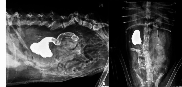
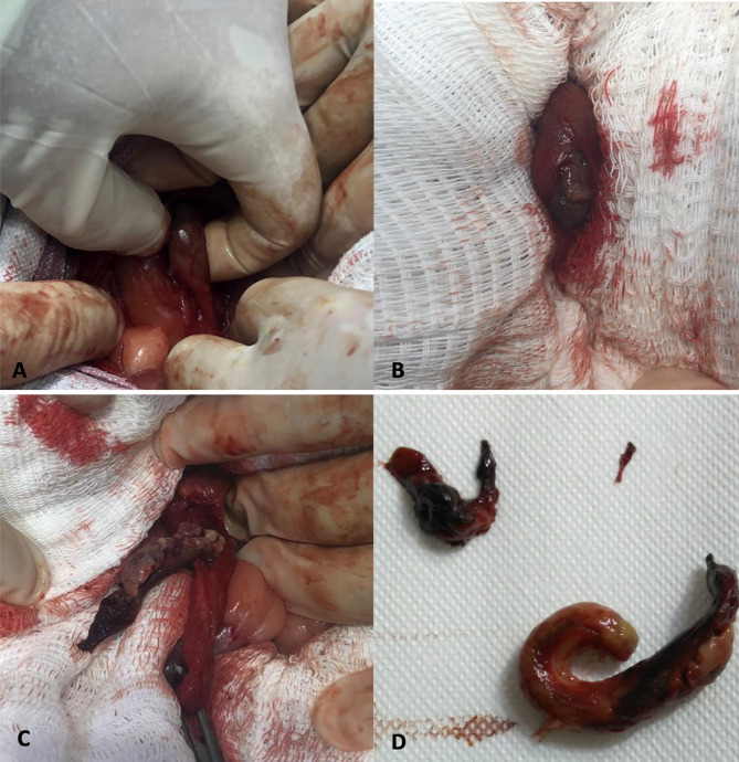
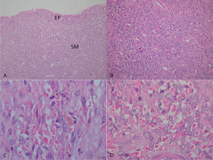
Case presentation: A 13-year-old, neutered male dog, weighing 14 kg, mixed-breed, presented with apathy, anorexia, acute-onset vomiting, and abdominal discomfort during the physical examination. Ultrasonography and pyelography revealed a right-sided dilation of the renal pelvis and ureter due to complete obstruction in the middle third of the ureter. A mass obstructing the lumen of the right ureter was completely resected, and ureteral suturing was performed, preserving the integrity of the involved structures. Histopathology confirmed primary ureteral hemangiosarcoma. Due to the local and non-invasive nature of the mass, chemotherapy was not initiated. The patient's survival was approximately two years, and normal renal function was preserved throughout this period.

Conclusions: Considering this type of tumor in the differential diagnosis of upper urinary tract obstructive disorders. Furthermore, the preservation of the ureter and kidney is a suitable therapeutic option after surgical resection of non-invasive tumors.

Keywords: Hydronephrosis; Pyelography; Ureteral neoplasia; Ureteral obstruction.


Conflict of interest statement

The authors declare no competing interests.

Figures

Fig. 1
Fig. 1
Ultrasound-guided percutaneous anterograde pyelography, in lateral (A) and ventrodorsal (B) projections. Radiographs were obtained immediately after injection of iodinated contrast, noting dilation of the right renal pelvis and ureter, obstruction of the contrast column in the middle third of the right ureter, as well as deviation of the bony axis of the lumbar spine, luxation of L3-L4, L5-L6, bone remodeling of L6 and articular processes of L3 and L4, due to a history of old trauma. Radiographic images were obtained after the administration of iodinated contrast at a dose of 600 mg/kg
Fig. 2
Fig. 2
Surgical Procedure. A) Identification of the affected ureter and isolation of the obstruction site (region between the surgeon’s fingers). B) Longitudinal incision of the ureter and appearance of the tissue obliterating the lumen. C) Dissection and exposure of the mass within the ureter lumen. D) Final appearance of the mass after complete removal. Only one mass was found and removed (fragmentation of the mass occurred after its manipulation)
Fig. 3
Fig. 3
Histopathological Findings. A) Fragment lined by well-differentiated urothelial epithelium (EP). Submucosa (SM) with diffuse malignant mesenchymal neoplasia. Hematoxylin and Eosin (HE) staining. Magnification 200x. B) Neoplastic cells with solid and diffuse organization. HE staining. Magnification 400x. C) Mitotic figure, cell in metaphase (arrow). HE staining. Magnification 1,000x. D) Malignant mesenchymal proliferation with the formation of irregular vascular spaces filled with erythrocytes. Neoplastic cells exhibit pleomorphic nuclei with vesicular appearance and prominent nucleoli. Mitotic figure, cells in prophase (arrow). HE staining. Magnification 1,000x

References

    1. Mullin C, Clifford CA. Histiocytic Sarcoma and Hemangiosarcoma Update. Veterinary Clin North America: Small Anim Pract. 2019;49(5):855–79. doi: 10.1016/j.cvsm.2019.04.009. - DOI - PubMed
    1. Griffin MA, Culp WTN, Rebhun RB. Canine and feline haemangiosarcoma. Vet Rec. 2021;189(9):585. doi: 10.1002/vetr.585. - DOI - PubMed
    1. Ward H, Fox LE, Calderwood-Mays MB, Hammer AS, Couto CG. Cutaneous hemangiosarcoma in 25 dogs: a retrospective study. J Vet Intern Med. 1994;8(5):345–8. doi: 10.1111/j.1939-1676.1994.tb03248.x. - DOI - PubMed
    1. Ware WA, Hopper DL. Cardiac tumors in dogs: 1982–1995. J Vet Intern Med. 1999;13(2):95–103. doi: 10.1111/j.1939-1676.1999.tb01136.x. - DOI - PubMed
    1. Mullin C, Clifford CA. Miscellaneous tumours: Hemangiosarcoma. In: Withrow SJ, Vail DM, Pag RL, editors. Withrow and MacEwen’s Small Animal Clinical Oncology. 5. St. Louis, MO, USA: Elsevier Saunders; 2020. pp. 773–8.

Publication types

LinkOut - more resources