. 2024 Jul 27:836:137881.
doi: 10.1016/j.neulet.2024.137881. Epub 2024 Jun 22.

Loss of Slc35a2 alters development of the mouse cerebral cortex

Affiliations

Loss of Slc35a2 alters development of the mouse cerebral cortex

Soad Elziny et al. Neurosci Lett. .

Abstract

Brain somatic variants in SLC35A2, an intracellular UDP-galactose transporter, are commonly identified mutations associated with drug-resistant neocortical epilepsy and developmental brain malformations, including focal cortical dysplasia type I and mild malformation of cortical development with oligodendroglial hyperplasia in epilepsy (MOGHE). However, the causal effects of altered SLC35A2 function on cortical development remain untested. We hypothesized that focal Slc35a2 knockout (KO) or knockdown (KD) in the developing mouse cortex would disrupt cortical development and change network excitability. Through two independent studies, we used in utero electroporation (IUE) to introduce CRISPR/Cas9/targeted guide RNAs or short-hairpin RNAs into the embryonic mouse brain at day 14.5-15.5 to achieve Slc35a2 KO or KD, respectively, from neural precursor cells. Slc35a2 KO or KD caused disrupted radial migration of electroporated neurons evidenced by heterotopic cells located in lower cortical layers and in the sub-cortical white matter. Slc35a2 KO in neurons did not induce changes in oligodendrocyte number, importantly suggesting that the oligodendroglial hyperplasia observed in MOGHE originates from distinct cell autonomous effects of Slc35a2 mutations. Adult KO mice were implanted with EEG electrodes for 72-hour continuous recording. Spontaneous seizures were not observed in focal Slc35a2 KO mice, but there was reduced seizure threshold following pentylenetetrazol injection. Here we demonstrate that focal Slc35a2 KO or KD in vivo disrupts corticogenesis through altered neuronal migration and that KO leads to reduced seizure threshold. Together these results demonstrate a direct causal role for SLC35A2 in cortical development.

Keywords: CRISPR; Cerebral cortex; Cortical dysplasia; Glycosylation; Short-hairpin RNA.


Conflict of interest statement

Declaration of competing interest The authors declare that they have no known competing financial interests or personal relationships that could have appeared to influence the work reported in this paper.

Figures

Fig. 1.
Fig. 1.. Slc35a2 KO alters neuronal migration.
A) Validation of CRISPR strategy targeting Slc35a2. Mouse N2A cells transfected with CRISPR/Cas9 plasmid DNA containing gRNAs targeting exons 2 and 3 of Slc35a2 show reduced SLC35A2 protein (western) and mRNA (RT-qPCR) expression compared to WT mice. N = 3 replicates per group. B) Plasmid DNA (EGFP) was delivered to the lateral ventricle of E14.5 mouse embryos by IUE to target neurons destined for the outer cortical layers. Coronal sections through the cortex showed appropriate laminar destination in layers II/III at PND 4. C) Slc35a2 KO resulted in significantly more malpositioned neurons (number of neurons outside layer II/III divided by total EGFP-positive cell count) located in the deep cortical layers and subcortical white matter (heterotopic neurons). Cortical layers are visualized by Satb2 (outer cortical layers) and Ctip2 (layer V) co-immunolabeling. D) Olig2 DAB IHC was compared for IUE KO and EGFP mice to assess Olig2 + cell density consistent with the MOGHE phenotype seen in human SLC35A2 patients. Olig2 + cells were manually counted within a standard sized ROI in the subcortical white matter for each hemisphere directly below the population of electroporated neurons. No differences were observed in Olig2-positive cell counts between Slc35a2 KO and control mice. Panels display adjacent sections showing Slc35a2 KO (left) and Olig2 expression (right, DAB staining). N = 5–6 mice per group and 1–3 sections per brain. Scale bars = 100 μm. * P < 0.05.
Fig. 2.
Fig. 2.. Slc35a2 KD is sufficient to disrupt cortical lamination.
A) Validation of shRNA strategy targeting Slc35a2. Mouse NIH/3T3 cells were transfected with plasmid DNA targeting exons 3 and 4 to assess KD efficiency. Slc35a2 mRNA expression was significantly reduced with shRNA treatment compared to scrambled control shRNA transfection. B) As seen in Fig. 1, plasmid DNA (EGFP) was delivered to the lateral ventricle of E15.5 mouse embryos by IUE and electroporated neurons were observed in layers II/III. C) Distribution of EGFP-positive cells in the mouse cortex at PND 1 was markedly altered in the KD conditions with more EGFP-positive cells observed in the deeper cortical layers and white matter. D) Slc35a2 KD resulted in significantly more neurons located in the deep cortical layers and subcortical white matter, as quantified by percent of EGFP-positive area in each of five bins spanning from the cortical plate (CP) to ventricular zone (VZ) defined based on DAPI staining. N = 3–8 mice per group and 1–3 sections per brain. Scale bars = 100 μm. Mean ± SEM. ** P < 0.01, ** P < 0.001.
Fig. 3.
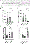
Fig. 3.. Slc35a2 KO results in a lower seizure threshold.
A) Representative EEG recording following PTZ injection. Left portion of EEG recording shows baseline activity, line depicts the onset of a Racine score 4 seizure with clear onset of high amplitude activity. Scale bar = 60 μV by 500 msec. B) Slc35a2 KO mice recorded at PND 60 and C) PND 120 exhibited reduced seizure thresholds as measured by latency to first electrographic seizure when compared to EGFP electroporated and WT control groups. D) Slc35a2 KO mice recorded at PND 60 and E) PND 120 reached higher maximum Racine scores during the recording period. (N = 6–12 mice per group. *P < 0.05.).

Update of

References

    1. Blumcke I, Aronica E, Miyata H, et al., International recommendation for a comprehensive neuropathologic workup of epilepsy surgery brain tissue: A consensus Task Force report from the ILAE Commission on Diagnostic Methods, Epilepsia 57 (3) (2016. Mar) 348–358. - PubMed
    1. Winawer MR, Griffin NG, Samanamud J, et al., Somatic SLC35A2 variants in the brain are associated with intractable neocortical epilepsy, Ann. Neurol 83 (6) (2018. Jun) 1133–1146. - PMC - PubMed
    1. Lai D, Gade M, Yang E, et al., Somatic variants in diverse genes leads to a spectrum of focal cortical malformations, Brain 145 (8) (2022. Aug 27) 2704–2720. - PMC - PubMed
    1. Bedrosian TA, Miller KE, Grischow OE, et al., Detection of brain somatic variation in epilepsy-associated developmental lesions, Epilepsia 63 (8) (2022. Aug) 1981–1997. - PubMed
    1. Bonduelle T, Hartlieb T, Baldassari S, et al., Frequent SLC35A2 brain mosaicism in mild malformation of cortical development with oligodendroglial hyperplasia in epilepsy (MOGHE), Acta Neuropathol. Commun 9 (1) (2021. Jan 6) 3. - PMC - PubMed

Substances