

Utility of Thin-slice Fat-suppressed Single-shot T2-weighted MR Imaging with Deep Learning Image Reconstruction as a Protocol for Evaluating the Pancreas
- PMID: 38910138
- PMCID: PMC12406158
- DOI: 10.2463/mrms.mp.2024-0017
Utility of Thin-slice Fat-suppressed Single-shot T2-weighted MR Imaging with Deep Learning Image Reconstruction as a Protocol for Evaluating the Pancreas
Abstract
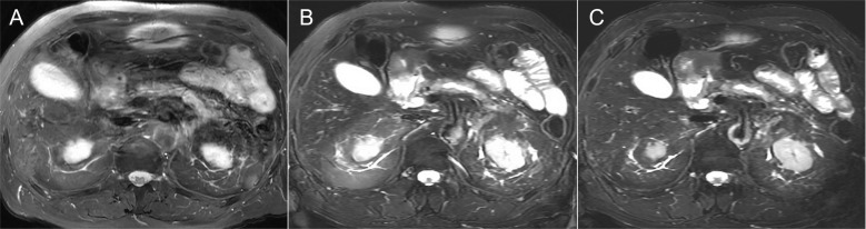
Purpose: To compare the utility of thin-slice fat-suppressed single-shot T2-weighted imaging (T2WI) with deep learning image reconstruction (DLIR) and conventional fast spin-echo T2WI with DLIR for evaluating pancreatic protocol.
Methods: This retrospective study included 42 patients (mean age, 70.2 years) with pancreatic cancer who underwent gadoxetic acid-enhanced MRI. Three fat-suppressed T2WI, including conventional fast-spin echo with 6 mm thickness (FSE 6 mm), single-shot fast-spin echo with 6 mm and 3 mm thickness (SSFSE 6 mm and SSFSE 3 mm), were acquired for each patient. For quantitative analysis, the SNRs of the upper abdominal organs were calculated between images with and without DLIR. The pancreas-to-lesion contrast on DLIR images was also calculated. For qualitative analysis, two abdominal radiologists independently scored the image quality on a 5-point scale in the FSE 6 mm, SSFSE 6 mm, and SSFSE 3 mm with DLIR.
Results: The SNRs significantly improved among the three T2-weighted images with DLIR compared to those without DLIR in all patients (P < 0.001). The pancreas-to-lesion contrast of SSFSE 3 mm was higher than those of the FSE 6 mm (P < 0.001) and tended to be higher than SSFSE 6 mm (P = 0.07). SSFSE 3 mm had the highest image qualities regarding pancreas edge sharpness, pancreatic duct clarity, and overall image quality, followed by SSFSE 6 mm and FSE 6 mm (P < 0.0001).
Conclusion: SSFSE 3 mm with DLIR demonstrated significant improvements in SNRs of the pancreas, pancreas-to-lesion contrast, and image quality more efficiently than did SSFSE 6 mm and FSE 6 mm. Thin-slice fat-suppressed single-shot T2WI with DLIR can be easily implemented for pancreatic MR protocol.
Keywords: T2-weighted; artificial intelligence; magnetic resonance imaging; pancreas.
Figures


References
-
- Lee SS, Byun JH, Hong HS, et al. Image quality and focal lesion detection on T2-weighted MR imaging of the liver: Comparison of two high-resolution free-breathing imaging techniques with two breath-hold imaging techniques. J Magn Reson Imaging 2007; 26:323–330. - PubMed
-
- Klessen C, Asbach P, Kroencke TJ, et al. Magnetic resonance imaging of the upper abdomen using a free-breathing T2-weighted turbo spin echo sequence with navigator triggered prospective acquisition correction. J Magn Reson Imaging 2005; 21:576–582. - PubMed
-
- Schreiber-Zinaman J, Rosenkrantz AB. Frequency and reasons for extra sequences in clinical abdominal MRI examinations. Abdom Radiol (NY) 2017; 42:306–311. - PubMed
-
- Hirokawa Y, Isoda H, Maetani YS, Arizono S, Shimada K, Togashi K. Evaluation of motion correction effect and image quality with the periodically rotated overlapping parallel lines with enhanced reconstruction (PROPELLER) (BLADE) and parallel imaging acquisition technique in the upper abdomen. J Magn Reson Imaging 2008; 28:957–962. - PubMed
-
- Rosenkrantz AB, Patel JM, Babb JS, Storey P, Hecht EM. Liver MRI at 3 T using a respiratory-triggered time-efficient 3D T2-weighted technique: Impact on artifacts and image quality. AJR Am J Roentgenol 2010; 194:634–641. - PubMed