. 2024 Jun 23;14(1):14438.
doi: 10.1038/s41598-024-64815-w.

Neferine inhibits BMECs pyroptosis and maintains blood-brain barrier integrity in ischemic stroke by triggering a cascade reaction of PGC-1α

Affiliations

Neferine inhibits BMECs pyroptosis and maintains blood-brain barrier integrity in ischemic stroke by triggering a cascade reaction of PGC-1α

Zi-Jian Zheng et al. Sci Rep. .

Abstract

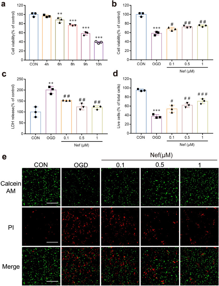
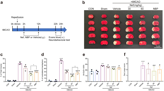
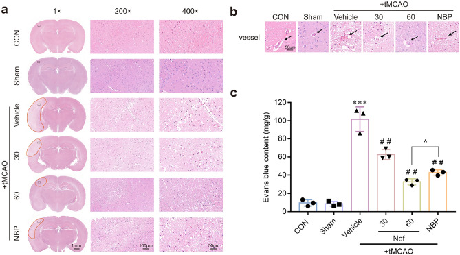
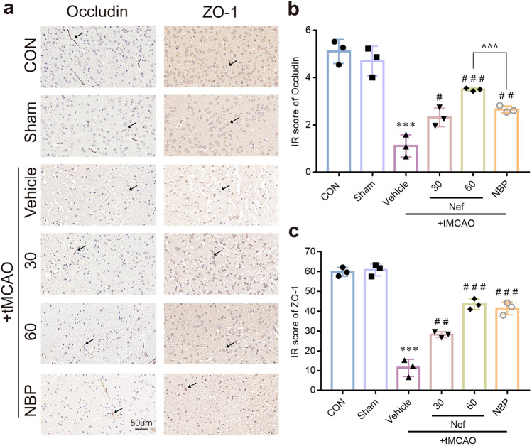
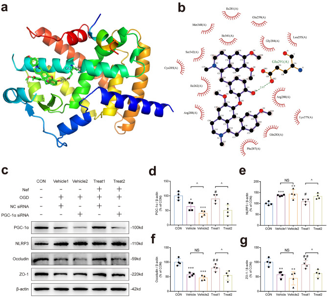
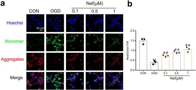
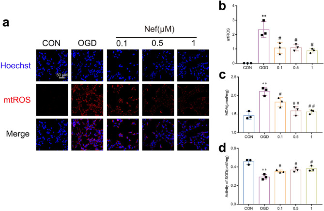
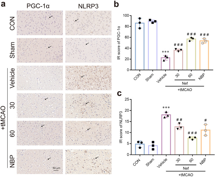
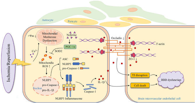
Blood-brain barrier disruption is a critical pathological event in the progression of ischemic stroke (IS). Most studies regarding the therapeutic potential of neferine (Nef) on IS have focused on neuroprotective effect. However, whether Nef attenuates BBB disruption during IS is unclear. We here used mice underwent transient middle cerebral artery occlusion (tMCAO) in vivo and bEnd.3 cells exposed to oxygen-glucose deprivation/reoxygenation (OGD/R) injury in vitro to simulate cerebral ischemia. We showed that Nef reduced neurobehavioral dysfunction and protected brain microvascular endothelial cells and BBB integrity. Molecular docking, short interfering (Si) RNA and plasmid transfection results showed us that PGC-1α was the most binding affinity of biological activity protein for Nef. And verification experiments were showed that Nef upregulated PGC-1α expression to reduce mitochondrial oxidative stress and promote TJ proteins expression, further improves the integrity of BBB in mice. Intriguingly, our study showed that neferine is a natural PGC-1α activator and illustrated the mechanism of specific binding site. Furthermore, we have demonstrated Nef reduced mitochondria oxidative damage and ameliorates endothelial inflammation by inhibiting pyroptosis to improve BBB permeability through triggering a cascade reaction of PGC-1α via regulation of PGC-1α/NLRP3/GSDMD signaling pathway to maintain the integrity of BBB in ischemia/reperfusion injury.

Keywords: Blood–Brain Barrier; Ischemic stroke; Neferine; Oxygen and glucose-deprivation/reperfusion; PGC-1α; Transient middle cerebral artery occlusion.


Conflict of interest statement

The authors declare no competing interests.

Figures

Figure 1
Figure 1
Effects of Nef on the cell viability of bEnd.3 cells under OGD/R conditions. Cell viability were measured after exposed to OGD for 4 to 10 h (a). Effect of Nef treatment (0.1 μM: 0.5 μM: 1 μM) on cell viability (b). The LDH release was measured after being treated with Nef (0.1 μM: 0.5 μM: 1 μM) during 9 h of OGD by a commercial kit (c). The live cells proportion was measured after being treated with Nef (0.1 μM: 0.5 μM: 1 μM) during 9 h of OGD by Live/Dead assay kit (d,e). scale bar: 200 μm. The experiments were performed in triplicate and repeated at least three times in different days. **P < 0.01: ***P < 0.001νs. CON: #P < 0.05: ##P < 0.01: ###P < 0.001νs. OGD.
Figure 2
Figure 2
Effects of Nef on cerebral infarct volume: brain swelling volume: brain water content and neuroethology in mice induced by tMCAO. Experimental design of tMCAO model (a). Representative photographs of mice brain tissue stained with 0.5% TTC solution (n = 3) (b). Quantification of cerebral infarction volume (n = 3) (c). Quantification of brain swelling volume (n = 3) (d). Quantification of brain water content (n = 3) (e). Evaluation of neural injury in mice by using Zea Longa Neuroethology score (n = 12) (f). Scare bar: 1 cm. **P < 0.01: ***P < 0.001νs. Sham: #P < 0.05: ##P < 0.01: ###P < 0.001νs. Vehicle: ^P < 0.05 νs. NBP.
Figure 3
Figure 3
Effects of Nef on brain morphology of tMCAO mice. Examination of H&E stained sections provided insight into the morphological characteristics of mouse brains and cerebral vessels following tMCAO. Normal tissues were characterized by a purple-red coloration with rounded: full nuclei and a compact structure: while damaged tissue appeared white with pyknotic nuclei and the presence of numerous lacunae (n = 3) (a). Representative images of cerebral vessels (n = 3) (b). Quantification of brain Evans blue content (n = 3) (c). Scare bar: 50 μm. ***P < 0.001νs. Sham: ##P < 0.01νs. Vehicle: ^P < 0.05 νs. NBP (n = 3).
Figure 4
Figure 4
Effects of Nef on TJ proteins expression in mice underwent tMCAO. Representative micrographs of IHC staining (a). Quantitative analysis of the immunohistochemistry (b,c). Scare bar: 50 μm. ***P < 0.001νs. Sham: #P < 0.05: ##P < 0.01: ###P < 0.001νs. Vehicle: ^^^P < 0.001 between treatment groups: (n = 3).
Figure 5
Figure 5
Effects of Nef on tube formation and endothelial barrier integrity under OGD/R condition. Representative Western blot bands of Occludin and ZO-1 (a). Quantitative analysis of the expressions of proteins(b,c). Relative mRNA expression quantification analysis (d,e). Quantitative analysis of tube branch length (f). Representative pictures depicting the formation of tubular structure (g). The TEER assay was employed to evaluate the barrier permeability of the bEnd.3 monolayer (h: j). EBA flux analysis was employed to evaluate the barrier permeability of the bEnd.3 monolayer (i: k). Scale bar: 200 μm. The experiments were performed in triplicate and repeated at least three times in different days. *P < 0.05: **P < 0.01: ***P < 0.001νs. Con: #P < 0.05: ##P < 0.01: ###P < 0.001νs. OGD.
Figure 6
Figure 6
The protective effects of Nef in bEnd.3 cells is involved in promoting PGC-1α expression. Prediction of the 3D binding site and interactions was visualized using Pymol(a). Prediction of the 2D binding site and interactions was visualized using LigPlot + . Hydrogen bonds are indicated by lines colored in cyan. (b). PGC-1α knockdown significantly reduced Nef's inhibitory effect on NLRP3 protein and reduced the expression of TJ proteins elevated by Nef (c). Quantitative analysis of the relative proteins(dg). *P < 0.05: **P < 0.01: ***P < 0.001 νs. CON: #P < 0.05: ##P < 0.01 νs. Vehicle1: ^P < 0.05 between NC or PGC-1α siRNA transfection groups: (n = 4).
Figure 7
Figure 7
Effects of Nef on MMP in bEnd. 3 cells under OGD/R condition. Representative immunofluorescence staining of MMP in bEnd.3 cells (a). MMP as estimated by JC-1 red/green fluorescence intensity rate (FIR) (b). Scare bar: 50 μm. The experiments were performed in triplicate and repeated at least three times in different days. **P < 0.01 νs. CON: ##P < 0.01 νs. OGD.
Figure 8
Figure 8
Effects of Nef on mtROS and intracellular oxidative stress in bEnd. 3 cells under OGD/R condition. Representative immunofluorescence of mtROS in bEnd.3 cells (a). Quantification of mtROS levels(b). Quantification of MDA and SOD represents cellular oxidative stress level (c,d). Scare bar = 50 μm. The experiments were performed in triplicate and repeated at least three times in different days. **P < 0.01 νs. CON: #P < 0.05: ##P < 0.01 νs. OGD.
Figure 9
Figure 9
Effects of Nef on PGC-1α and NLRP3 proteins expression in mice underwent tMCAO. Representative micrographs of IHC staining (a). Quantitative analysis of the immunohistochemistry (b,c). Scare bar: 50 μm. ***P < 0.001νs. Sham: #P < 0.05: ##P < 0.01: ###P < 0.001νs. Vehicle: ^^^P < 0.001 between treatment groups: (n = 3).
Figure 10
Figure 10
The regulatory effects of Nef on PGC-1α/NLRP3/GSDMD signaling pathway. Expression of PGC-1α and NLRP3 signaling pathway associated proteins (ASC: AIM2: NLRP3: Caspase-1: Cleaved caspase-1: IL-1β: IL-18: GSDMD) in bEnd.3 cells (aj). *P < 0.05: **P < 0.01: ***P < 0.001νs. Con: #P < 0.05: ##P < 0.01: ###P < 0.001νs. OGD: (n = 3).
Figure 11
Figure 11
A schematic diagram is presented to illustrate the proposed mechanisms underlying the beneficial effects of Neferine in BMECs and its anti-ischemic activities. Nef activates PGC-1α: increases intracellular SOD activity and reduces the aggradation of mtROS: thereby inhibiting the assembly of the NLRP3 inflammasome and suppressing the cleavage and release of IL-1β and IL-18: further improving TJ disruption and BMECs pyroptosis. This effect of Nef leads to protect BMECs: BBB integrity and ischemic brain injury induced by ischemic stroke.

References

    1. He Z, Ning N, Zhou Q, Khoshnam SE, Farzaneh M. Mitochondria as a therapeutic target for ischemic stroke. Free Radic. Biol. Med. 2020;146:45–58. doi: 10.1016/j.freeradbiomed.2019.11.005. - DOI - PubMed
    1. Jia J, Jin H, Nan D, Yu W, Huang Y. New insights into targeting mitochondria in ischemic injury. Apoptosis. 2021 doi: 10.1007/s10495-021-01661-5. - DOI - PubMed
    1. Abu SO, Arroum T, Morris S, Busch KB. PGC-1α Is a master regulator of mitochondrial lifecycle and ROS stress response. Antioxidants. 2023 doi: 10.3390/antiox12051075. - DOI - PMC - PubMed
    1. Xu Y, et al. The PGC-1α Activator ZLN005 Ameliorates Ischemia-Induced Neuronal Injury In Vitro and In Vivo. Cell. Mol. Neurobiol. 2018 doi: 10.1007/s10571-017-0567-0. - DOI - PMC - PubMed
    1. Chen S-D, et al. Activation of calcium/calmodulin-dependent protein kinase IV and peroxisome proliferator-activated receptor γ coactivator-1α signaling pathway protects against neuronal injury and promotes mitochondrial biogenesis in the hippocampal CA1 subfield after transient global ischemia. J. Neurosci. Res. 2010 doi: 10.1002/jnr.22469. - DOI - PubMed

MeSH terms

Substances