. 2024 Jun 23;30(1):95.
doi: 10.1186/s10020-024-00864-1.

β-hydroxybutyrate resensitizes colorectal cancer cells to oxaliplatin by suppressing H3K79 methylation in vitro and in vivo

Affiliations

β-hydroxybutyrate resensitizes colorectal cancer cells to oxaliplatin by suppressing H3K79 methylation in vitro and in vivo

Meng Deng et al. Mol Med. .

Abstract

Background: Ketone β-hydroxybutyrate (BHB) has been reported to prevent tumor cell proliferation and improve drug resistance. However, the effectiveness of BHB in oxaliplatin (Oxa)-resistant colorectal cancer (CRC) and the underlying mechanism still require further proof.

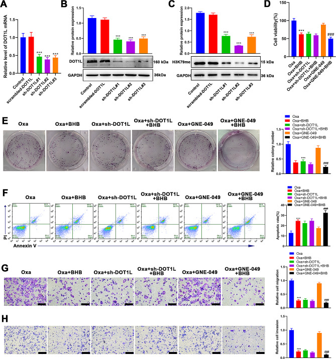
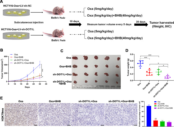
Methods: CRC-Oxa-resistant strains were established by increasing concentrations of CRC cells to Oxa. CRC-Oxa cell proliferation, apoptosis, invasion, migration, and epithelial-mesenchymal transition (EMT) were checked following BHB intervention in vitro. The subcutaneous and metastasis models were established to assess the effects of BHB on the growth and metastasis of CRC-Oxa in vivo. Eight Oxa responders and seven nonresponders with CRC were enrolled in the study. Then, the serum BHB level and H3K79me, H3K27ac, H3K14ac, and H3K9me levels in tissues were detected. DOT1L (H3K79me methyltransferase) gene knockdown or GNE-049 (H3K27ac inhibitor) use was applied to analyze further whether BHB reversed CRC-Oxa resistance via H3K79 demethylation and/or H3K27 deacetylation in vivo and in vitro.

Results: Following BHB intervention based on Oxa, the proliferation, migration, invasion, and EMT of CRC-Oxa cells and the growth and metastasis of transplanted tumors in mice were suppressed. Clinical analysis revealed that the differential change in BHB level was associated with drug resistance and was decreased in drug-resistant patient serum. The H3K79me, H3K27ac, and H3K14ac expressions in CRC were negatively correlated with BHB. Furthermore, results indicated that H3K79me inhibition may lead to BHB target deletion, resulting in its inability to function.

Conclusions: β-hydroxybutyrate resensitized CRC cells to Oxa by suppressing H3K79 methylation in vitro and in vivo.

Keywords: Colorectal cancer; Drug resistance; H3K79me; Oxaliplatin; Β-hydroxybutyrate.


Conflict of interest statement

The authors declare that they have no competing interests.

Figures

Fig. 1
Fig. 1
BHB reversed the acquired resistance of CRC cells to Oxa. A: CCK-8 determination of the cell viability of HCT-116 and LoVo parental and drug-resistant cells treated with Oxa at a series of concentrations (0, 0.1, 0.5, 1, 2, 5, 10, 20, 40, and 80 µM) for 24 h, and then, the IC50 value was calculated. B: Chemical structural formula of BHB. C: CCK-8 determination of the cell viability of HCT-116 and LoVo parental and drug-resistant cells treated with BHB at a series of concentrations (0, 1, 2, 4, 8, 16, 20, and 40 mM) for 24 h. D: CCK-8 determination of the cell viability of HCT-116-Oxa and LoVo-Oxa cells incubated with Oxa (10 µM) and/or BHB (4 mM) for 24 h. E: Clone formation determination of the cell proliferation of HCT-116-Oxa and LoVo-Oxa cells incubated with Oxa (10 µM) and/or BHB (4 mM) for 24 h. F: Annexin V-FITC/PI double staining determination of cell apoptosis of HCT-116-Oxa and LoVo-Oxa cells incubated with Oxa (10 µM) and/or BHB (4 mM) for 24 h. G: Western blotting determination of Bax and Bcl-2 protein levels in HCT-116-Oxa and LoVo-Oxa cells incubated with Oxa (10 µM) and/or BHB (4 mM) for 24 h. H–I: Transwell assay determination of the invasion and migration ability of HCT-116-Oxa and LoVo-Oxa cells incubated with Oxa (10 µM) and/or BHB (4 mM) for 24 h. J: Western blotting determination of E-cadherin, N-cadherin, and vimentin protein levels in HCT-116-Oxa and LoVo-Oxa cells incubated with Oxa (10 µM) and/or BHB (4 mM) for 24 h. Data: Mean ± SEM. **P < 0.01, ***P < 0.001 vs. Control group. ###P < 0.001 vs. Oxa group (n = 3)
Fig. 2
Fig. 2
BHB resensitized CRC to Oxa in vivo.A: The animal experiment process. The tumor-bearing nude mice were randomly divided into four groups (n = 6): control (saline), Oxa (5 mg/kg/day), BHB (40 mg/mice/day), and Oxa + BHB. B: From the first day of administration, the weight of nude mice was measured once every 2 days. C: The tumor growth curve of each group. D: Representative diagram of tumors in each group. E: The tumor weight of each group. F: Immunohistochemical determination of the Ki67 (day 30) and cleaved-caspase3 (day 15) expressions in tumor tissues of subcutaneous tumor model mice after injecting Oxa (5 mg/kg/day) and/or BHB (40 mg/mice/day), and the immunohistochemical score was calculated. G: Western blotting determination of the E-cadherin, N-cadherin, and vimentin expression levels in tumor tissues of subcutaneous tumor model mice after injecting Oxa (5 mg/kg/day) and/or BHB (40 mg/mice/day). H: Liver metastasis histograms and HE staining sections of nude mice in each group. I: Statistics of liver metastatic nodules of nude mice in each group. Data: Mean ± SEM. *P < 0.05, ***P < 0.001 vs. Control group. ###P<0.001 vs. Oxa group (n = 6)
Fig. 3
Fig. 3
Serum BHB level is associated with Oxa resistance in patients with CRC. A: The serum of eight Oxa-sensitive and seven drug-resistant patients was collected after Oxa chemotherapy, and the BHB content was analyzed by β-hydroxybutyrate (ketone body) colorimetric assay kit. B: Immunohistochemical determination of the H3K79me, H3K27ac, H3K14ac, and H3K9me expressions in tumor tissues of 15 patients with CRC, including eight Oxa-sensitive (responder) and seven nonresponder patients. C: Correlation analysis between H3K79me tissue score and serum BHB level. D: Correlation analysis between H3K27ac tissue score and serum BHB level. E: Correlation analysis between the H3K14ac tissue score and serum BHB level. Data: Mean ± SEM. *P < 0.05, ***P < 0.001 vs. the responder group
Fig. 4
Fig. 4
Resensitizing CRC cells to Oxa by BHB mainly relied on H3K79 methylation suppression. A: RT-qPCR determination of the mRNA level of DOT1L in HCT-116-Oxa cells, which transfected with scrambled-DOT1L, sh-DOT1L#1, sh-DOT1L#2, and sh-DOT1L#3. B: Western blotting determination of the protein level of DOT1L in HCT-116-Oxa cells, which transfected with scrambled-DOT1L, sh-DOT1L#1, sh-DOT1L#2, and sh-DOT1L#3. C: CCK-8 determination of the cell viability of HCT-116-Oxa cells, which inhibited H3K79 methylation or H3K27 acetylation and/or BHB intervention. D: Clone formation determination of the cell proliferation of HCT-116-Oxa cells, which inhibited H3K79 methylation or H3K27 acetylation and/or BHB intervention. E: Annexin V-FITC/PI double staining determination of the cell apoptosis of HCT-116-Oxa cells, which inhibited H3K79 methylation, H3K27 acetylation, and/or BHB intervention. F: Transwell assay determination of the cell migration of HCT-116-Oxa cells, which inhibited H3K79 methylation, H3K27 acetylation, and/or BHB intervention. G: Transwell assay determination of the cell invasion of HCT-116-Oxa cells, which inhibited H3K79 methylation, H3K27 acetylation, and/or BHB intervention. Data: Mean ± SEM. ***P < 0.001 vs. Oxa group. ###P < 0.001 vs. Oxa + GNE-409 group (n = 3)
Fig. 5
Fig. 5
BHB reversed Oxa resistance by suppressing H3K79 methylation in vivo. A: The animal experiment process. The tumor-bearing nude mice were randomly divided into four groups (n = 6): Oxa (5 mg/kg/day), Oxa + BHB (40 mg/mice/day), sh-DOT1L + Oxa, and sh-DOT1L + Oxa + BHB. B: The tumor growth curve of each group. C: Representative diagram of tumors in each group. D: The tumor weight of each group. E: Immunohistochemical determination of the expression of H3K79me in tumor tissues of subcutaneous tumor model mice, and the immunohistochemical score was calculated. Data: Mean ± SEM. ***P < 0.001 vs. Oxa group (n = 6)

References

    1. Baidoun F, Elshiwy K, Elkeraie Y, Merjaneh Z, Khoudari G, Sarmini MT, et al. Colorectal Cancer epidemiology: recent trends and Impact on outcomes. Curr Drug Targets. 2021;22:998–1009. doi: 10.2174/18735592MTEx9NTk2y. - DOI - PubMed
    1. Bajbouj K, Al-Ali A, Ramakrishnan RK, Saber-Ayad M, Hamid Q. Histone modification in NSCLC: Molecular mechanisms and therapeutic targets. Int J Mol Sci. 2021;22:11701. doi: 10.3390/ijms222111701. - DOI - PMC - PubMed
    1. Bannister AJ, Kouzarides T. Regulation of chromatin by histone modifications. Cell Res. 2011;21:381–95. doi: 10.1038/cr.2011.22. - DOI - PMC - PubMed
    1. Barrea L, Caprio M, Tuccinardi D, Moriconi E, Di Renzo L, Muscogiuri G, et al. Could ketogenic diet starve cancer? Emerging evidence. Crit Rev Food Sci Nutr. 2022;62:1800–21. doi: 10.1080/10408398.2020.1847030. - DOI - PubMed
    1. Byun WS, Lee GH, Park HG, Lee SK. Inhibition of DOT1L by half-selenopsammaplin A Analogs suppresses Tumor Growth and EMT-Mediated metastasis in Triple-negative breast Cancer. Pharmaceuticals (Basel) 2020;14:18. doi: 10.3390/ph14010018. - DOI - PMC - PubMed

MeSH terms