. 2024 Sep 4;32(9):3114-3127.
doi: 10.1016/j.ymthe.2024.06.029. Epub 2024 Jun 22.

Overcoming effector T cell exhaustion in ovarian cancer ascites with a novel adenovirus encoding for a MUC1 bispecific antibody engager and IL-2 cytokine

Affiliations

Overcoming effector T cell exhaustion in ovarian cancer ascites with a novel adenovirus encoding for a MUC1 bispecific antibody engager and IL-2 cytokine

Saru Basnet et al. Mol Ther. .

Erratum in

Abstract

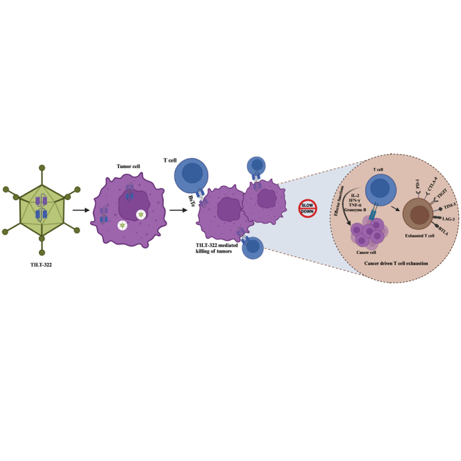
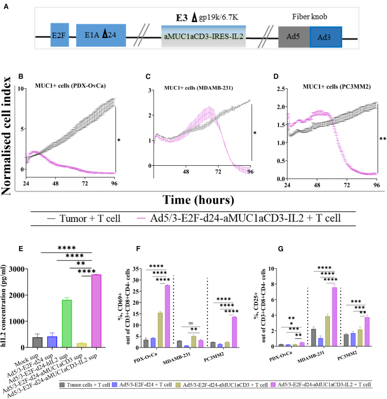
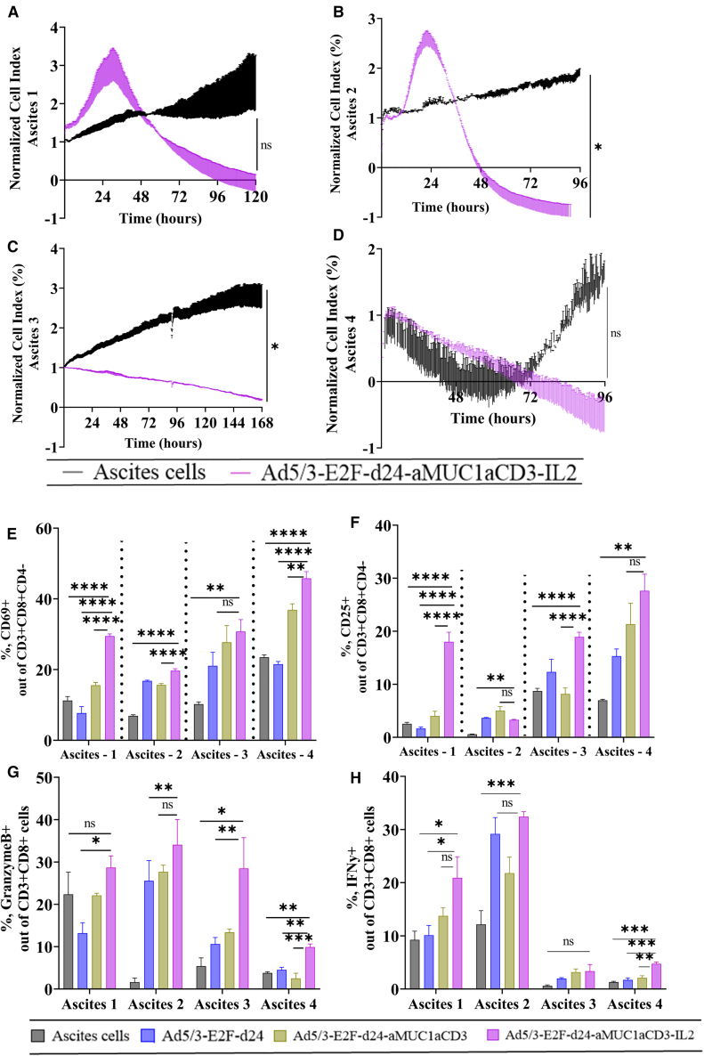
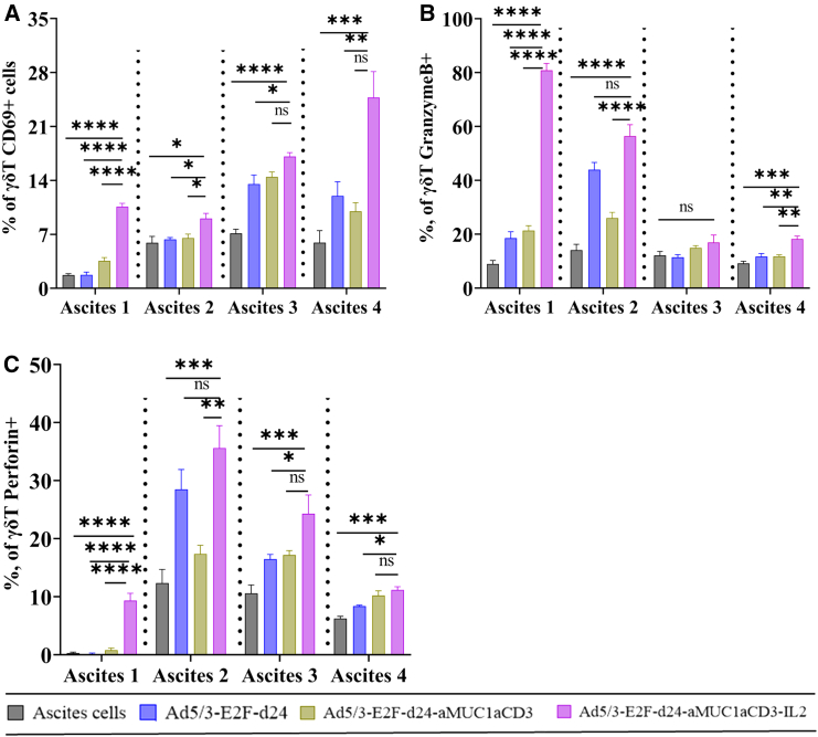
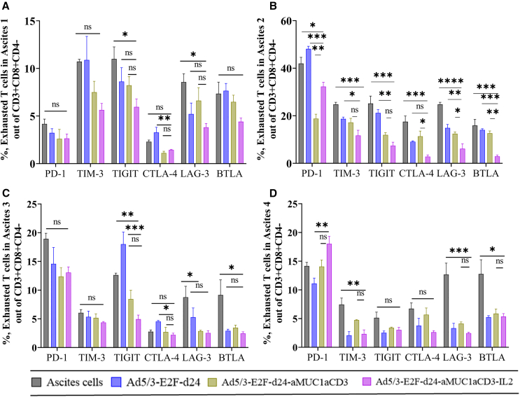
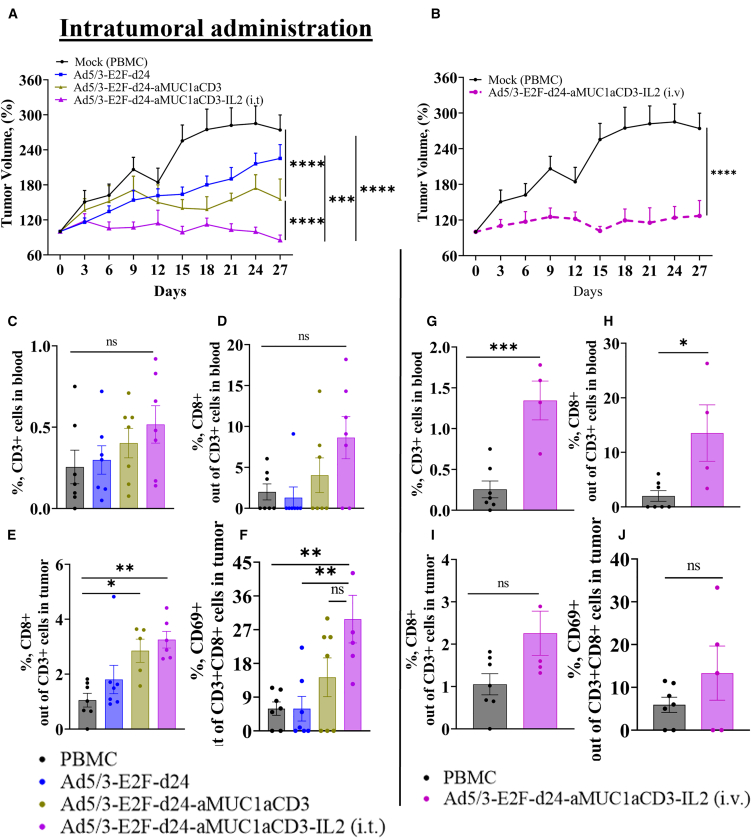
T cell-focused cancer immunotherapy including checkpoint inhibitors and cell therapies has been rapidly evolving over the past decade. Nevertheless, there remains a major unmet medical need in oncology generally and immuno-oncology specifically. We have constructed an oncolytic adenovirus, Ad5/3-E2F-d24-aMUC1aCD3-IL-2 (TILT-322), which is armed with a human aMUC1aCD3 T cell engager and IL-2. TILT-322 treatment stimulated T cell cytotoxicity through the increased presence of granzyme B, perforin, and interferon-gamma. Additional immune profiling indicated TILT-322 increased gamma delta T cell activation and impacted other cell types such as natural killer cells and natural killer-like T cells that are traditionally involved in cancer immunotherapy. TILT-322 treatment also decreased the proportion of exhausted CD8+ T cells as demarked by immune checkpoint expression in ovarian ascites samples. Overall, our data showed that TILT-322 treatment led to an enhanced T cell activation and reversed T cell exhaustion translating into high antitumor efficacy when given locally or intravenously. The analysis of blood and tumors isolated from an in vivo patient-derived ovarian cancer xenograft model suggested TILT-322 mediated tumor control through improved T cell functions. Therefore, TILT-322 is a promising novel anti-tumor agent for clinical translation.

Keywords: IL-2; Mucin1; T cell exhaustion; bispecific T cell engager; oncolytic adenovirus.


Conflict of interest statement

Declaration of interests A.H. is a shareholder of Circio Holdings ASA. (Norway). A.H., J.C., J.M.S., and D.Q. are employees and shareholders of TILT Biotherapeutics, Ltd.

Figures

None
Graphical abstract
Figure 1
Figure 1
Ad5/3-E2F-d24-aMUC1aCD3-IL2/TILT-322 schematic view and its oncolytic activity in vitro (A) Schematic presentation of chimeric oncolytic adenovirus with E2F promoter; 24-base-pair deletion in E1A; aMUC1aCD3 IRES-IL2 inserted in the E3 region; and an Ad3 serotype knob in the Ad5 fiber. (B–D) Real-time xCELLigence-based cytotoxicity assay showing the lysis of MUC1-positive PDX-OvCa (C) MDAMB-231 and, (D) PC3MM2 tumor cells in the presence of virus and T cells at E:T ratio of 1:1 for up to 120 h. The mean ± SEM of duplicates is shown. (E) Level of IL2 in the infected cancer cell line (A549) supernatants collected after 48 h of infection. Effect of Ad5/3-E2F-d24-aMUC1aCD3-IL2 treatment on T cell activation. (F and G) Increased activation of CD69+ and, (G) CD25+ out of CD3+CD4CD8+ cells cultured for 3 days at E:T ratio of 5:1 with virus and MUC1+ tumor cells assessed by flow cytometry. Cells were collected, washed, stained with antibodies, and analyzed by flow cytometry afterward. All experiments were run in quadruplicates, and the resulting data are presented as mean ± SEM. The statistical difference among different groups of iCElligence assay was calculated by using Welch’s t test and one-way ANOVA with Tukey’s post hoc test was used to compare more than two groups in the activation assay. Statistical significance is represented as ∗p < 0.05, ∗∗p < 0.01, ∗∗∗p < 0.001, and ∗∗∗∗p < 0.0001. ns, non-significant.
Figure 2
Figure 2
TILT-322 virus exerts an improved cytotoxic effect in OvCa ascites samples and stimulates T lymphocyte activation (A–D) Ascites cells were seeded using Matrigel and infected with the virus. Real-time xCELLigence-based cytotoxicity assay showing TILT-322 mediated killing of tumor cells that are present in (A) ascites 1, (B) ascites 2, (C) ascites 3, and (D) ascites 4 patient samples. (E) Increased levels of positive CD69+ and (F) CD25+ T cells out of CD3+CD8+CD4 obtained after treating ascites samples with TILT-322 at 500 VP/cell for 8 days. (G) Detection of increased expression of granzyme B-positive cells in CD8+ T cells. (H) Higher expression of interferon-gamma positive cells presents in CD8+ T cells followed by TILT-322 infection. The resulting data are presented as mean ± SEM of quadruplets. One-way ANOVA with Tukey’s post hoc test was used to run statistical significance and is represented as ∗p < 0.05, ∗∗∗p < 0.001, ∗∗∗∗p < 0.0001. ns, non-significant.
Figure 3
Figure 3
Activation of γδ T cells present in ascites samples by TILT-322 treatment The graph shows the percentage of (A) CD69+ γδ T cells (B) granzyme B + γδ T cells, and (C) perforin + γδ T cells in four different ascites samples. Ascites cells were incubated with TILT-322 virus at 1,000 VP for 7 days followed by the cells staining for flow cytometry analysis. The resulting data are presented as mean ± SEM of quadruplet. Statistical significance was run using One-way ANOVA with Tukey’s post hoc test and is represented as ∗p < 0.05, ∗∗p < 0.01, ∗∗∗p < 0.001, ∗∗∗∗p < 0.0001. ns, non-significant.
Figure 4
Figure 4
Reduction of T cell exhaustion in ovarian ascites followed by TILT-322 treatment Ovarian ascites samples were treated with the TILT-322 virus for 5 days. Cells were then stained and analyzed by flow cytometry to access the expression of exhausted T cells. Percentage of Expression of inhibitory receptors such as PD-1+, TIM-3+, TIGIT+, CTLA-4+, LAG-3+, and BTLA+ cells (A–D) out of CD3+CD8+CD4 T cell. The resulting data are presented as mean ± SEM of quadruplet. One-way ANOVA with Tukey’s post hoc test was run to determine statistical significance and the result is represented as ∗∗∗p < 0.001, ∗∗∗∗p < 0.0001. ns, non-significant.
Figure 5
Figure 5
In vivo efficacy of Ad5/3-E2F-d24-aMUC1aCD3-IL2 virus in an OvCa PDX model (A–D) (A) Enhanced anti-tumor efficacy of TILT-322 achieved by the intratumoral and (B) intravenous administration in vivo. Tumor volumes were measured every day until day 27 and the tumor growth curve was generated by normalizing against day 0 tumor volume. PDX-OvCa tumors were implanted subcutaneously in 4-week-old immunodeficient female mice. PBMCs were administered intraperitoneally, and viruses were injected either intratumorally or intravenously. The mock (PBMC) group received an intratumor injection of PBS during treatment days. On the day of euthanization, mice blood and tumor samples were collected and stained for flow cytometry. Immune cell analysis of blood samples from mice receiving intratumoral treatment showing positive percentage of (C) CD3+ and, (D) CD8+ T cells. (E and F) (E) CD8+ and (F) CD8+CD69+ T cells in tumor samples from mice receiving intratumoral treatment. (G–H) Similarly, T cell expression in blood samples from mice receiving intravenous treatment is shown. Activated tumor infiltrating (I) CD8+ and, (J) CD8+CD69+ lymphocytes in tumor (intravenous) were assessed by flow cytometry. The resulting data are presented as mean ± SEM (n = 7, per group). One-way ANOVA with Tukey’s post hoc test was used to run statistical significance of flow cytometry results. The result is represented as ∗p < 0.05, ∗∗p = 0.01, ∗∗∗p < 0.001, ∗∗∗∗p < 0.0001. ns, non-significant. Tumor growth curves were compared using a two-way ANOVA mixed-model analysis with the Tukey multiple comparisons test. Results were considered statistically significant at p < 0.05. Data are presented as mean ± standard error of the mean (SEM).

References

    1. Havunen R., Siurala M., Sorsa S., Grönberg-Vähä-Koskela S., Behr M., Tähtinen S., Santos J.M., Karell P., Rusanen J., Nettelbeck D.M., et al. Oncolytic Adenoviruses Armed with Tumor Necrosis Factor Alpha and Interleukin-2 Enable Successful Adoptive Cell Therapy. Mol. Ther. Oncolytics. 2017;4:77–86. doi: 10.1016/j.omto.2016.12.004. - DOI - PMC - PubMed
    1. Liikanen I., Basnet S., Quixabeira D.C.A., Taipale K., Hemminki O., Oksanen M., Kankainen M., Juhila J., Kanerva A., Joensuu T., et al. Oncolytic adenovirus decreases the proportion of TIM-3 + subset of tumor-infiltrating CD8 + T cells with correlation to improved survival in patients with cancer. J. Immunother. Cancer. 2022;10:e003490. doi: 10.1136/jitc-2021-003490. - DOI - PMC - PubMed
    1. Basnet S., Santos J.M., Quixabeira D.C.A., Clubb J.H.A., Grönberg-Vähä-Koskela S.A.M., Arias V., Pakola S., Kudling T.V., Heiniö C., Havunen R., et al. Oncolytic adenovirus coding for bispecific T cell engager against human MUC-1 potentiates T cell response against solid tumors. Mol. Ther. Oncolytics. 2023;28:59–73. doi: 10.1016/j.omto.2022.12.007. - DOI - PMC - PubMed
    1. Acres B., Limacher J.-M. MUC1 as a target antigen for cancer immunotherapy. Expert Rev. Vaccin. 2005;4:493–502. doi: 10.1586/14760584.4.4.493. - DOI - PubMed
    1. Jing X., Liang H., Hao C., Yang X., Cui X. Overexpression of MUC1 predicts poor prognosis in patients with breast cancer. Oncol. Rep. 2019;41:801–810. doi: 10.3892/or.2018.6887. - DOI - PMC - PubMed

MeSH terms