

A Case Report of Uterine Torsion in a Postmenopausal Female with a Large Leiomyoma
- PMID: 38910588
- PMCID: PMC11188770
- DOI: 10.3941/jrcr.v18i1.5035
A Case Report of Uterine Torsion in a Postmenopausal Female with a Large Leiomyoma
Abstract
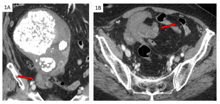
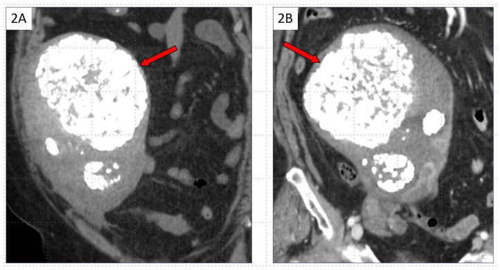
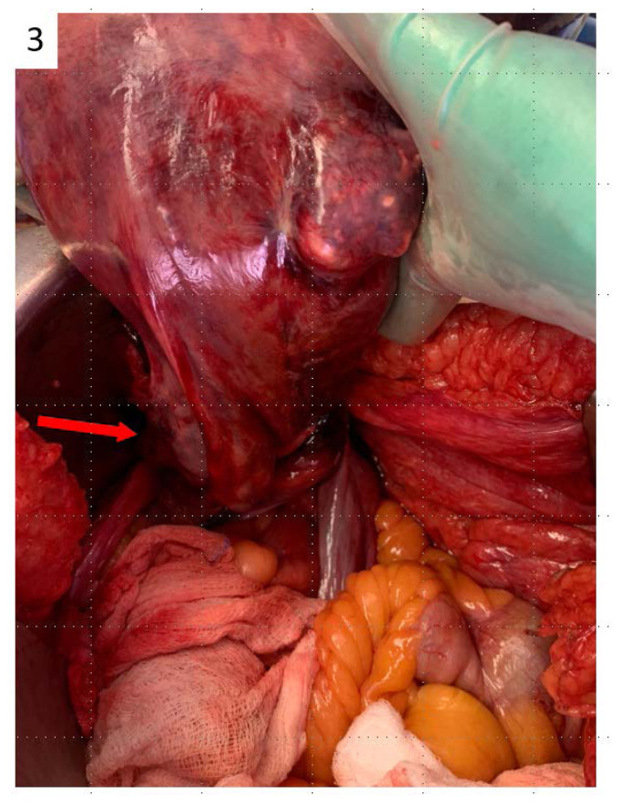
This case report discusses a diagnosis of uterine torsion in an 84-year-old woman who presented with five days of right lower quadrant abdominal pain, nausea, vomiting, constipation, and poor intake. Computed tomography (CT) imaging demonstrated a whorled configuration at the junction of the cervix and lower uterine segment, with the left gonadal vein crossing midline, and two previously known right leiomyomas now appearing on the left. These findings were consistent with the diagnosis of uterine torsion. She then underwent an urgent exploratory laparotomy, and the uterus was found to be dextroverted 270 degrees, with dark mottled purple tissue and engorged vessels. A supracervical hysterectomy and bilateral salpingo-oopherectomy were performed. Final pathology demonstrated extensive necrosis. This case reviews the classic presentation and imaging findings for the rare diagnosis of uterine torsion and options for management of both non-gravid and gravid patients.
Keywords: Gynecologic Emergency; Leiomyoma; Pelvic CT; Pelvic MRI; Uterine Torsion; Whirlpool Sign; X-Shaped.
Copyright Journal of Radiology Case Reports.
Figures
Similar articles
-
Uterine torsion with necrosis of bilateral adnexa in a postmenopausal woman.BMJ Case Rep. 2019 Jun 18;12(6):e229311. doi: 10.1136/bcr-2019-229311. BMJ Case Rep. 2019. PMID: 31217212 Free PMC article.
-
Torsion of a uterine leiomyoma - a rare cause of hemoperitoneum; a case report and review of the literature.Med Ultrason. 2019 Feb 17;21(1):77-82. doi: 10.11152/mu-1784. Med Ultrason. 2019. PMID: 30779835 Review.
-
Torsion of pedunculated subserous uterine leiomyoma: A rare complication of a common disease.Taiwan J Obstet Gynecol. 2018 Apr;57(2):300-303. doi: 10.1016/j.tjog.2018.02.021. Taiwan J Obstet Gynecol. 2018. PMID: 29673677
-
Bilateral massive leiomyomas in a bicornuate uterus, with torsion of the right horn.BMJ Case Rep. 2025 Mar 25;18(3):e264361. doi: 10.1136/bcr-2024-264361. BMJ Case Rep. 2025. PMID: 40132934 Free PMC article.
-
Torsion of a non-gravid leiomyomatous uterus in a patient with myotonic dystrophy complaining of acute urinary retention: anaesthetic management for total abdominal hysterectomy.Clin Exp Obstet Gynecol. 2003;30(2-3):147-50. Clin Exp Obstet Gynecol. 2003. PMID: 12854863 Review.
Cited by
-
Asymptomatic Cervical Amputation Caused by Uterine Torsion in a Non-Gravid Woman.J Clin Med. 2024 Dec 3;13(23):7356. doi: 10.3390/jcm13237356. J Clin Med. 2024. PMID: 39685814 Free PMC article.
References
-
- Collinet P, Narducci F, Stien L. Torsion of a nongravid uterus: an unexpected complication of an ovarian cyst. Eur J Obstet Gynecol Reprod Biol. 2001;98(2):256–257. - PubMed
-
- Ramseyer AM, Whittington JR, Resendez VA, Whitcombe DD, Magann EF. Torsion in the Gravid and Nongravid Uterus: A Review of the Literature of an Uncommon Diagnosis. Obstet Gynecol Surv. 2020;75(4):243–252. - PubMed
-
- Yap FY, Radin R, Tchelepi H. Torsion, infarction, and rupture of a nongravid uterus: a complication of a large ovarian cyst. Abdom Radiol (NY) 2016;41(12):2359–2363. - PubMed