. 2024 Jun 24;9(12):e172959.
doi: 10.1172/jci.insight.172959.

Histone demethylase JARID1C/KDM5C regulates Th17 cells by increasing IL-6 expression in diabetic plasmacytoid dendritic cells

Affiliations

Histone demethylase JARID1C/KDM5C regulates Th17 cells by increasing IL-6 expression in diabetic plasmacytoid dendritic cells

Christopher O Audu et al. JCI Insight. .

Abstract

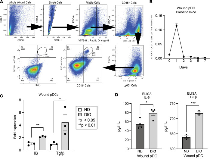
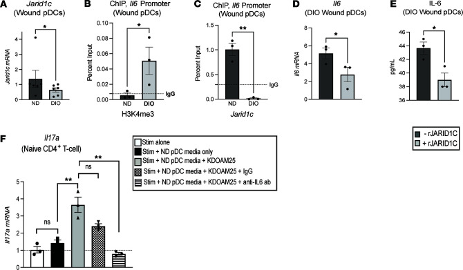
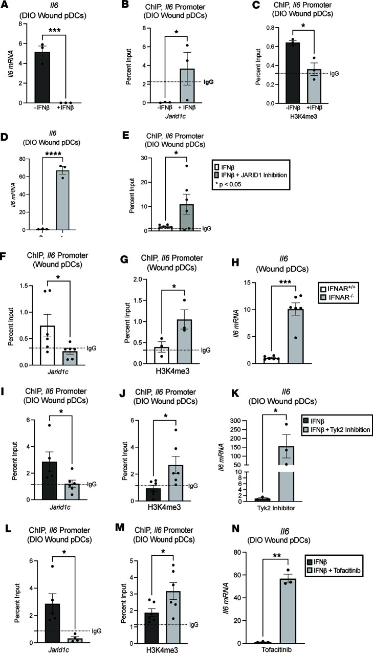
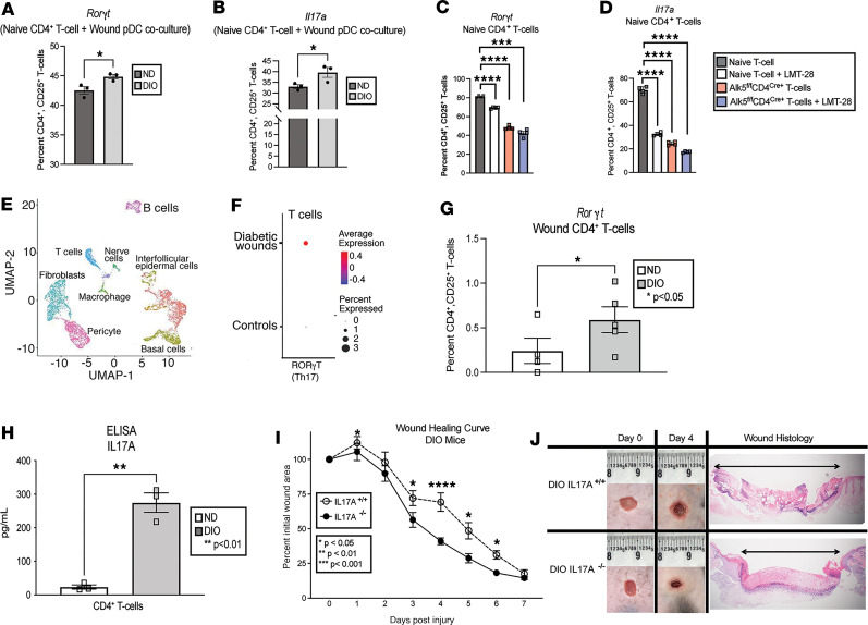
Plasmacytoid dendritic cells (pDCs) are first responders to tissue injury, where they prime naive T cells. The role of pDCs in physiologic wound repair has been examined, but little is known about pDCs in diabetic wound tissue and their interactions with naive CD4+ T cells. Diabetic wounds are characterized by increased levels of inflammatory IL-17A cytokine, partly due to increased Th17 CD4+ cells. This increased IL-17A cytokine, in excess, impairs tissue repair. Here, using human tissue and murine wound healing models, we found that diabetic wound pDCs produced excess IL-6 and TGF-β and that these cytokines skewed naive CD4+ T cells toward a Th17 inflammatory phenotype following cutaneous injury. Further, we identified that increased IL-6 cytokine production by diabetic wound pDCs is regulated by a histone demethylase, Jumonji AT-rich interactive domain 1C histone demethylase (JARID1C). Decreased JARID1C increased IL-6 transcription in diabetic pDCs, and this process was regulated upstream by an IFN-I/TYK2/JAK1,3 signaling pathway. When inhibited in nondiabetic wound pDCs, JARID1C skewed naive CD4+ T cells toward a Th17 phenotype and increased IL-17A production. Together, this suggests that diabetic wound pDCs are epigenetically altered to increase IL-6 expression that then affects T cell phenotype. These findings identify a therapeutically manipulable pathway in diabetic wounds.

Keywords: Adaptive immunity; Dendritic cells; Epigenetics; Immunology; Inflammation.


Conflict of interest statement

Conflict of interest: The authors have declared that no conflict of interest exists.

Figures

Figure 1
Figure 1. Diabetic wound pDCs respond early to tissue damage and produce IL-6 and TGF-β.
(A) Representative flow cytometry gating schematic to isolate pDCs (CD45+, Ly6C+, CD11c+, PDCA1+) from murine diabetic wounds. (B) Kinetic plot of diabetic wound pDCs from 6 mm wounds over time (N = 5/group, pooled and repeated in triplicate) determined by flow cytometry. (C) mRNA fold expression of Il6 and Tgfb in isolated wound pDCs from diabetic and nondiabetic control mice. (N = 3–5/group, pooled, repeated in triplicate.) (D) Protein expression of IL-6 and TGF-β in isolated wound pDCs from diabetic and nondiabetic control mice. (N = 3–5/group, pooled, repeated in triplicate.) Wound pDCs were isolated using EasySep magnetic bead pDC negative-selection kit, according to manufacturer instructions. *P < 0.05, **P < 0.01, ***P < 0.001. Data are presented as the mean ± SEM and were analyzed using 2-tailed Student’s t test.
Figure 2
Figure 2. Histone demethylase JARID1C regulates IL-6 in diabetic wound pDCs and affects T cell Il17a expression.
(A) Jarid1c expression in ND and DIO wound pDCs. (N = 6/group, pooled, repeated in triplicate.) (B) ChIP of the H3K4me3 mark on the Il6 promoter in wound pDCs. (N = 6/group, pooled, repeated in triplicate.) (C) ChIP of Jarid1c at the Il6 promoter in wound pDCs. (N = 3–5/group, pooled, repeated in triplicate.) (D) Fold expression of Il6 in diabetic wound pDCs with and without recombinant JARID1C (rJARID1C, 10 nM, 24 hours; N = 3–5/group, pooled, repeated in triplicate). (E) Protein expression determined by ELISA of IL-6 in diabetic wound pDCs with and without rJARID1C (10 nM, 24 hours; N = 3–5/group, pooled, repeated in triplicate). (F) Fold expression of stimulated CD4+ T cell Il17a following incubation with supernatant from nondiabetic wound pDCs with and without JARID1 inhibition for 24 hours (KDOAM25, 500 nM; N = 3/group, pooled, repeated in triplicate) in the presence and absence of anti–IL-6 antibody. For each experiment, wound pDCs were harvested on day 1 after injury and isolated using EasySep pDC negative-selection magnetic bead kit. *P < 0.05, **P < 0.01. All data are presented as mean ± SEM. Data in F were statistically analyzed using 1-way ANOVA with Holm-Šidák multiple-comparison test. For all other panels, data were analyzed using 2-tailed Student’s t test once normality was assessed.
Figure 3
Figure 3. IFN-β regulates JARID1C via TYK2/JAK1,3 signaling.
(A) Il6 expression in DIO wound pDCs with and without recombinant IFN-β. (B) ChIP analysis of Jarid1c on the Il6 promoter in diabetic wound pDCs with and without IFN-β stimulation. (C) ChIP of the H3K4me3 mark on the Il6 promoter in DIO wound pDCs with and without IFN-β stimulation. (D) Il6 expression in DIO wound pDCs with IFN-β stimulation only with and without JARID1 inhibition. (E) ChIP of the H3K4me3 mark on the Il6 promoter in DIO wound pDCs with IFN-β stimulation, with and without JARID1 inhibition. The graphs are color-coded according to the key, which denotes the cell treatment. (F) ChIP of Jarid1c at the Il6 promoter in IFNAR–/– mouse wound pDCs compared with their age-matched littermate controls. (G) ChIP of the H3K4me3 mark on the Il6 promoter in IFNAR–/– mouse wound pDCs, compared with their age-matched littermate controls. (H) Il6 expression in wound pDCs from IFNAR–/– mice and their age-matched littermate controls. (I) ChIP of Jarid1c at the Il6 promoter in DIO mouse wound pDCs with a TYK2 inhibitor. (J) ChIP of the H3K4me3 mark at the Il6 promoter in DIO mouse wound pDCs with TYK2 inhibition. (K) Il6 expression in DIO wound pDCs following TYK2 inhibition. (L) ChIP of Jarid1c on the Il6 promoter in DIO wound PDCs following JAK1,3 inhibition. (M) ChIP of the H3K4me3 mark on the Il6 promoter in DIO mouse wound pDCs following JAK1,3 inhibition. (N) Il6 expression in DIO wound pDCs after JAK1,3 inhibition. *P < 0.05, **P < 0.01, ***P < 0.001. All experiments were conducted with N = 3–6 mice/group, pooled and repeated in triplicate. Murine wound pDCs were harvested on day 1 after injury and isolated using EasySep pDC negative-selection kit. Data are presented as the mean ± SEM. Data were first analyzed for normal distribution, and if data passed the normality test, 2-tailed Student’s t test was used.
Figure 4
Figure 4. Diabetic wound pDCs promote T cell differentiation toward Th17 phenotype.
(A) Flow cytometry analysis of RORγt expression in naive CD4+ T cells cocultured for 48–72 hours with DIO and ND wound pDCs. (B) Flow cytometry analysis of IL-17A in CD4+ T cells cocultured for 48–72 hours with DIO and ND wound pDCs. (C) RORγt expression in CD4+ T cells after 48- to 72-hour coculture with DIO wound pDCs under several conditions — naive CD4+ T cells (control), naive CD4+ T cells with IL-6 preinhibition (LMT-28, 200 nM, 1 hour), TGF-β receptor–deficient naive CD4+ T cells (Alk5fl/flCD4Cre+ T cells), and Alk5fl/flCD4Cre+ CD4+ T cells with IL-6 receptor preinhibition (LMT-28, 200 nM, 1 hour). (D) IL-17A expression in CD4+ T cells after 48- to 72-hour coculture with DIO wound pDCs under various conditions, as detailed in C. For these coculture experiments, each mouse received 3–4 wounds, and N = 3–5 mice/group, pooled and repeated in triplicate. (E) Cluster uniform manifold approximation and projection (UMAP) of scRNA-Seq from human T2D and non-T2D wounds showed 10 unique cell clusters (representative). (F) scRNA-Seq of human wound T-cell population demonstrating RORγt expression in T2D versus non-T2D controls (N = 42). Dot size corresponds to proportion of cells within the group expressing RORγt, while dot color corresponds to expression level. (G) Intracellular flow cytometry quantifying intracellular RORγt in ND and DIO wound CD4+ T cells. (N = 3–5 mice/group, pooled and repeated in triplicate.) (H) Protein expression by ELISA of IL-17A in ND versus DIO wound CD4+ T cells (N = 5/group, pooled and repeated in triplicate; day 5 wounds). (I) Wound healing curve in global knockout, diabetic IL17A–/– mice compared with age-matched, littermate controls (N = 3/group, pooled and repeated in triplicate). (J) Representative wound healing images and histology. Data are presented as the mean ± SEM. For C and D, data were analyzed using 1-way ANOVA with Holm-Šídák multiple-comparison test. Data for I were analyzed using 2-way repeated measures ANOVA. For all other panels, data were analyzed using 2-tailed Student’s t test once normality was assessed.

References

    1. Boniakowski AE, et al. Macrophage-mediated inflammation in normal and diabetic wound healing. J Immunol. 2017;199(1):17–24. doi: 10.4049/jimmunol.1700223. - DOI - PubMed
    1. Falanga V. Wound healing and its impairment in the diabetic foot. Lancet. 2005;366(9498):1736–1743. doi: 10.1016/S0140-6736(05)67700-8. - DOI - PubMed
    1. Rodrigues M, et al. Wound healing: a cellular perspective. Physiol Rev. 2019;99(1):665–706. doi: 10.1152/physrev.00067.2017. - DOI - PMC - PubMed
    1. Velnar T, et al. The wound healing process: an overview of the cellular and molecular mechanisms. J Int Med Res. 2009;37(5):1528–1542. doi: 10.1177/147323000903700531. - DOI - PubMed
    1. Galicia G, Gommerman JL. Plasmacytoid dendritic cells and autoimmune inflammation. Biol Chem. 2014;395(3):335–346. doi: 10.1515/hsz-2013-0213. - DOI - PubMed