. 2024 Aug 1;213(3):339-346.
doi: 10.4049/jimmunol.2400145.

Reappraising the Role of T Cell-Derived IFN-γ in Restriction of Mycobacterium tuberculosis in the Murine Lung

Affiliations

Reappraising the Role of T Cell-Derived IFN-γ in Restriction of Mycobacterium tuberculosis in the Murine Lung

Karolina Maciag et al. J Immunol. .

Abstract

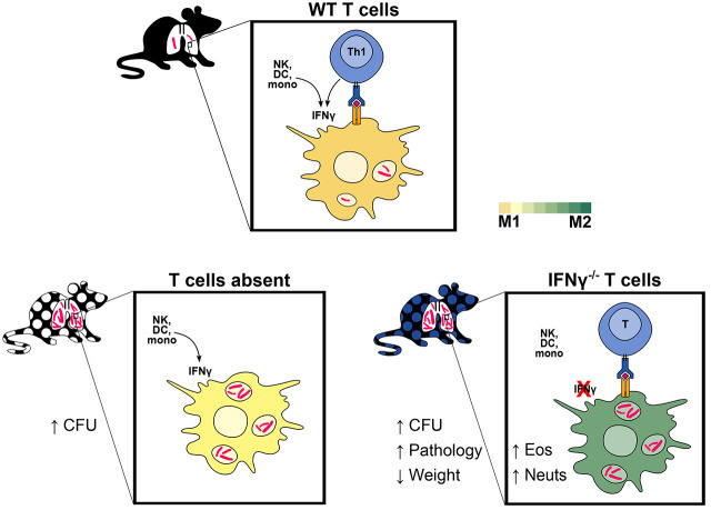
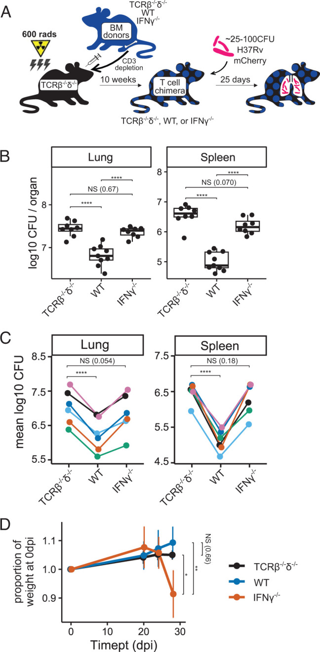
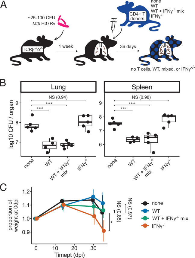
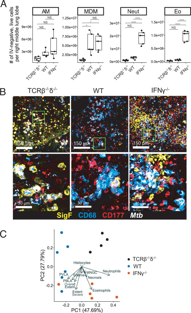
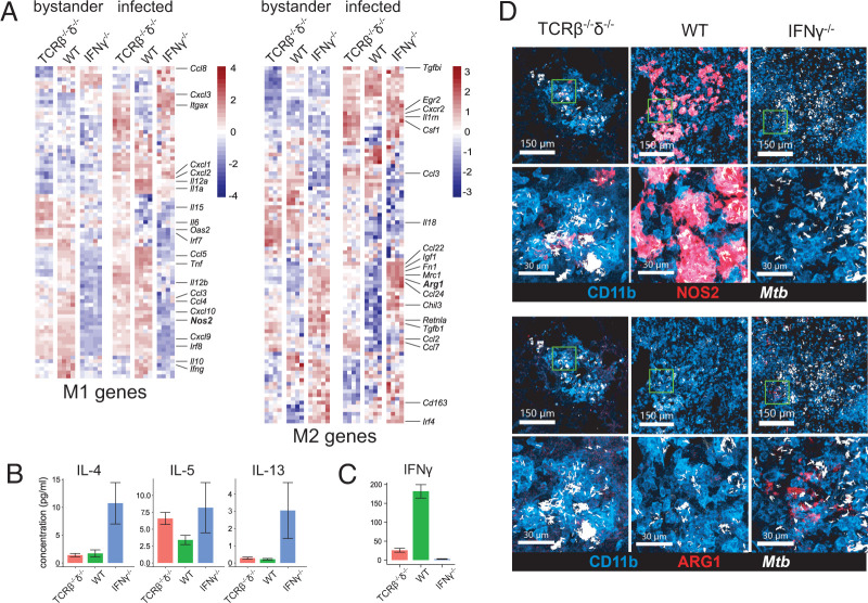
T cells producing IFN-γ have long been considered a stalwart for immune protection against Mycobacterium tuberculosis (Mtb), but their relative importance to pulmonary immunity has been challenged by murine studies that achieved protection by adoptively transferred Mtb-specific IFN-γ-/- T cells. Using IFN-γ-/- T cell chimeric mice and adoptive transfer of IFN-γ-/- T cells into TCRβ-/-δ-/- mice, we demonstrate that control of lung Mtb burden is in fact dependent on T cell-derived IFN-γ, and, furthermore, mice selectively deficient in T cell-derived IFN-γ develop exacerbated disease compared with T cell-deficient control animals, despite equivalent lung bacterial burdens. Deficiency in T cell-derived IFN-γ skews infected and bystander monocyte-derived macrophages to an alternative M2 phenotype and promotes neutrophil and eosinophil influx. Our studies support an important role for T cell-derived IFN-γ in pulmonary immunity against tuberculosis.


Conflict of interest statement

The authors have no financial conflicts of interest.

Figures

None
Graphical abstract
FIGURE 1.
FIGURE 1.
IFN-γ−/− T cells do not reduce Mtb bacterial burden, and they exacerbate disease in T cell chimeric mice. (A) Schematic of the preparation of TCRβ−/−δ−/−, WT, and IFN-γ−/− T cell chimeric mice, followed by infection with aerosolized Mtb. (B and C) Bacterial burden in lungs and spleens of Mtb-infected T cell chimeric mice at 25 dpi in one representative experiment (B), as well as group means from six independent experiments (C). Total n = 35 TCRβ−/−δ−/−, n = 34 WT, and n = 33 IFN-γ−/− T cell chimeric mice. (D) Weight trends of Mtb-infected T cell chimeric mice through 29 dpi for n = 45 TCRβ−/−δ−/−, n = 3 WT, and n = 6 IFN-γ−/− T cell chimeric mice in one representative experiment. Statistical significance is shown for weights at 29 dpi. Statistical significance was determined by Tukey’s range test (B and D) and paired t test (C). *p ≤ 0.05, **p ≤ 0.01, ****p ≤ 0.0001. BM, bone marrow.
FIGURE 2.
FIGURE 2.
Adoptively transferred IFN-γ−/− CD4+ T cells do not reduce Mtb burden, and they exacerbate disease in TCRβ−/−δ−/− mice. (A) Schematic of adoptive transfer of zero (none) or 3 × 10⁶ WT, 50%/50% mixed WT+IFN-γ−/−, or IFN-γ−/− CD4+ T cells to T cell–deficient host mice postinfection with aerosolized Mtb. (B) Bacterial burden in lungs and spleens of Mtb-infected adoptive transfer mice at 36 dpi. Results are representative of two independent experiments. (C) Weight trends of Mtb-infected adoptive transfer mice through 36 dpi. Statistical significance is shown for weights at 36 dpi. n = 5 none, n = 4 WT, n = 5 WT+IFN-γ−/−, and n = 5 IFN-γ−/− adoptive transfer mice. Results are representative of two independent experiments. Statistical significance was determined by Dunnett’s test using “none” as the control group. *p ≤ 0.05, ***p ≤ 0.001, ****p ≤ 0.0001.
FIGURE 3.
FIGURE 3.
IFN-γ−/− T cells promote neutrophil and eosinophil recruitment to pulmonary lesions in T cell chimeric mice infected with Mtb. (A) Absolute number of each indicated cell type among live, parenchymal (IV-) cells in the right lung of T cell chimeric mice at 25 dpi. Results are representative of six independent experiments. Statistical significance was determined by Tukey’s range test. *p ≤ 0.05, ****p ≤ 0.0001. AM, alveolar macrophage; Eo, eosinophil; Neut, neutrophil. (B) Representative confocal microscopy demonstrating AMs (Siglec F+, CD68+), MDMs (Siglec F, CD68+), neutrophils (CD177+), and eosinophils (Siglec F+, CD68) in TB lesions in T cell chimeric mice. (C) Principal component analysis of 15 histopathologic features assessed in representative sections of fixed and H&E-stained lung of T cell chimeric mice at 25 dpi. MNGC, multinucleated giant cells; PBLA, peribronchial lymphoid aggregates; PVLA, perivascular lymphoid aggregates.
FIGURE 4.
FIGURE 4.
IFN-γ−/− T cells drive a type 2 cytokine milieu and alternative activation of MDMs in T cell chimeric mice infected with Mtb. (A) Relative expression of classical (M1) and alternative (M2) genes in FACS-sorted bystander and Mtb-infected MDMs in lungs of T cell chimeric mice at 25 dpi. Results are representative of two independent experiments. (B) Concentration of type 2 cytokines in lung lysates from Mtb-infected T cell chimeric mice at 25 dpi, as measured by cytokine bead array. n = 4 TCRβ−/−δ−/−, n = 5 WT, and n = 5 IFN-γ−/− T cell chimeric mice. Results are representative of three independent experiments. (C) Concentration of IFN-γ in lung lysates from Mtb-infected T cell chimeric mice at 25 dpi, as measured by Luminex assay. n = 5 TCRβ−/−δ−/−, n = 5 WT, and n = 6 IFN-γ−/− T cell chimeric mice. Results are representative of three independent experiments. (D) Representative confocal microscopy demonstrating expression of iNOS and ARG1 in lungs of Mtb-infected T cell chimeric mice at 25 dpi.

Update of

References

    1. Caruso, A. M., Serbina N., Klein E., Triebold K., Bloom B. R., Flynn J. L.. 1999. Mice deficient in CD4 T cells have only transiently diminished levels of IFN-gamma, yet succumb to tuberculosis. J. Immunol. 162: 5407–5416. - PubMed
    1. Casanova, J.-L., Abel L.. 2002. Genetic dissection of immunity to mycobacteria: the human model. Annu. Rev. Immunol. 20: 581–620. - PubMed
    1. Esmail, H., Riou C., Du Bruyn E., Lai R. P.-J., Harley Y. X. R., Meintjes G., Wilkinson K. A., Wilkinson R. J.. 2018. The immune response to Mycobacterium tuberculosis in HIV-1-coinfected persons. Annu. Rev. Immunol. 36: 603–638. - PubMed
    1. Flynn, J., Chan J., Triebold K., Dalton D., Stewart T., Bloom B.. 1993. An essential role for interferon gamma in resistance to Mycobacterium tuberculosis infection. J. Exp. Med. 178: 2249–2254. - PMC - PubMed
    1. Pearl, J. E., Saunders B., Ehlers S., Orme I. M., Cooper A. M.. 2001. Inflammation and lymphocyte activation during mycobacterial infection in the interferon-γ-deficient mouse. Cell. Immunol. 211: 43–50. - PubMed

Publication types

LinkOut - more resources