. 2024 Jun 24;15(1):4566.
doi: 10.1038/s41467-024-48816-x.

Prefrontal coding of learned and inferred knowledge during REM and NREM sleep

Affiliations

Prefrontal coding of learned and inferred knowledge during REM and NREM sleep

Kareem Abdou et al. Nat Commun. .

Abstract

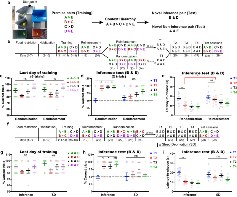
Idling brain activity has been proposed to facilitate inference, insight, and innovative problem-solving. However, it remains unclear how and when the idling brain can create novel ideas. Here, we show that cortical offline activity is both necessary and sufficient for building unlearned inferential knowledge from previously acquired information. In a transitive inference paradigm, male C57BL/6J mice gained the inference 1 day after, but not shortly after, complete training. Inhibiting the neuronal computations in the anterior cingulate cortex (ACC) during post-learning either non-rapid eye movement (NREM) or rapid eye movement (REM) sleep, but not wakefulness, disrupted the inference without affecting the learned knowledge. In vivo Ca2+ imaging suggests that NREM sleep organizes the scattered learned knowledge in a complete hierarchy, while REM sleep computes the inferential information from the organized hierarchy. Furthermore, after insufficient learning, artificial activation of medial entorhinal cortex-ACC dialog during only REM sleep created inferential knowledge. Collectively, our study provides a mechanistic insight on NREM and REM coordination in weaving inferential knowledge, thus highlighting the power of idling brain in cognitive flexibility.


Conflict of interest statement

S.M. owns equity in a company, Gene Therapy Research Institution, that commercializes the use of AAV vectors for gene therapy applications. To the extent that the work in this manuscript increases the value of these commercial holdings, S.M. has a conflict of interest. The other authors declare no competing interests.

Figures

Fig. 1
Fig. 1. The emergence of inference requires sleep after training.
a Representative photo of the arena and the five different contexts with an arrow representing the starting point (left). The four premise pairs with the contexts’ hierarchy (right); premise pairs were consistently color-coded across all figures to facilitate tracking each pair without confusion. b The behavioral schedule used to establish the transitive inference paradigm in mice. The contexts’ hierarchy was as follows: A > B > C > D > E. The order of presentation of the premise pairs during the randomization stage is different across animals within the same group (See “Methods” section; Table 1). c Performance during the last day of training (day 14 for A > B and B > C; day 18 for C > D and D > E) for each premise pair was calculated as the percent of correct trials out of the total number of trials (5 trials). d, e Performance during test sessions; the percent of correct trials in the inference test (2 trials) (d), the latency time to choose in the inference test (e). f Behavioral protocol for the transitive inference paradigm; SD, sleep deprivation. g Percent of correct trials during the last day of training (day 14 for A > B and B > C; day 18 for C > D and D > E) for each premise pair. h, i Performance during test sessions; percent of correct trials during the inference test (h), latency time to choose during the inference test (i). T1, test 1; T2, test 2; T3, test 3; T4, test 4. n = 10 mice/group; n = 8 mice for the sleep deprivation (SD) group. Statistical comparisons were made using a two-way repeated-measures ANOVA with Tukey’s multiple comparison test (ce, g, h) and Holm–Sidak’s test between groups (e). *P < 0.05; **P < 0.01; ****P < 0.0001; ns, not significant (P > 0.05). Data are presented as the mean ± standard error of the mean (s.e.m.). Experiments were independently repeated four times. Source data are provided as a Source Data file. Detailed statistics are shown in Supplementary Data 1.
Fig. 2
Fig. 2. ACC computations during sleep are crucial for the emergence of inference.
a Labeling excitatory neurons of the ACC with ArchT (left), and the expression of ArchT-eYFP (green) in the ACC (right). Blue, 4′,6-diamidino-2-phenylindole (DAPI) staining. Scale bar, 100 µm. b Behavioral schedule used to manipulate ACC activity during sleep and awake periods after test sessions. The order of presentation of the premise pairs during the randomization stage is different across animals within the same group (See “Methods” section; Table 1). c Diagram showing the state-specific manipulation (top) and representative electroencephalogram (EEG) and electromyography (EMG) (bottom) traces. Scale bar, 15 min. d Performance during the last day of training (day 14 for A > B and B > C; day 18 for C > D and D > E) for each premise pair. e, f Performance during test sessions; percent of correct trials during the inference test (e), latency time to choose during the inference test (f). T1, test 1; T2, test 2; T3, test 3; T4, test 4. n = 9 mice for the awake group; n = 8 mice for the non-rapid eye movement (NREM) sleep group; n = 7 mice for the rapid eye movement (REM) sleep group. Statistical comparisons were made using two-way repeated-measures analysis of variance (ANOVA) with Tukey’s multiple comparison test (df). In (e), the statistical significance denotes the comparison between performance relative to the chance level (50%). *P < 0.05; **P < 0.01; ***P < 0.001; ns, not significant (P > 0.05). Data are presented as the mean ± standard error of the mean (s.e.m.). The Experiment was independently repeated six times. Source data are provided as a Source Data file. Detailed statistics are shown in Supplementary Data 1.
Fig. 3
Fig. 3. Identifying inference-related ensembles that emerge gradually and become evident during REM sleep.
a Labeling of the ACC with GCaMP7 (left), and the expression of GCaMP7 (green) in the ACC (right). Blue, 4′,6-diamidino-2-phenylindole (DAPI) staining. Scale bar, 100 µm. b Diagram showing the state-specific imaging. c The behavioral schedule used to capture the Ca2+ transients across the task. On day 10, Ca2+ transients were recorded from 3 imaging sessions; habituation, NREM 1, and REM 1. On day 24, Ca2+ transients were recorded from 4 imaging sessions; T1, awake in home cage, NREM 2, and REM 2. During the training stage, Ca2+ transients were collected for each premise pair on a separate day (days 13, 14, 17, 18) to extract specific representations for a particular premise pair without interfering with the other pair. The order of presentation of the premise pairs during the randomization stage is different across animals within the same group (See “Methods” section; Table 1). d Performance during the last day of training (day 13 for A > B; day 14 for B > C; day 17 for C > D; day 18 for D > E) for each premise pair (left). Performance during test sessions (right). T1, test 1; T2, test 2; T3, test 3; T4, test 4. e Total number of detected cells in each mouse, n = 5 mice (d, e). f Example of ACC coactivity pattern (inference-related pattern) detected in T2 session. The pattern is represented as a vector containing the contribution (weight) of each neuron’s spiking to the coactivity defining that pattern. Neurons with a weight above 2 s.d. of the mean were referred to as members (Red) (top). The temporal appearance of the pattern with the behavior signature (just before the correct choice) (bottom). gi Activation strength (z-scored) of inference-related patterns (g, i), of stable training-related patterns (h, i). j Number of neurons constituting both inference patterns and stable training patterns, n = 5 inference-related patterns (g, i, j); n = 10 Stable training-related patterns (h, i, j). Hab, habituation session; NREM 1, NREM sleep after last habituation session; REM 1, REM sleep after last habituation session; NREM 2, NREM sleep after T1; REM 2, REM sleep after T1. Statistical comparisons were made using one-way repeated-measures analysis of variance (ANOVA) with Dunnett’s multiple comparison test (g, h). *P < 0.05; **P < 0.01; ns not significant (P > 0.05). Data are presented as the mean ± standard error of the mean (s.e.m.). The Experiment was independently repeated four times. Source data are provided as a Source Data file. Detailed statistics are shown in Supplementary Data 1.
Fig. 4
Fig. 4. NREM and REM sleep coordinate to build synchronized inference representation with learned knowledge representation.
a Explanation of the coactivity calculation method. b Examples of raster plots showing coactivity analysis. This analysis calculates coactivity by normalizing the number of synchronizations every 200 ms among different neuronal subpopulations representing different sessions. Coactivity analysis was done for neurons belonging to different patterns representing 2 sessions (Pairwise coactivity, left), representing 4 sessions (Quadruple coactivity, middle), and representing 5 sessions (Quintuple, right). Each color denotes neurons representing a specific session. The equation used to calculate the coactivity index (bottom). ce Coactivity index between 2 sessions (c), 4 sessions (d), and 5 sessions (e) with the shuffled data; n = 5 mice. Statistical comparisons were made using paired t-test (ce). *P < 0.05; **P < 0.01; ns not significant (P > 0.05). Data are presented as the mean ± standard error of the mean (s.e.m.). Source data are provided as a Source Data file. Detailed statistics are shown in Supplementary Data 1.
Fig. 5
Fig. 5. MEC→ACC crosstalk during only REM sleep is sufficient to inspire inference from inadequate training.
a The strategy for labeling MEC neurons and targeting their terminals in the ACC (left), and expression of oChIEF-tdTomato (red) in MEC neurons (middle) and their terminals in the ACC (right). Blue, 4′,6-diamidino-2-phenylindole (DAPI) staining. Scale bars, 100 µm. b The behavioral schedule used to manipulate the MEC→ACC circuit during different sleep stages. c Diagram showing the sleep stage-specific manipulation. Scale bar, 15 min. d Performance during the last day of training (day 14 for A > B and B > C; day 18 for C > D and D > E) for each premise pair. e, f Performance during test sessions; the percent of correct trials during the inference test (e), the latency time to choose during the inference test (f). T1, test 1; T2, test 2; T3, test 3; T4, test 4. n = 10 mice for the light-off group; n = 8 mice for the non-rapid eye movement (NREM) sleep group; n = 8 mice for the rapid eye movement (REM) sleep group. Statistical comparisons were made using a two-way repeated-measures analysis of variance (ANOVA) with Tukey’s multiple comparison test (df). *P < 0.05; **P < 0.01; ***P < 0.001; ns not significant (P > 0.05). Data are presented as the mean ± standard error of the mean (s.e.m.). The Experiment was independently repeated six times. Source data are provided as a Source Data file. Detailed statistics are shown in Supplementary Data 1.

References

    1. Behrens TEJ, et al. What is a cognitive map? Organizing knowledge for flexible behavior. Neuron. 2018;100:490–509. doi: 10.1016/j.neuron.2018.10.002. - DOI - PubMed
    1. Wagner U, Gais S, Haider H, Verleger R, Born J. Sleep inspires insight. Nature. 2004;427:352–355. doi: 10.1038/nature02223. - DOI - PubMed
    1. Ellenbogen JM, Hu PT, Payne JD, Titone D, Walker MP. Human relational memory requires time and sleep. Proc. Natl Acad. Sci. USA. 2007;104:7723–7728. doi: 10.1073/pnas.0700094104. - DOI - PMC - PubMed
    1. Brunamonti E, et al. Neuronal modulation in the prefrontal cortex in a transitive inference task: evidence of neuronal correlates of mental schema management. J. Neurosci. 2016;36:1223–1236. doi: 10.1523/JNEUROSCI.1473-15.2016. - DOI - PMC - PubMed
    1. Spalding KN, et al. Ventromedial prefrontal cortex is necessary for normal associative inference and memory integration. J. Neurosci. 2018;38:3767–3775. doi: 10.1523/JNEUROSCI.2501-17.2018. - DOI - PMC - PubMed