. 2024 Jun 24;10(1):300.
doi: 10.1038/s41420-024-02067-3.

Curcumol ameliorates neuroinflammation after cerebral ischemia-reperfusion injury via affecting microglial polarization and Treg/Th17 balance through Nrf2/HO-1 and NF-κB signaling

Affiliations

Curcumol ameliorates neuroinflammation after cerebral ischemia-reperfusion injury via affecting microglial polarization and Treg/Th17 balance through Nrf2/HO-1 and NF-κB signaling

Ying Liu et al. Cell Death Discov. .

Abstract

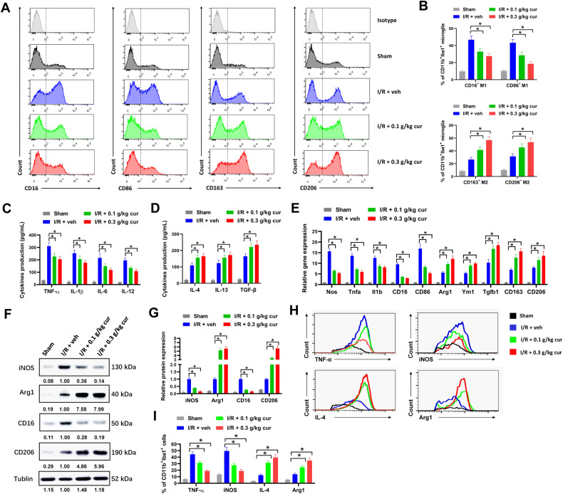
Neuroinflammation caused by microglia and other immune cells plays pivotal role in cerebral ischemia/reperfusion injury and recovery. Modulating microglial polarization or Treg/Th17 balance from pro-inflammatory phenotype to anti-inflammatory phenotype are promising strategies for the treatment of cerebral ischemia. Curcumol has potential to fight against oxidative stress and inflammation, but whether it has protective effect in cerebral ischemia is uncertain. In the present study, cerebral ischemia was induced in C57BL/6 mice via middle cerebral artery occlusion (MCAO). MCAO mice were treated with curcumol for 7 days, then post-stroke ischemic injury, neurological deficits, microglial polarization and brain leukocyte infiltration were evaluated by TTC staining, behavioural tests, flow cytometry, western blot and immunofluorescence. We found that poststroke administration of curcumol reduced infarct volume, attenuated neuronal damage and inflammation, and improved motor function recovery of MCAO mice. Curcumol skewed microglial polarization toward anti-inflammatory phenotype in MCAO mice in vivo or after oxygen-glucose deprivation and reoxygenation (OGD/R) in vitro. In addition, curcumol reduced local T cell infiltration in ischemic brain of MCAO mice and impaired Treg/Th17 balance. Curcumol inhibited ROS production and regulated Nrf2/HO-1 and NF-κB signaling in microglia. Finally, inhibiting Nrf2/HO-1 signaling or activating NF-κB signaling abrogated the influence of curcumol on microglial polarization. In conclusion, curcumol treatment reduced brain damage and neuroinflammation via modulating anti-inflammatory microglial polarization and Treg/Th17 balance through Nrf2/HO-1 and NF-κB signaling. Curcumol might be a promising treatment strategy for stroke patients.


Conflict of interest statement

The authors declare no competing interests.

Figures

Fig. 1
Fig. 1. Poststroke administration of curcumol reduces infarct volume and improves motor function recovery in MCAO mice.
A, B MCAO mice were treated with 0.1 g/kg curcumol (I/R + 0.1 g/kg cur group), 0.3 g/kg curcumol (I/R + 0.3 g/kg cur group) and equal volume of PBS (I/R + veh group and Sham group) daily for 7 d post-MCAO. Representative images of TTC staining (A) and quantification of infarct volume (B) were shown. C Neurological deficit score was evaluated by mNSS at day 1 and day 7 after MCAO. D-E, the time of latency to fall (D) in rotarod test and time to fall (E) in inverted wire mesh grid grip test were shown. FH Motion trajectory (F), distance moved (G), and mean velocity (H) of mice were evaluated by open field test at day 1 and day 7 after MCAO. IM modified Garcia scores were used to evaluate sensorimotor deficits. The scores for body proprioception (I), limb symmetry (J), lateral turning (K), forelimb walking (L), and vibrissae touch (M) were shown. *P < 0.05.
Fig. 2
Fig. 2. Curcumol ameliorate poststroke neuronal damage and neuroinflammation in MCAO mice.
A, B histopathological changes of brain tissues of MCAO mice were evaluated by H&E staining (A). Denatured cell index was shown (B). Scale bar = 200 μm. C, D apoptotic cells in brain tissues of MCAO mice were evaluated by TUNEL staining (C). Relative number of TUNEL positive cells were shown (D). Arrows indicated TUNEL-positive cells. Scale bar = 100 μm. E the levels of pro-inflammatory cytokines in brain tissues of MCAO mice were evaluated by ELISA assay. *P < 0.05.
Fig. 3
Fig. 3. Curcumol skews microglial polarization toward anti-inflammatory phenotype in MCAO mice.
A, B representative histogram (A) and percentages of CD16+, CD86+, CD163+ and CD206+ cells (B) in CD11b+Iba1+ microglia of MCAO mice were shown. C, D the levels of proinflammatory cytokines TNF-α, IL-1β, IL-6 and IL-12 (C) and anti-inflammatory cytokines IL-4, IL-13 and TGF-β (D) in primary microglia of MCAO mice were evaluated by ELISA assay. E the mRNA expression of proinflammatory marker genes (Nos, Tnfa, Il1b, CD16 and CD86) and anti-inflammatory marker genes (Arg1, Ym1, Tgfb1, CD163 and CD206) in primary microglia of MCAO mice was evaluated by RT-qPCR. F, G the protein expression of indicated genes was evaluated by western blot (F). Relative protein expression was shown (G). H, I the expression levels of M1 phenotype markers (TNF-α and iNOS) and M2 phenotype markers (IL-4 and Arg1) were detected by flow cytometry. Representative histogram (H) and percentages of TNF-α+, iNOS+, IL-4+ and Arg1+ cells were shown (I). *P < 0.05.
Fig. 4
Fig. 4. Curcumol reduces local T cell infiltration and impairs Treg/Th17 balance in brains of MCAO mice.
A representative gating strategies in flow cytometry were shown. B the percentages of CD45+ Leukocytes, CD3+ T cells, CD3+CD4+ Th cells, CD3+CD8+ Cyto T cells, CD3CD19+CD20+ B cells and CD3NK1.1+ NK cells in ischemic brain tissues of MCAO mice were evaluated by flow cytometry. CF representative histogram (C, E) and the percentages of IFN-γ+IL-4 Th1, IFN-γIL-4+ Th2 cells, IL-17A+ Th17 cells and CD25+Foxp3+ Treg cells (D, F) in CD3+CD4+ Th cells of ischemic brain tissues were evaluated by flow cytometry. *P < 0.05.
Fig. 5
Fig. 5. Curcumol-treated microglia modulates Treg/Th17 balance in vitro.
Primary microglia were treated with 0.1 g/L or 0.3 g/L curcumol or equal volume of PBS (vehicle) after OGD/R, then co-cultured with CFSE-labelled splenic T cells for 7 days. AD representative plots (A, C) and percentages of CD4+IL-17A+ Th17 cells and CD25+Foxp3+ Treg cells (B, D) in CFSE-labelled splenic T cells were shown. EH representative histogram (E, G) and percentages of CFSE negative proliferative Th17 and Treg cells (F, H) were shown. *P < 0.05.
Fig. 6
Fig. 6. Curcumol inhibits ROS production and Nrf2/HO-1/NF-κB signaling in microglia.
A, B primary microglia from MCAO mice were incubated with DCF-DA, then oxidized DCF was analyzed by flow cytometry. Representative histogram (A) and percentages of oxidized DCF cells (B) were shown. C, D relative MDA (C) and GSH (D) levels in primary microglia from MCAO mice were shown. E, F primary microglia treated with OGD/R were incubated with DCF-DA, then oxidized DCF was analyzed by flow cytometry. Representative histogram (E) and percentages of oxidized DCF cells (F) were shown. G, H relative MDA (G) and GSH (H) levels of primary microglia treating with OGD/R were shown. IL protein levels of indicated genes in primary microglia of MCAO mice or post OGD/R were evaluated by western blot (I, K). Relative protein levels (J, L) were shown. *P < 0.05.
Fig. 7
Fig. 7. Inhibiting of Nrf2/HO-1/NF-κB signaling abrogates the influence of curcumol on microglial polarization in vivo.
Mice were treated with 0.3 g/kg curcumol (cur), 30 mg/kg ML385 or equal volume of DMSO (veh) intraperitoneally for 7 days after MCAO as indicated. A, B protein levels of indicated genes in primary microglia of MCAO mice were evaluated by western blot (A). Relative protein levels (B) were shown. C, D representative histogram (C) and percentages of CD86+ and CD206+ cells (D) in CD11b+Iba1+ microglia of MCAO mice were shown. E, F the levels of proinflammatory cytokines TNF-α, IL-1β, IL-6 and IL-12 (E) and anti-inflammatory cytokines IL-4, IL-13 and TGF-β (F) in primary microglia of MCAO mice were evaluated by ELISA assay. G the mRNA expression of proinflammatory marker genes (Nos, Tnfa, Il1b, CD16 and CD86) and anti-inflammatory marker genes (Arg1, Ym1, Tgfb1, CD163 and CD206) in primary microglia of MCAO mice was evaluated by RT-qPCR. HM post-OGD/R primary microglia were transfected with sh-NRF2, sh-IκBα or SCR as indicated, then treated with 0.3 g/L curcumol (cur) or equal volume of DMSO (veh) for 72 h. Collected cell lysates for western blot (H, I). Percentages of CD86+ and CD206+ cells in CD11b+Iba1+ microglia were evaluated by flow cytometry (J, K). The levels of proinflammatory cytokines TNF-α, IL-1β, IL-6 and IL-12 (L) and anti-inflammatory cytokines IL-4, IL-13 and TGF-β (M) were evaluated by ELISA assay. *P < 0.05.

References

    1. Katan M, Luft A. Global burden of stroke. Semin Neurol. 2018;38:208–11. doi: 10.1055/s-0038-1649503. - DOI - PubMed
    1. Lallukka, T, Ervasti, J, Lundstrom, E, Mittendorfer-Rutz, E, Friberg, E, Virtanen, M et al. Trends in diagnosis-specific work disability before and after stroke: a longitudinal population-based study in Sweden. J Am Heart Assoc 2018;7:e006991. - PMC - PubMed
    1. Feigin VL, Forouzanfar MH, Krishnamurthi R, Mensah GA, Connor M, Bennett DA, et al. Global and regional burden of stroke during 1990-2010: findings from the global burden of disease study 2010. Lancet. 2014;383:245–54. doi: 10.1016/S0140-6736(13)61953-4. - DOI - PMC - PubMed
    1. Li L, Scott CA, Rothwell PM, Oxford Vascular S. Trends in stroke incidence in high-income countries in the 21st century: population-based study and systematic review. Stroke. 2020;51:1372–80. doi: 10.1161/STROKEAHA.119.028484. - DOI - PMC - PubMed
    1. Krishnamurthi RV, Feigin VL, Forouzanfar MH, Mensah GA, Connor M, Bennett DA, et al. Global and regional burden of first-ever ischaemic and haemorrhagic stroke during 1990-2010: findings from the Global Burden of Disease Study 2010. Lancet Glob Health. 2013;1:e259–281. doi: 10.1016/S2214-109X(13)70089-5. - DOI - PMC - PubMed