[Preprint]. 2024 Jun 11:2024.06.11.598425.
doi: 10.1101/2024.06.11.598425.

Neural crest and periderm-specific requirements of Irf6 during neural tube and craniofacial development

Affiliations

Neural crest and periderm-specific requirements of Irf6 during neural tube and craniofacial development

Shannon H Carroll et al. bioRxiv. .

Update in

Abstract

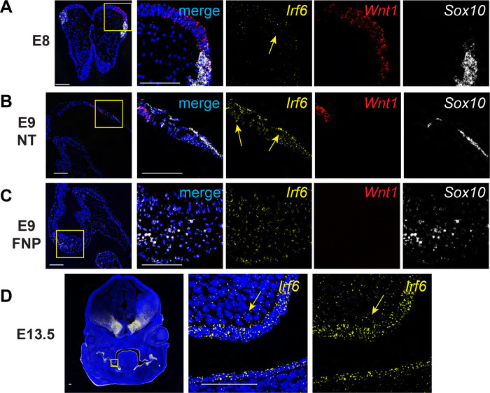
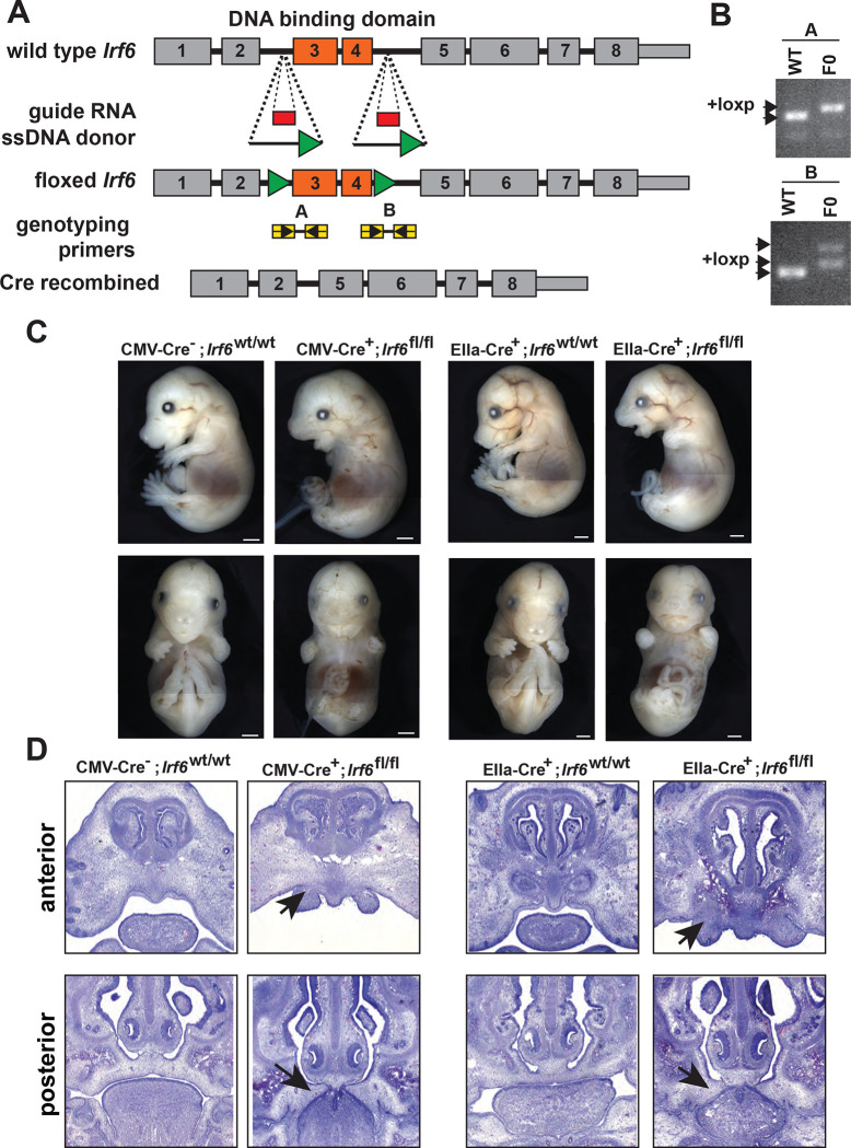
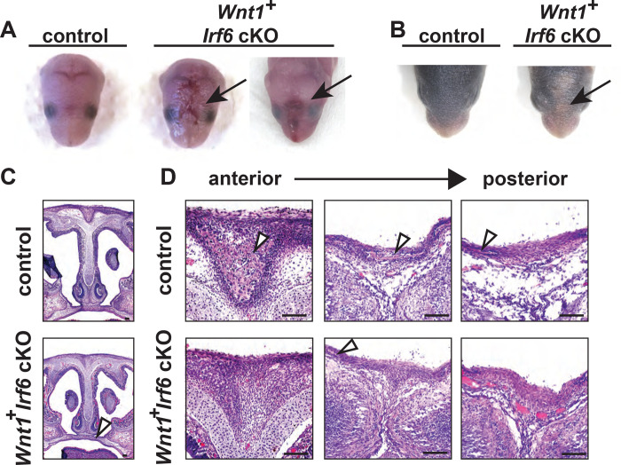
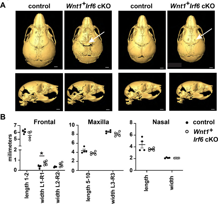
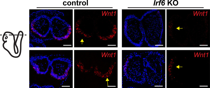
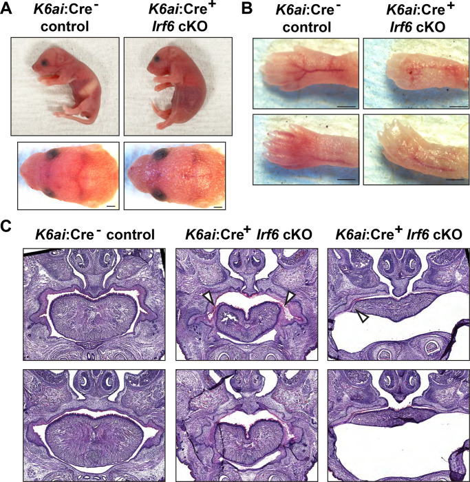
IRF6 is a key genetic determinant of syndromic and non-syndromic cleft lip and palate. The ability to interrogate post-embryonic requirements of Irf6 has been hindered, as global Irf6 ablation in the mouse causes neonatal lethality. Prior work analyzing Irf6 in mouse models defined its role in the embryonic surface epithelium and periderm where it is required to regulate cell proliferation and differentiation. Several reports have also described Irf6 gene expression in other cell types, such as muscle, and neuroectoderm. However, analysis of a functional role in non-epithelial cell lineages has been incomplete due to the severity and lethality of the Irf6 knockout model and the paucity of work with a conditional Irf6 allele. Here we describe the generation and characterization of a new Irf6 floxed mouse model and analysis of Irf6 ablation in periderm and neural crest lineages. This work found that loss of Irf6 in periderm recapitulates a mild Irf6 null phenotype, suggesting that Irf6-mediated signaling in periderm plays a crucial role in regulating embryonic development. Further, conditional ablation of Irf6 in neural crest cells resulted in an anterior neural tube defect of variable penetrance. The generation of this conditional Irf6 allele allows for new insights into craniofacial development and new exploration into the post-natal role of Irf6.

Keywords: Irf6; Van der Woude Syndrome; cleft palate; neural crest; neural tube; periderm.


Figures

Fig. 1.
Fig. 1.
Irf6 is expressed with neural crest cell markers Wnt1 and Sox10 in neural folds and neural tube during early embryogenesis. In situ hybridization of Irf6 (yellow), Wnt1 (red), and Sox10 (white) RNA transcripts. A. Coronal section of E8 mouse embryo (dorsal to top) showing the neural fold. In situ hybridization shows RNA expression domains of Irf6, Wnt1, and Sox10, where Irf6 and Wnt1 transcripts are found in the same regions of the neural tube, highlighted by yellow arrow. Box indicates area of higher magnification to the right. B. Sagittal section of E9 mouse embryo (cranial to left). Box indicates a magnified portion of the neural tube. Irf6 is expressed in the neuroectoderm and overlaps with Wnt1 and Sox10 expression (yellow arrows). C. Sagittal section of E9 mouse embryo (cranial to left). Box indicates a magnified portion of frontonasal prominence (FNP). Irf6 is expressed in the FNP mesenchyme, along with the migratory NCC marker Sox10. D. Coronal section of E13.5 embryo (dorsal to top). Box indicates higher magnification of palate shelf epithelium and mesenchyme. Irf6 is highly expressed in the basal epithelium and periderm and the palate mesenchyme (yellow arrow). Blue is dapi. Scale: 100 uM.
Fig. 2.
Fig. 2.
Generation and validation of a conditional Irf6 null mouse model. A. Schematic of gene targeting strategy. Introns flanking Irf6 exons 3 and 4 were targeted for CRISPR-Cas9-directed homologous recombination with each donor ssDNA containing loxP sequences (green triangles). Insertion of loxP sites into Irf6 was confirmed by PCR. B. and Sanger sequencing. C. Cre-mediated recombination was validated using the ubiquitous Cre expressing lines CMV-Cre and EIIa-Cre. CMV-Cre+;Irf6fl/fl and EIIa-Cre+; Irf6fl/fl mice phenocopied the Irf6 global KO while Cre;Irf6fl/fl and Cre+;Irf6wt/wt littermates were normal. D. Hematoxylin and Eosin staining of coronal sections of E15 CMV-Cre or EIIa-Cre knockout embryos and littermate controls. Top row is a relatively anterior section while the bottom row is relatively posterior. CMV-Cre and EIIa-Cre Irf6 KO embryos phenocopy the dysmorphic alveolar bone and the cleft palate with oral adhesions of the total Irf6 knockout mouse (arrows).
Fig. 3.
Fig. 3.
Wnt1-Cre-dependent Irf6 ablation causes cranial defects. A. Representative images of littermate control and Wnt1-Cre, Irf6 cKO pups at P0. At parturition, Wnt1-Cre+;Irf6fl/fl cKO mice display midline lesions of varying penetrance (arrow). B. Representative images of littermate control and Wnt1-Cre+;Irf6fl/fl cKO pups at P6. As the mouse neonate develops, these frontal lesions resolve but remain evident with deficient or delayed fur growth (arrow). C. Hematoxylin and eosin staining of coronal sections through the palate of E16 Wnt1-Cre+;Irf6fl/fl cKO and littermate control embryos shows normal development (arrow). D. Hematoxylin and eosin staining of coronal sections through the nasal and frontal bones of Wnt1-Cre+;Irf6fl/fl cKO and littermate control. Sections move anterior to posterior from left to right. Bone tissue is indicated with arrows. Wnt1-Cre+;Irf6fl/fl cKO mice have a lack of cranial bone development and suture formation at the midline (bone tissue indicated by arrows). Scale: 100 μM.
Fig. 4.
Fig. 4.
Cranial bone development is impaired in Wnt1-Cre Irf6 cKO mice. A. Representative microCT reconstructions of P10 Wnt1-Cre+;Irf6fl/fl cKO mice and littermate sex-matched controls. Wnt1-Cre+;Irf6fl/fl cKO mice have decreased formation or mineralization of the cranial bones at the midline with variable penetrance (arrows). Scale: 1 mm. B. MicroCT reconstructions were utilized for cranial bone measurements. The space between the left and right frontal bones of Wnt1-Cre+;Irf6fl/fl cKO mice was significantly wider than controls (L1-R1, *p<0.05) and the frontal bones tended to have decreased total length (length 1–2). Maxilla of Wnt1-Cre+;Irf6fl/fl cKO mice tended to be smaller (lower length and width measurements) and the frontal bone of Wnt1-Cre+;Irf6fl/fl cKO mice tended to be shorter, however, these differences were not significantly different. N=4.
Fig. 5.
Fig. 5.
Irf6 ablation in the neuroectoderm and neural crest changes Wnt1 expression domains within the neural folds. A. RNAscope in situ hybridization of transverse sections of Wnt1-Cre+;Irf6fl/fl cKO and littermate control E8 embryos. Rows represent 2 individuals of each genotype. Whereas Wnt1 expression (red) is localized to the caudal-dorsal neural folds in the control embryos, Wnt1 expression in Wnt1-Cre+;Irf6fl/fl cKO embryos is displaced laterally (arrows). Blue is dapi. Scale: 100 μM
Fig. 6.
Fig. 6.
Periderm-specific ablation of Irf6 results in a comparable but mild form of the global Irf6 KO phenotype. Krt6ai-Cre+;Irf6fl/fl and littermate control neonates were collected at P1. A. Lateral and caudal representation of neonates comparing control Krt6ai-Cre;Irf6fl/fl with Krt6ai-Cre+;Irf6fl/fl cKO. B. Krt6ai-Cre;Irf6fl/fl exhibit normal skin and digits; however Krt6ai-Cre+;Irf6fl/fl reveal abnormal skin and fused digits phenotype. Scale: 500 μM. C. Hematoxylin and Eosin staining of coronal sections through vomeronasal and primary palate of neonates. Krt6ai-Cre;Irf6fl/fl mice show normal septum and palate. Krt6ai-Cre+;Irf6fl/fl mice reveal abnormal septum and adhesions of the tongue.

Similar articles

References

    1. Bailey C.M., Abbott D.E., Margaryan N.V., Khalkhali-Ellis Z., and Hendrix M.J. (2008). Interferon regulatory factor 6 promotes cell cycle arrest and is regulated by the proteasome in a cell cycle-dependent manner. Molecular and cellular biology 28, 2235–2243. - PMC - PubMed
    1. Bertol J.W., Johnston S., Ahmed R., Xie V.K., Hubka K.M., Cruz L., Nitschke L., Stetsiv M., Goering J.P., Nistor P., et al. (2022). TWIST1 interacts with beta/delta-catenins during neural tube development and regulates fate transition in cranial neural crest cells. Development 149. - PMC - PubMed
    1. Biggs L.C., Rhea L., Schutte B.C., and Dunnwald M. (2012). Interferon regulatory factor 6 is necessary, but not sufficient, for keratinocyte differentiation. The Journal of investigative dermatology 132, 50–58. - PMC - PubMed
    1. Botti E., Spallone G., Moretti F., Marinari B., Pinetti V., Galanti S., De Meo P.D., De Nicola F., Ganci F., Castrignano T., et al. (2011). Developmental factor IRF6 exhibits tumor suppressor activity in squamous cell carcinomas. Proceedings of the National Academy of Sciences of the United States of America 108, 13710–13715. - PMC - PubMed
    1. Carroll S.H., Macias Trevino C., Li E.B., Kawasaki K., Myers N., Hallett S.A., Alhazmi N., Cotney J., Carstens R.P., and Liao E.C. (2020). An Irf6-Esrp1/2 regulatory axis controls midface morphogenesis in vertebrates. Development 147. - PMC - PubMed

Publication types