[Preprint]. 2024 Jun 12:2023.03.03.531011.
doi: 10.1101/2023.03.03.531011.

A nucleosome switch primes Hepatitis B Virus infection

Affiliations

A nucleosome switch primes Hepatitis B Virus infection

Nicholas A Prescott et al. bioRxiv. .

Update in

  • A nucleosome switch primes hepatitis B virus infection.
    Prescott NA, Biaco T, Mansisidor A, Bram Y, Rendleman J, Faulkner SC, Lemmon AA, Lim C, Tiersky R, Salataj E, Garcia-Martinez L, Borges RL, Morey L, Hamard PJ, Koche RP, Risca VI, Schwartz RE, David Y. Prescott NA, et al. Cell. 2025 Apr 17;188(8):2111-2126.e21. doi: 10.1016/j.cell.2025.01.033. Epub 2025 Feb 20. Cell. 2025. PMID: 39983728

Abstract

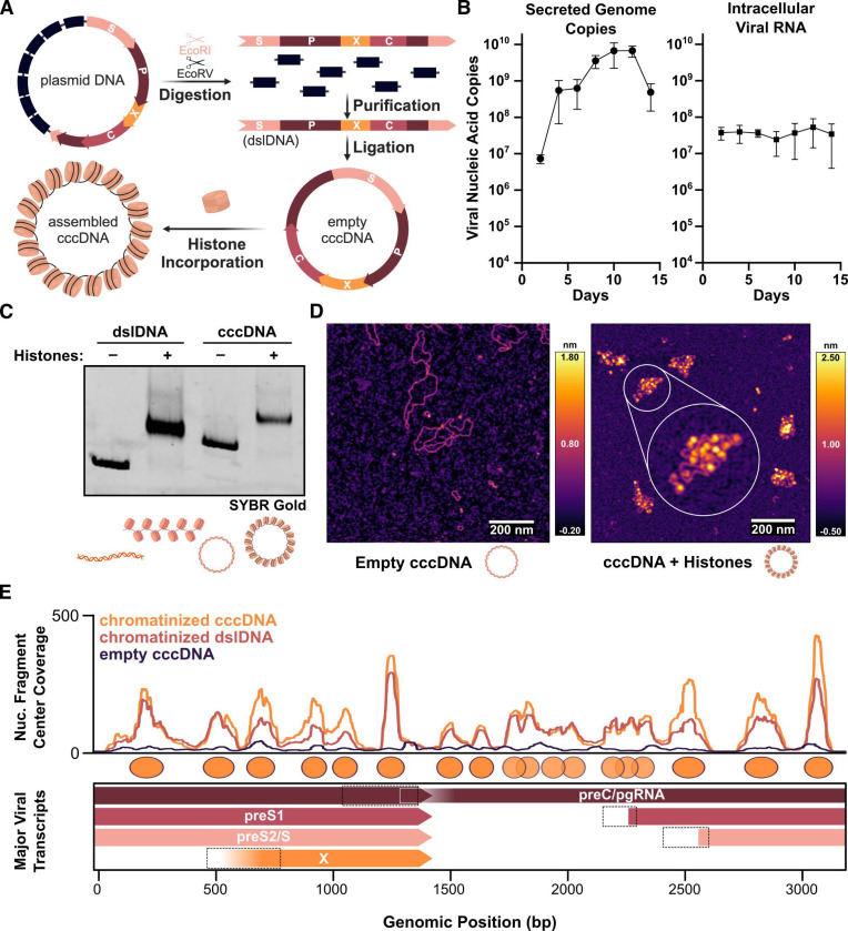
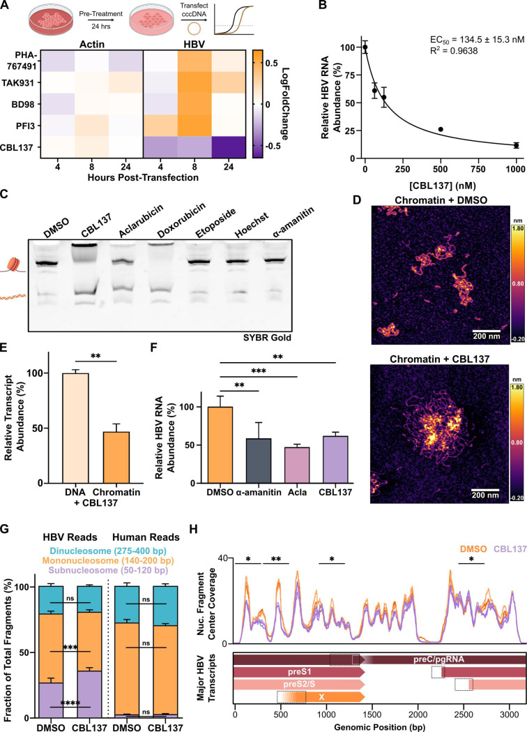
Chronic hepatitis B virus (HBV) infection is an incurable global health threat responsible for causing liver disease and hepatocellular carcinoma. During the genesis of infection, HBV establishes an independent minichromosome consisting of the viral covalently closed circular DNA (cccDNA) genome and host histones. The viral X gene must be expressed immediately upon infection to induce degradation of the host silencing factor, Smc5/6. However, the relationship between cccDNA chromatinization and X gene transcription remains poorly understood. Establishing a reconstituted viral minichromosome platform, we found that nucleosome occupancy in cccDNA drives X transcription. We corroborated these findings in cells and further showed that the chromatin destabilizing molecule CBL137 inhibits X transcription and HBV infection in hepatocytes. Our results shed light on a long-standing paradox and represent a potential new therapeutic avenue for the treatment of chronic HBV infection.

Keywords: Hepatitis B Virus; chromatin; epigenetics; nucleosome stability; transcription.


Conflict of interest statement

Declaration of interests R.E.S. is on the scientific advisory boards of Miromatrix Inc. and Lime Therapeutics and is a speaker and consultant for Alnylam Inc. All other authors declare that they have no competing interests.

Figures

Figure 1.
Figure 1.. Recombinant cccDNA robustly assembles into minichromosomes in vitro.
(A) Scheme illustrating minichromosome reconstitution approach. (B) Quantification of secreted HBV DNA (left) and intracellular total HBV RNA (right) following transfection of recombinant cccDNA into HepG2 cells. Data represent means ± SD of 3 biological replicates. (C) Electrophoretic mobility shift of linear (dslDNA) and circular (cccDNA) HBV DNA upon incorporation of recombinant human histone octamers. (D) Representative atomic force micrographs of empty (left) or chromatinized (right) cccDNA. (E) Coverage of centers of mononucleosome-length (140–200 bp) DNA fragments arising from MNase digestion of chromatinized cccDNA (orange), dslDNA (red), or empty cccDNA (navy) aligned to the HBV genome and smoothed with a 50 bp window chosen to show partially overlapping nucleosome occupancy peaks that may indicate underlying heterogeneity in nucleosome positioning, overlaid above a schematic of the four major HBV transcripts. Dotted boxes indicate promoter regions for each transcript, in genomic coordinate order: enhancer I/X promoter, enhancer II/basal core promoter, S1 promoter, S2 promoter.
Figure 2.
Figure 2.. Chromatinization of reconstituted cccDNA impacts viral transcription.
(A) Quantification of relative abundance of HBV RNA produced via in vitro transcription (IVT) of empty or chromatinized cccDNA in HepG2 nuclear extract. (B) Representative AFM micrographs of empty, undersaturated, intermediately, or fully saturated cccDNA minichromosomes. (C) Quantification of chromatin fiber volume (top) and volume-surface area ratio, a proxy for compaction, (bottom) for n = 49 (undersaturated), 70 (intermediate), or 96 (saturated) chromatin fibers pooled from multiple sample preparations on multiple days. (D) Quantification of transcript abundance for each of the four major HBV transcript following IVT of empty, undersaturated, intermediately, or fully saturated cccDNA species in HeLa nuclear extract. (E) Schematic depiction (top) and representative atomic force micrograph (bottom) of chimeric X-601 chromatin array comprised of the X promoter and gene body fused upstream of six 601 repeats. (F) Quantification of X transcript produced following IVT of empty or chromatinized chimeric X-601 template DNA, relative to an internal control, in HepG2 nuclear extract. (G) IVT as in E, with 1 μM synthetic LANA peptide added to all reactions. Data represent means of 3 replicates ± SD and were analyzed by Welch’s t-test (A, C, F, G) and 2-way ANOVA with Dunnett’s multiple comparison T3 test (D). *P < 0.05, **P < 0.01, ***P < 0.001, ****P <0.0001.
Figure 3.
Figure 3.. Early X gene transcription is linked to the established cccDNA nucleosome landscape
(A) Mononucleosome-sized fragment center coverage over the HBV genome following digestion of cccDNA chromatin arrays prepared as in Fig. 2B with different levels of histone octamer saturation (B) Coverage of the HBV genome by centers of unique molecular identifier (UMI)-deduplicated mononucleosome-sized fragments from MNase-seq of HepG2.2.15 (blue) and HepAD38 (green) cells. (C) Coverage of the HBV genome by centers of unique molecular identifier (UMI)-deduplicated mononucleosome-sized fragments from MNase-seq 4 (orange) and 24 hours (plum) after transfection of HEK293T cells with recombinant cccDNA. (D) HBV genomic coordinates of RNA-seq reads mapped at all time points measured up to 24 hours after cccDNA transfection into HEK 293T cells (top) and zoomed in on 4 and 8 hour time points (bottom). Dotted boxes are as in Fig. 1. (E) Quantification of total number of aligned reads to HBV or human genomes at various RNA-seq time points (left), and of the fraction of HBV-aligned reads mapping to the HBx ORF (right) for 3 biological replicates.
Figure 4.
Figure 4.. Nucleosome destabilizing drugs inhibit viral transcription in cells and disrupt cccDNA integrity in vitro.
(A) Heatmap depicting log-fold change of HBV or β-actin mRNA levels, normalized against GAPDH, in cccDNA-transfected HEK 293T cells treated for 24 hours with the indicated molecules (1μM PHA-767491, 300 nM TAK-931, 2.5 μM BD98, 1 μM PFI-3, 500 nM CBL137). Data represent 2 biological replicates. (B) Dose-dependence of HBV RNA levels in HEK293T cells 24-hours after cccDNA transfection and concurrent treatment with the indicated concentration of CBL137. (C) Native PAGE gel stained with SYBR Gold to assess the stability of recombinant mononucleosomes following incubation with the indicated molecules (all at 10 μM concentrations). (D) Representative atomic force micrographs of recombinant HBV dslDNA chromatin arrays treated with CBL137 or a vehicle control prior to AFM imaging. (E) Quantification of X transcript produced following in vitro transcription of empty or chromatinized chimeric X-601 template DNA using HepG2 nuclear extract, relative to an internal control from an unchromatinized template under control of the CMV promoter, in reactions where 1 μM CBL137 was added. (F) Quantification of relative HBV RNA 24 hours after transfecting HEK293T cells with recombinant cccDNA and concurrent treatment with DMSO (vehicle), 500 nM α-amanitin (positive control), 125 nM CBL137, or 50 nM aclarubicin. (G) Quantification of the fraction of total subnucleosome- (50–120 bp), mononucleosome- (140–200 bp), or dinucleosome-sized (275–400 bp) DNA fragments mapping to either the HBV or human genomes following MNase-seq of HepAD38 nuclei after 48 hour treatment with 500 nM CBL137 or a vehicle control. (H) Coverage of centers of mononucleosome-length DNA fragments mapping to the HBV genome from G, where indicated 300 bp regions show a significant change in fragment counts between mock- and CBL137-treated samples. Data represent 2 (A-D), 3 (E, G-H), or 4 (F) independent experiments/biological replicates as means ± SD and were analyzed by 1-way ANOVA with Dunnett’s multiple comparison test (F), Welch’s t-test (E), and 2-way ANOVA with Tukey’s multiple comparison test (G), and two-tailed two-sample t-test (H). *P < 0.05, **P < 0.01, ***P < 0.001.
Figure 5.
Figure 5.. CBL137 inhibits viral transcription, antigen secretion, and genome replication in hepatocyte models of HBV infection.
(A) Quantification of relative total HBV RNA from HepG2 cells 4- or 24-hours after transfection with recombinant cccDNA and concurrent treatment with DMSO or 500 nM CBL137. (B) Quantification of the proportion of total HBV RNA comprised by each of the four major viral transcripts from samples in A. (C) Quantification of secreted HBV surface antigen (HBsAg), secreted antigen (HBeAg), intracellular total RNA, secreted rcDNA, or intracellular cccDNA from HepG2-NTCP cells following infection and either 24 hour pre-infection treatment or 5 day post-infection treatment with 125 nM CBL137. (D) Dose-dependence of total HBV RNA abundance in primary human hepatocytes infected with HBV for 5 days and subsequently treated with the indicated concentrations of CBL137 for 2 days. (E) Dose-dependence of intracellular cccDNA abundance in primary hepatocytes cultured and treated as in D. (F) Proposed model illustrating recognition of the chromatinized X gene promoter as a driving force behind transcription of the X gene during the genesis of infection. All data are means ± SD of 3 biological replicates and were analyzed by Welch’s t-test (A) and 2-way ANOVA with Tukey’s (B) or Dunnett’s (C) multiple comparisons tests. *P < 0.05, **P < 0.01, ***P < 0.001, ****P <0.0001.

References

    1. Yuen M.-F., Chen D.-S., Dusheiko G.M., Janssen H.L.A., Lau D.T.Y., Locarnini S.A., Peters M.G., and Lai C.-L. (2018). Hepatitis B virus infection. Nature Reviews Disease Primers 4, 18035. 10.1038/nrdp.2018.35. - DOI - PubMed
    1. Levrero M., and Zucman-Rossi J. (2016). Mechanisms of HBV-induced hepatocellular carcinoma. Journal of Hepatology 64, S84–S101. 10.1016/j.jhep.2016.02.021. - DOI - PubMed
    1. Yan H., Zhong G., Xu G., He W., Jing Z., Gao Z., Huang Y., Qi Y., Peng B., Wang H., et al. (2012). Sodium taurocholate cotransporting polypeptide is a functional receptor for human hepatitis B and D virus. eLife 2012, 1–28. 10.7554/eLife.00049. - DOI - PMC - PubMed
    1. Rabe B., Vlachou A., Panté N., Helenius A., and Kann M. (2003). Nuclear import of hepatitis B virus capsids and release of the viral genome. Proceedings of the National Academy of Sciences 100, 9849–9854. 10.1073/pnas.1730940100. - DOI - PMC - PubMed
    1. Yang R., Ko Y.-H., Li F., Lokareddy R.K., Hou C.-F.D., Kim C., Klein S., Antolínez S., Marín J.F., Pérez-Segura C., et al. (2024). Structural basis for nuclear import of hepatitis B virus (HBV) nucleocapsid core. Science Advances 10, eadi7606. 10.1126/sciadv.adi7606. - DOI - PMC - PubMed

Publication types

LinkOut - more resources