[Preprint]. 2024 Jun 12:2024.06.12.598725.
doi: 10.1101/2024.06.12.598725.

PTBP1 mediates Sertoli cell actin cytoskeleton organization by regulating alternative splicing of actin regulators

Affiliations

PTBP1 mediates Sertoli cell actin cytoskeleton organization by regulating alternative splicing of actin regulators

Yuexi Wang et al. bioRxiv. .

Update in

Abstract

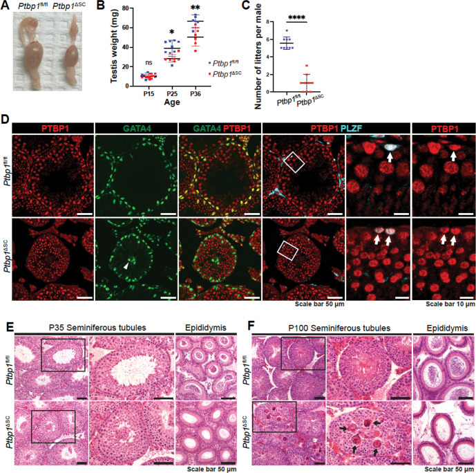
Spermatogenesis is a biological process within the testis that produces haploid spermatozoa for the continuity of species. Sertoli cells are somatic cells in the seminiferous epithelium that orchestrate spermatogenesis. Cyclic reorganization of Sertoli cell actin cytoskeleton is vital for spermatogenesis, but the underlying mechanism remains largely unclear. Here, we report that RNA-binding protein PTBP1 controls Sertoli cell actin cytoskeleton reorganization by programming alternative splicing of actin cytoskeleton regulators. This splicing control enables ectoplasmic specializations, the actin-based adhesion junctions, to maintain the blood-testis barrier and support spermatid transport and transformation. Particularly, we show that PTBP1 promotes actin bundle formation by repressing the inclusion of exon 14 of Tnik, a kinase present at the ectoplasmic specialization. Our results thus reveal a novel mechanism wherein Sertoli cell actin cytoskeleton dynamics is controlled post-transcriptionally by utilizing functionally distinct isoforms of actin regulatory proteins, and PTBP1 is a critical regulatory factor in generating such isoforms.


Conflict of interest statement

DECLARATION OF INTERESTS The authors declare no competing interests.

Figures

Figure 1.
Figure 1.. Sertoli cell-specific PTBP1 deletion disrupted spermatogenesis.
(A-B) shows the size difference between the testes from Ptbp1f/f and Ptbp1ΔSC mice. (C) shows the number difference between the litters generated by Ptbp1f/f and Ptbp1ΔSC mice. Data in B and C are presented as mean ± SD. NS: not significant. ***P<0.001, **** p < 0.0001. Each symbol in B and C represents the value of each mouse. (D) Double immunofluorescence staining using antibodies against PTBP1, GATA4 (a marker for Sertoli cells), or PLZF (a marker for undifferentiated spermatogonia stem cells) shows the specific and efficient PTBP1 deletion in Sertoli cells of Ptbp1ΔSC mice. The arrowhead indicates mislocalized Sertoli cells in Ptbp1ΔSC mice. Arrowheads indicate the PLZF-positive cells in which PTBP1 expression is intact in Ptbp1ΔSC mice. (E-F) Hematoxylin and eosin (H&E)-stained testis and cauda epididymis sections from Ptbp1fl/fl and Ptbp1ΔSC mice at P35 and P100. Boxed regions were magnified in the middle panel. Arrows in F point to multinucleated giant cells in the seminiferous tubules of Ptbp1ΔSC mice. Note very few spermatozoa are present in the cauda region.
Figure 2.
Figure 2.. Spermiogenesis is arrested in Ptbp1ΔSC mice
(A-B) The PAS and hematoxylin staining shows seminiferous cycles in the testes of Ptbp1fl/fl and Ptbp1ΔSC mice at P35 and P100. Regions in yellow boxes were magnified in the right panels. Yellow arrows in A point to sloughed germ cells at P35. Red arrows in B point to multinucleated giant cells at P100 in Ptbp1ΔSC mice. Yellow arrowheads point to acrosome caps in step 8 spermatids. (C-D) Ultrastructural images of differentiating spermatids in Ptbp1fl/fl and Ptbp1ΔSC mice at P100. Red arrowheads in C point to the differentiating spermatids with abnormal head morphologies. Spermatid cytoplasm regions were pseudo-colored in yellow (D).
Figure 3.
Figure 3.. The transcriptome changes in the testis of Ptbp1ΔSC mice
(A) A Venn diagram shows the number of genes that are differentially expressed or alternatively spliced in Ptbp1ΔSC testes. Three genes(Gm28356, Porcn, and Il17re3) displayed changes in both gene expression and splicing. (B) Breakdown of 501 alternatively spliced events into various event categories. A3SS: alternative 3’ splice site, A5SS: Alternative 5’ splice site, MXE: Mutually exclusive exon, RI: Retained intron, SE: Skipped exon. (C) Violin plots demonstrate ΔPSI (Inclusion level difference) distributions of significantly altered splicing events in Ptbp1ΔSC testes. (D) Metagene analysis of alternatively spliced exons detected in knockout mice by their positions on mRNA transcripts. Distribution along the transcript bins is shown on top, and the breakup of events into relative transcript regions is shown at the bottom. (E) Effect of alternatively spliced events induced by PTBP1 deficiency on the transcripts. Categories indicate if the exon inclusion preserves the open reading frame (ORF) and if the inclusion transcript is subjected to Non-sense Mediated Decay (NMD). (F) Exon ontology-based distribution of skipped exons, either increase or decrease in inclusion, based on encoded protein features. PTM: post-translational modification. (G) shows the relative enrichment of [CT]-rich motif near cassette exons (represented in green) that displayed a significant increase (red curve) or decrease (blue curve) in inclusion in Ptbp1ΔSC testes. Alternatively spliced exons were identified using rMATS, and a motif map was constructed using RMAPs (with a 50-nucleotide sliding window). The set of background cassette exons is represented in black. (H) Gene ontology analysis demonstrates the top 8 biological processes enriched among differentially expressed genes.
Figure 4.
Figure 4.. PTBP1 deficiency induced aberrant splicing of Sertoli cell-enriched actin regulators
(A) The heatmap shows the PSI values of exons in 8 actin regulators enriched in Sertoli cells. (B-C) PCR-based splicing assays validate 5 alternatively spliced events in 5 actin regulatory genes. Representative gel images are shown in B, and the quantifications of PSI are shown in C. 3 Ptbp1ΔSC mice and 3 sibling littermate Ptbp1fl/fl mice were assessed. Data are presented as mean±SD, n=3. *P<0.05, **P<0.01, ***P<0.001.
Figure 5.
Figure 5.. Ptbp1ΔSC mice had disorganized F-actin at the ES and impaired BTB integrity
(A) Phalloidin staining shows F-actin organization. Apical ES (yellow boxes) and basal ES (red boxes) are magnified in insets. Note that Sertoli cells of Ptbp1ΔSC mice exhibit abnormal actin assembly at basal ES (arrows) and apical ES (arrowheads). (B) Immunofluorescence staining using the antibody against ESPN shows impaired basal ES (arrows) and apical ES (arrowheads). (C) TEM images show the basal ES. Arrowheads indicate the packed F-actin bundles in the ES of adjoining Sertoli cells. The asterisk points to the region where F-actin bundles are missing in the Sertoli cells of Ptbp1ΔSC mice. (D and E) Immunofluorescence staining using the antibodies against GATA4, ZO-1, or CLDN11 shows the distribution of tight junction proteins. Boxed regions are magnified on the right. Arrows show the normal distribution of ZO-1 and CLDN11 at the basal region of Sertoli cells in Ptbp1fl/fl mice. Arrowheads show the mislocalized ZO-1 and CLDN11 in Ptbp1ΔSC mice. (F) Sulfo-NHSLC-biotin staining shows impaired BTB integrity.
Figure 6.
Figure 6.. PTBP1 deficiency increases the inclusion of Tnik exon 14 and alters TNIK protein distribution.
(A-B) PCR-based splicing assays demonstrate the splicing patterns of Tnik exon 14 at different ages. Representative gel images in A show the presence of exon 14-included and -excluded Tnik isoforms in the testes at different ages. Quantifications of PSI are shown in B. 3 Ptbp1ΔSC mice and 3 sibling littermate Ptbp1fl/fl mice were assessed at each age. Data are presented as mean±SD, n=3. **P<0.01, ***P<0.001, ****P<0.0001. ns, not significant. (C) Double immunofluorescence staining using antibodies against TNIK and TUBB3 shows TNIK expression in the Sertoli cell cytoplasm. Boxed regions were magnified in the right panels. (D) Double immunofluorescence staining using antibodies against TNIK and ESPN shows TNIK expression in the apical ES (blue boxes) and basal ES (yellow boxes). Boxed regions are magnified in insets. PTBP1 deficiency altered TNIK distribution at both the apical ES (arrowheads) and basal ES (arrows).
Figure 7.
Figure 7.. PTBP1 represses the inclusion of Tnik exon14 to promote F-actin assembly in Sertoli cells.
(A) Double fluorescence staining of PTBP1 and F-actin shows reduced F-actin bundle formation in PTBP1-deficient Sertoli cells cultured in vitro. Boxed regions are magnified in the right panels. (B) A schematic diagram shows the action of Tnik splicing-inhibiting ASO in blocking Tnik exon 14 inclusion. Tnik ASO binds to the 3’end of Tnik exon 14 and the adjacent 3’ splice site in the intron region, which prevents the recognition of 3’ splice site by the spliceosome. This leads to the skipping of exon 14. (C and D) PCR-based splicing assays show the Tnik ASO treatment reduced the inclusion of Tnik exon 14 in PTBP1-deficient Sertoli cells. The data shown are representative of three independent experiments. (E) Phalloidin staining shows F-actin bundles in Tnik ASO- or vehicle-treated Sertoli cells derived from Ptbp1fl/fl and Ptbp1ΔSC mice. Sertoli cells from Ptbp1fl/fl were used as a reference for normal F-actin bundle formation. Boxed regions are magnified in the bottom panels. Note F-actin bundles in Tnik ASO-treated PTBP1-deficient Sertoli cells are much thicker than those in vehicle-treated ones.
Figure 8.
Figure 8.. A proposed model of PTBP1 in regulating actin cytoskeleton organization in Sertoli cells
PTBP1 controls AS of actin cytoskeleton regulators in Sertoli cells to support the apical and basal ES function, which is essential for BTB integrity, germ cell transport, adhesion, and differentiation. Of particular, PTBP1 represses the inclusion of Tnik exon 14 to promote the F-actin bundle formation.

References

    1. Cheng C.Y. and Mruk D.D. (2010) The biology of spermatogenesis: the past, present and future. Philos Trans R Soc Lond B Biol Sci, 365, 1459–1463. - PMC - PubMed
    1. Griswold M.D. (2018) 50 years of spermatogenesis: Sertoli cells and their interactions with germ cells. Biol Reprod, 99, 87–100. - PMC - PubMed
    1. Vogl A.W., Vaid K.S. and Guttman J.A. (2008) The Sertoli cell cytoskeleton. Adv Exp Med Biol, 636, 186–211. - PubMed
    1. Mruk D.D. and Cheng C.Y. (2015) The Mammalian Blood-Testis Barrier: Its Biology and Regulation. Endocr Rev, 36, 564–591. - PMC - PubMed
    1. Smith B.E. and Braun R.E. (2012) Germ cell migration across Sertoli cell tight junctions. Science, 338, 798–802. - PMC - PubMed

Publication types