An official website of the United States government
The .gov means it’s official.
Federal government websites often end in .gov or .mil. Before
sharing sensitive information, make sure you’re on a federal
government site.
The site is secure.
The https:// ensures that you are connecting to the
official website and that any information you provide is encrypted
and transmitted securely.
Ben-Simon Y, Hooper M, Narayan S, Daigle TL, Dwivedi D, Way SW, Oster A, Stafford DA, Mich JK, Taormina MJ, Martinez RA, Opitz-Araya X, Roth JR, Alexander JR, Allen S, Amster A, Arbuckle J, Ayala A, Baker PM, Bakken TE, Barcelli T, Barta S, Bendrick J, Bertagnolli D, Bielstein C, Bishwakarma P, Bowlus J, Boyer G, Brouner K, Casian B, Casper T, Chakka AB, Chakrabarty R, Chance RK, Chavan S, Clark M, Colbert K, Collman F, Daniel S, Departee M, DiValentin P, Donadio N, Dotson N, Egdorf T, Fliss T, Gabitto M, Garcia J, Gary A, Gasperini M, Gloe J, Goldy J, Gore BB, Graybuck L, Greisman N, Haeseleer F, Halterman C, Haradon Z, Hastings SD, Helback O, Ho W, Hockemeyer D, Huang C, Huff S, Hunker A, Johansen N, Jones D, Juneau Z, Kalmbach B, Kannan M, Khem S, Kussick E, Kutsal R, Larsen R, Lee C, Lee AY, Leibly M, Lenz GH, Li S, Liang E, Lusk N, Madigan Z, Malloy J, Malone J, McCue R, Melchor J, Mollenkopf T, Moosman S, Morin E, Newman D, Ng L, Ngo K, Omstead V, Otto S, Oyama A, Pena N, Pham T, Phillips E, Pom CA, Potekhina L, Ransford S, Ray PL, Rette D, Reynoldson C, Rimorin C, Rocha D, Ruiz A, Sanchez REA, Sawyer L, Sedeno-Cortes A, Sevigny JP, Shapovalova N, Shepard N, Shulga L, Sigler…See abstract for full author list ➔Ben-Simon Y, et al.Cell. 2025 May 29;188(11):3045-3064.e23. doi: 10.1016/j.cell.2025.05.002. Epub 2025 May 21.Cell. 2025.PMID: 40403729
Abstract
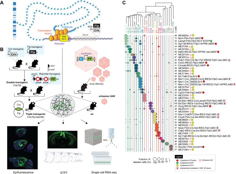
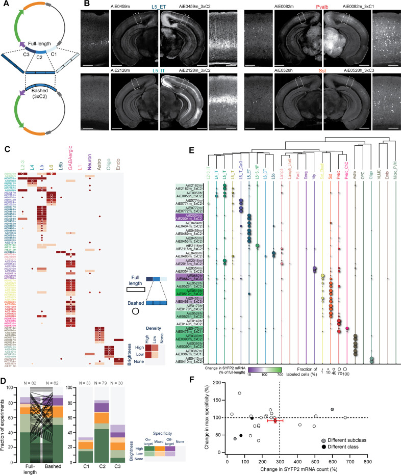
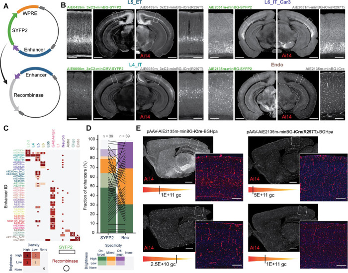
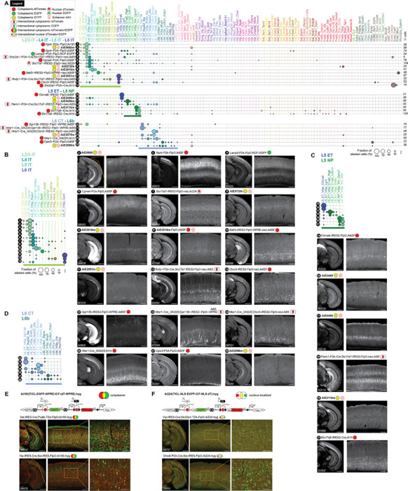
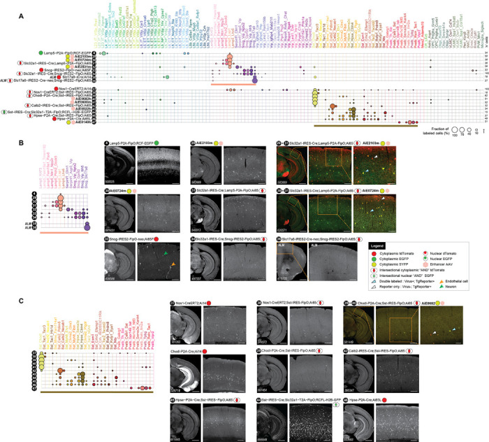
The mammalian cortex is comprised of cells classified into types according to shared properties. Defining the contribution of each cell type to the processes guided by the cortex is essential for understanding its function in health and disease. We used transcriptomic and epigenomic cortical cell type taxonomies from mouse and human to define marker genes and putative enhancers and created a large toolkit of transgenic lines and enhancer AAVs for selective targeting of cortical cell populations. We report evaluation of fifteen new transgenic driver lines, two new reporter lines, and >800 different enhancer AAVs covering most subclasses of cortical cells. The tools reported here as well as the scaled process of tool creation and modification enable diverse experimental strategies towards understanding mammalian cortex and brain function.
Figure 1:. Cell-type-specific genetic tool design and characterization.
A. A schematic illustration of gene regulation…
Figure 1:. Cell-type-specific genetic tool design and characterization.
A. A schematic illustration of gene regulation and location for recombinase insertions for knock-in transgenic mouse generation. B. Diagrams describing two strategies for genetic tool generation and characterization. Left: knock-in transgenic mouse lines are generated by insertions of recombinases into cell-type-specific differentially expressed genes. Generation of experimental animals requires one or more crosses to other recombinase lines and reporters. Bottom: The characterization of tool expression in brains of experimental animals is performed by three modalities: epifluorescence on select brain sections, serial-two-photon tomography (STPT) on whole brain, and single-cell RNA-seq on the visual cortex. Right: viral vectors utilize enhancers to achieve tool specificity. Generation of experimental animals requires retroorbital, intracerebroventricular or stereotaxic virus delivery to the animal. C. Single-cell RNA-seq data for some of the best tools reported in the study. Cortical taxonomy at the subclass level is on top and fraction of cells labeled per tool is represented by circles. Total number of cells per experiment (n cells) is represented in front of each tool’s name. Viruses are represented by pink hexagons. Other tools are transgenes.
Figure 2:. Selection of putative enhancer sequences.
A. Mouse and human single-nucleus transcriptomes obtained from…
Figure 2:. Selection of putative enhancer sequences.
A. Mouse and human single-nucleus transcriptomes obtained from single-nucleus multiomes represented in a transcriptomic UMAP and labeled according to cortical cell subclasses. Mouse nuclei were collected from the primary visual (VISp), somatosensory (SSp) and motor (MOp) cortices; human nuclei were collected from the middle temporal gyrus (MTG). Numbers of nuclei included in the analysis are represented by ‘n’. B. Simplified representation of the unified mouse-human taxonomy of cortical cell subclasses, along with the cluster-level taxonomy for mouse only. C. Summary of all ‘native’ putative enhancer sequences tested (n = 682), divided by the genome of origin, followed by cross-species conservation of sequence and accessibility (left). The modified sequences produced by concatenation are not included. The ‘Original’ and ‘Liftover’ plots show relative accessibility of all individual sequences in each subclass in their respective species, alongside the relative accessibility of its orthologous liftover sequence in the other species, respectively. Correlation between the original and liftover accessibility data is shown as a green/purple heatmap in the middle of these plots. Black arcs on the very right indicate instances where orthologs from both species were tested.
Figure 3:. A pipeline for enhancer AAV…
Figure 3:. A pipeline for enhancer AAV screening in the mouse brain.
A. Graphical summary…
Figure 3:. A pipeline for enhancer AAV screening in the mouse brain.
A. Graphical summary of the enhancer screening pipeline. B. Representative images of the visual cortex, showing examples of 11 categories of visually distinguishable cortical populations used to evaluate the labeling pattern of each enhancer AAV. Individual enhancer IDs are shown beneath each image (left), and a scoring matrix for evaluating brightness of the fluorescent signal and the labeling density, compared to the density expected for each category (right). C. Representative tracks of chromatin accessibility for four individual enhancers targeting across the cortical subclasses, demonstrating differential accessibility in L2–3_IT, alongside representative epifluorescence image sets, showing the resulting labeling pattern, and the score given to each in VISp. The closest L2–3_IT marker gene is shown above each set of tracks, along with the distance from the enhancer to its TSS. Scale bars below the tracks represent 100 bp (horizontal) and 0.3 RPKM/cell (vertical). D. Schematic describing the approach used to determine target specificity, according to the alignment between the TCP and LCP (top left) and a matrix used for classifying all enhancers, based on a combination of their target specificity and signal brightness (bottom left). E. Summary plot showing performance of all tested enhancers (n = 682) according to the categories (right). > 50% of enhancers (n = 376) exhibited signal in the cortex, ~43% were putatively on-target or had mixed (on- and off-target) labeling, and ~30% were putatively on-target (202/682). F. Proportion of enhancers in each of the categories specified in (D), according to their genome of origin (top, n = 150 for human and 532 for mouse) and TCP. Numerical values represent the number of tested enhancers in each column. For images, scale bars for full section and expanded views are 1.0 and 0.2 mm, respectively.
Figure 4:. Secondary validation of target specificity,…
Figure 4:. Secondary validation of target specificity, with scRNA-seq and whole-brain imaging.
A. Schematic of…
Figure 4:. Secondary validation of target specificity, with scRNA-seq and whole-brain imaging.
A. Schematic of methods for secondary validation. B. scRNA-seq analysis using SSv4, of FACS-sorted SYFP2+ cells from the mouse visual cortex, following RO administration of the enhancer AAV. The fraction of cells mapped to each cortical subclass corresponds to circle size, and the median SYFP2 mRNA count in each experiment, relative to the hSyn1 promoter, is denoted by a purple-to-green color gradient. The number of sequenced cells for each experiment is shown to the right of the table. Total number of enhancers examined is n = 149. Asterisks denote the top performing enhancers for each subclass, i.e., the ones with highest proportion of cells mapping to the subclass of interest. C. Box plot showing for each cortical population, all enhancer AAVs for which that population was the main enriched target population. The thick black bars represent medians, color-coded boxes represent top and bottom 25%, and whiskers represent top and bottom 10%. Data for individual enhancers is shown as superimposed black circles. D. Representative STPT images for five enhancers, with an expanded view of VISp displayed to the right. Dashed arrows connect each image set to its corresponding SSv4 data. In the case of AiE2543m, which labels L2–3_IT cells and Lamp5 cells, pink arrows point to sparse, yet brightly labeled non-L2–3 neurons, which are likely the Lamp5 interneurons. These are overrepresented in SSv4 (B), likely due to the stringent gating strategy in FACS focusing on the highly fluorescent cells. For images, scale bars for the hemisphere and the VISp magnified view are 1.0 and 0.2 mm, respectively.
Figure 5:. Optimization of enhancer activity through…
Figure 5:. Optimization of enhancer activity through core bashing.
A. Schematic representation of the core…
Figure 5:. Optimization of enhancer activity through core bashing.
A. Schematic representation of the core bashing approach for enhancer optimization (C = Core). B. Representative STPT images of coronal hemisections, showing labeling pattern for four individual full-length enhancers (left) and their best bashed version (right), which was selected according to the combination of brightness and specificity. A magnified view of VISp is shown alongside each hemisection. Scale bars for full section and expanded view are 1.0 and 0.2 mm, respectively. C. Heatmap showing the scoring results of epifluorescence image sets of the full-length enhancer (rectangles) alongside its best bashed version (circles); n = 82 pairs. D. Summary of the scoring data in (C), sorted according to brightness and specificity of the full-length vs. the bashed enhancer (left) and for the different cores tested (right). E. Dot plot of SSv4 data for full-length enhancers and their bashed counterpart shown in pairs, with circle size denoting the fraction of cells mapped to each of the cortical subclasses in each experiment. The color overlaying each pair name corresponds to the relative change in SYFP2 transcript count of the bashed relative to the full-length enhancer; n = 24 pairs. F. Change in specificity vs. change in SYFP2 transcript count for all enhancer pairs in (E). Average and SEM for all experiments corresponds to the red dot with error bars. Pairwise comparisons for individual enhancers correspond to white dots if no change in specificity if observed. If the bashed version preferentially labeled a different subclass or class compared to the corresponding full-length enhancer, the dots are grey or black, respectively.
Figure 6:. Recombinase-expressing enhancer-AAVs.
A. Schematic representation…
Figure 6:. Recombinase-expressing enhancer-AAVs.
A. Schematic representation of the vector design. B. Representative STPT images…
Figure 6:. Recombinase-expressing enhancer-AAVs.
A. Schematic representation of the vector design. B. Representative STPT images of coronal hemisections, showing labeling pattern for four individual enhancers expressing SYFP2 delivered to a wild-type mouse (left) and the same enhancers driving iCre(R297T), delivered to the Ai14 reporter mouse (right). A magnified view of VISp is shown alongside each hemisection. C. Heatmap showing the scoring of epifluorescence imaging data of the enhancers driving SYFP2 (rectangles) or a recombinase (circles); n = 39 pairs. D. Summary plot of the scoring data in (C), comparing brightness and specificity of the SYFP or recombinase expression. E. Specificity of Cre-dependent recombination in endothelial cells with the Ai2135m enhancer is reduced in iCre compared to the mutated version iCre(R297T) and is more sensitive to the viral dose (gc, genome copies). Scale bars in (B) and (E), 1.0 and 0.2 mm for full section and expanded view, respectively.
Figure 7:. Characterization of new transgenic driver…
Figure 7:. Characterization of new transgenic driver lines, preferentially targeting glutamatergic subclasses and clusters.
A. …
Figure 7:. Characterization of new transgenic driver lines, preferentially targeting glutamatergic subclasses and clusters.
A. scRNA-seq (SSv4) data showing distribution of labeled cells mapped to the mouse VISp taxonomy at the cluster level. 28 different experimental conditions (tools numbered 1–28) were grouped into panels according to predominant cell types labeled. They may include transgenic drivers and reporters as indicated or may be wild-type animals that received a systemic delivery of enhancer viruses (marked by pink hexagons). B. Focused view of tools 1–13 that label IT neurons from Layer 2–3, L4, L5 and L6, including the previously reported enhancer AiE2016m (originally called mscRE16) expressing SYFP in a wild-type animal (9) and driving a FlpO recombinase in Ai65F (9*). C. Same as in (B) for tools 14–21 that label ET and NP neurons in L5. D. Same as in (B) for tools 22–28 that label L6_CT and the L6b neurons. E. Schematics and representative sections from STPT data for a new Flp-Cre:AND/OR reporter line, Ai193 (TICL-EGFP-WPRE-ICF-tdT-WPRE)-hyg. The line was tested in triple transgenic crosses with two recombinase lines. Cells that express the Cre recombinase are EGFP-positive (green) and those that express FlpO are tdTomato-positive (red). Cells that express both appear yellow. F. Same as in (E) for a new reporter line, Ai224 (TICL-NLS-EGFP-ICF-NLS-dT)-hyg, where fluorophores are nucleus-localized. Scale bars: 1.0 and 0.2 mm for full section and expanded view; 0.1 mm for further expanded view in (E) and (F).
Figure 8:. Characterization of new transgenic driver…
Figure 8:. Characterization of new transgenic driver lines, preferentially targeting GABAergic subclasses and clusters.
A. …
Figure 8:. Characterization of new transgenic driver lines, preferentially targeting GABAergic subclasses and clusters.
A. scRNA-seq (SSv4) data, same as in Figure 7A but for 20 lines (tools numbered 4, 29–34, 37–47) targeting GABAergic types grouped into panels according to predominant cell types labeled. B. Focused representation of the same data as in (A) for tools labeling clusters within Lamp5 and Sncg GABAergic cortical subclasses. All data (tools numbered 4, 29–34) are from VISp, except for tools 35 and 36 that were characterized in cortical area ALM. The tools 29+31 and 30+31 show expression of two different Lamp5-expressing viruses in the triple transgenic mouse that expresses highly specifically only in Lamp5 interneurons (Slc32a1-IRES-Cre;Lamp5-P2A-FlpO;Ai65 – see tool 31). C. Focused representation of the same data as in (A) for tools 37–47 labeling clusters in Sst and Pvalb GABAergic cortical subclasses. Scale bars: 1.0 and 0.2 mm for full section and expanded view; 0.1 mm for further expanded view for tools 29+31, 30+31, 39+40.
Zeng H. (2022). What is a cell type and how to define it? Cell 185, 2739–2755. 10.1016/j.cell.2022.06.031.
-
DOI
-
PMC
-
PubMed
DeNardo L., and Luo L. (2017). Genetic strategies to access activated neurons. Curr. Opin. Neurobiol. 45, 121–129.
-
PMC
-
PubMed
Luo L., Callaway E.M., and Svoboda K. (2018). Genetic dissection of neural circuits: a decade of progress. Neuron 98, 256–281. 10.1016/j.neuron.2018.03.040.
-
DOI
-
PMC
-
PubMed
Peng H., Xie P., Liu L., Kuang X., Wang Y., Qu L., Gong H., Jiang S., Li A., Ruan Z., et al. (2021). Morphological diversity of single neurons in molecularly defined cell types. Nature 598, 174–181.
-
PMC
-
PubMed
Rudy B., Fishell G., Lee S., and Hjerling-Leffler J. (2011). Three groups of interneurons account for nearly 100% of neocortical GABAergic neurons. Devel Neurobio 71, 45–61. 10.1002/dneu.20853.
-
DOI
-
PMC
-
PubMed