. 2024 Jun 25;35(1):37.
doi: 10.1007/s10856-024-06806-x.

Intraocular pressure control efficacy and safety of HA-Mg glaucoma drainage plate implantation in the anterior chamber of rabbit eyes

Affiliations

Intraocular pressure control efficacy and safety of HA-Mg glaucoma drainage plate implantation in the anterior chamber of rabbit eyes

Mingming Cai et al. J Mater Sci Mater Med. .

Abstract

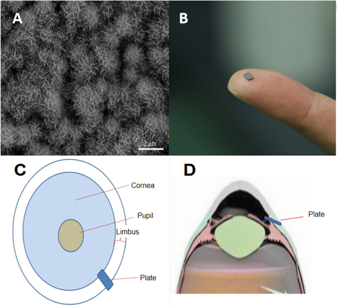
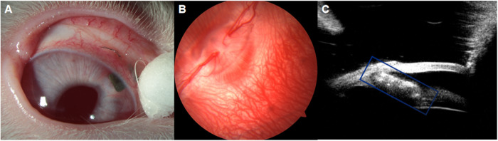
The current clinical application of glaucoma drainage devices is made of non-degradable materials. These non-degradable drainage devices often trigger inflammatory responses and scar proliferation, possibly leading to surgical failure. We developed a biodegradable material hydroxyapatite-coated magnesium (HA-Mg) as a glaucoma drainage device. Twelve New Zealand white rabbits were randomly assigned to three groups: HA-Mg drainage plate group (6 right eyes), trabeculectomy group (6 right eyes), and control group (12 left eyes). Results showed that all HA-Mg drainage plates were completely degraded ~4 months postoperatively. At the 5th month postoperatively, there was no statistical difference in the corneal endothelium density between the HA-Mg drainage plate group and the control group (p = 0.857). The intraocular pressure (IOP) level in the HA-Mg drainage plate implantation group was lower than in the other two groups. The trypan blue dye still drained from the anterior chamber to the subconjunctiva 5 months after HA-Mg drainage plate implantation. HE staining revealed the scleral linear aqueous humor drainage channel and anterior synechia were observed after drainage plate completely degraded, with no obvious infiltration with the inflammatory cells. This study showed the safety and efficacy of HA-Mg glaucoma drainage plate in controlling IOP after implantation into the anterior chamber of rabbit eyes.


Conflict of interest statement

The authors declare no competing interests.

Figures

None
We developed a biodegradable material hydroxyapatite-coated magnesium (HA-Mg) as a glaucoma drainage device. This study evaluated the safety and efficacy of HA-Mg glaucoma drainage plate in controlling intraocular pressure after implantation into the anterior chamber of rabbit eyes.
Fig. 1
Fig. 1
The gaps on hydroxyapatite (HA) coating surface and surgical approach of drainage plate. A Scanning electron microscope (SEM) images of HA coating surface morphology and drainage gaps at 20,000× magnification. B The appearance of HA-Mg glaucoma drainage plate. C The top view of the implanted plate in the eye. D The lateral view of the implanted plate
Fig. 2
Fig. 2
The anterior and posterior segments examination and the fixation of drainage plate. A The anterior segment of the eye 30 days post surgery. B The posterior segment of the eye 60 days post surgery. C The UBM image of the eye 30 days post surgery
Fig. 3
Fig. 3
The corneal endothelium density measurement. Five rabbits corneal endothelium density specular microscopy examination reports, OD: HA-Mg drainage plate group. OS: control group
Fig. 4
Fig. 4
The density of corneal endothelium. The p value of the different groups was no statistical difference (p = 0.857). Data were analyzed using a paired-sample T-test, N = 5
Fig. 5
Fig. 5
Surgery procedure and postoperative drainage channel measurement. AC Surgery procedure of HA-Mg glaucoma drainage plate implantation. A The appearance of HA-Mg glaucoma drainage plate and sclera tunnel. B HA-Mg glaucoma drainage plate implanted. C The appearance after surgery. DF Postoperative drainage channel measurement. D 1st month after trabeculectomy. E 1st month after HA-Mg glaucoma drainage plate implantation. F 5th month after HA-Mg glaucoma drainage plate implantation
Fig. 6
Fig. 6
Hematoxylin and eosin staining of HA-Mg glaucoma drainage plate implantation. Blue arrow: aqueous humor drainage channel. Red circle: anterior synechia. Blue rectangle: raised, microcystic filtering bubble

References

    1. Quigley HA, Broman AT. The number of people with glaucoma worldwide in 2010 and 2020. Br J Ophthalmol. 2006;90:262–7. - PMC - PubMed
    1. Dada T, Midha N, Shah P, Sidhu T, Angmo D, Sihota R. Innovations in glaucoma surgery from Dr. Rajendra Prasad Centre for Ophthalmic Sciences. Indian J Ophthalmol. 2017;65:103–8. - PMC - PubMed
    1. Singh AP. Medicinal leech therapy (hirudotherapy): a brief overview. Complement Ther Clin Pract. 2010;16:213–5. - PubMed
    1. Lin MK, Ge J. How to make the intraocular pressure after glaucoma filtering surgery more controllable. Chin J Ophthalmol. 2020;56:13–16. - PubMed
    1. Cipriano AF, Sallee A, Tayoba M, Cortez Alcaraz MC, Lin A, Guan RG, et al. Cytocompatibility and early inflammatory response of human endothelial cells in direct culture with Mg-Zn-Sr alloys. Acta Biomater. 2017;48:499–520. - PMC - PubMed