. 2024 Jun 25;134(16):e173690.
doi: 10.1172/JCI173690.

SLC44A2 regulates vascular smooth muscle cell phenotypic switching and aortic aneurysm

Affiliations

SLC44A2 regulates vascular smooth muscle cell phenotypic switching and aortic aneurysm

Tianyu Song et al. J Clin Invest. .

Abstract

Aortic aneurysm is a life-threatening disease with limited interventions that is closely related to vascular smooth muscle cell (VSMC) phenotypic switching. SLC44A2, a member of the solute carrier series 44 (SLC44) family, remains undercharacterized in the context of cardiovascular diseases. Venn diagram analysis based on microarray and single-cell RNA sequencing identified SLC44A2 as a major regulator of VSMC phenotypic switching in aortic aneurysm. Screening for Slc44a2 among aortic cell lineages demonstrated its predominant location in VSMCs. Elevated levels of SLC44A2 were evident in the aorta of both patients with abdominal aortic aneurysm and angiotensin II-infused (Ang II-infused) Apoe-/- mice. In vitro, SLC44A2 silencing promoted VSMCs toward a synthetic phenotype, while SLC44A2 overexpression attenuated VSMC phenotypic switching. VSMC-specific SLC44A2-knockout mice were more susceptible to aortic aneurysm under Ang II infusion, while SLC44A2 overexpression showed protective effects. Mechanistically, SLC44A2's interaction with NRP1 and ITGB3 activates TGF-β/SMAD signaling, thereby promoting contractile gene expression. Elevated SLC44A2 in aortic aneurysm is associated with upregulated runt-related transcription factor 1 (RUNX1). Furthermore, low-dose lenalidomide (LEN; 20 mg/kg/day) suppressed aortic aneurysm progression by enhancing SLC44A2 expression. These findings reveal that the SLC44A2-NRP1-ITGB3 complex is a major regulator of VSMC phenotypic switching and provide a potential therapeutic approach (LEN) for aortic aneurysm treatment.

Keywords: Cardiovascular disease; Signal transduction; Vascular biology.


Figures
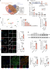
Figure 1
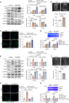
Figure 1. Aortic SLC44A2 expression is elevated in aortic aneurysm.
(A) Venn diagram showing the overlap between VSMC phenotype–related genes, differentially expressed markers for VSMCs, DEGs in human aortic aneurysm (GSE47472), and DEGs in VSMCs niched in mouse aortic aneurysm (GSE186865). (B) MASMCs were isolated from the whole abdominal aortas of saline- or Ang II–infused mice. The mRNA levels of Slc44a2, Uchl1, Dkk3, Anxa3, and Cryab were detected by qRT-PCR. n = 5. (C) Uniform manifold approximation and projection (UMAP) visualization of single cells from abdominal aortic tissue of mice (GSE152583). Cells were partitioned into 8 major lineages: vascular smooth muscle cells (VSMCs), fibroblasts (Fibro), endothelial cells (EC), macrophages (MΦ), T cells (T), B cells (B), erythrocytes (Eryth), and dendritic cells (DC). (D) Slc44a2 expression among distinct cellular populations. (E and F) Apoe–/– mice were infused with saline or Ang II for 28 days. (E) Immunofluorescent staining for SLC44A2 (red), ACTA2 (white), and staining with DAPI (blue) in the suprarenal abdominal aorta. Elastic fibers are green (autofluorescence). Arrowheads indicate elastin breaks. IgG was used as the isotype control. Scale bars: 200 μm. n = 5. (F) Slc44a2 mRNA level in aorta. n = 5. (G) Western blot analysis of SLC44A2 expression in the aorta from non-AAA groups and AAA patients. n = 6. (H) SLC44A2 mRNA level in the aorta from non-AAA groups and AAA patients. n = 6. (I) Immunofluorescent staining for SLC44A2 (red), ACTA2 (green), and staining with DAPI (blue) in the aortic media of non-AAA groups and AAA patients. IgG was used as the isotype control. L, lumen. Scale bars: 40 μm. n = 6. Differences were analyzed by unpaired, 2-tailed Student’s t test (B, F, and H), Welch’s ANOVA followed by Tamhane’s T2 multiple-comparison test or 1-way ANOVA followed by Tukey’s multiple-comparison test (E), Welch’s t test (G), or Welch’s ANOVA followed by Tamhane’s T2 multiple-comparison test (I).
Figure 2
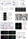
Figure 2. SLC44A2 maintains the contractile phenotype of VSMCs.
(AD) HASMCs were transfected with siRNA against SLC44A2 (siSLC44A2) or negative control (siNC), and then treated with Ang II (1 μM, 24 hours). (A) The synthetic and contractile markers were detected by Western blotting. n = 5. (B) Contraction of HASMCs grown in collagen discs was assessed and quantified by gel area. Scale bars: 5 mm. n = 5. (C) Immunofluorescence images of in situ zymography (DQ gelatin) in HASMCs. MMP activity was quantified by immunofluorescence intensity. RFU, relative fluorescence units. Scale bars: 40 μm. n = 5. (D) The activity of MMP2 and MMP9 in culture medium was measured by gel zymography. n = 6. (EH) HASMCs were infected with lentivirus containing empty vector or SLC44A2-encoding plasmids to overexpress SLC44A2 (SLC44A2OE), and then treated with Ang II (1 μM, 24 hours). (E) The synthetic and contractile markers were detected by Western blotting. n = 5. (F) Contraction of HASMCs grown in collagen discs was assessed and quantified by gel area. Scale bars: 5 mm. n = 5. (G) Immunofluorescence images of in situ zymography (DQ gelatin) in HASMCs. MMP activity was quantified by immunofluorescence intensity. Scale bars: 40 μm. n = 5. (H) The activity of MMP2 and MMP9 in culture medium was measured by gel zymography. n = 6. Differences were analyzed by 1-way ANOVA followed by Tukey’s multiple-comparison test (A, C, E, G, and H), unpaired, 2-tailed Student’s t test (B and F), or Welch’s ANOVA followed by Tamhane’s T2 multiple-comparison test or 1-way ANOVA followed by Tukey’s multiple-comparison test (D).
Figure 3
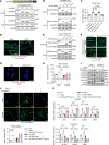
Figure 3. VSMC overexpression of SLC44A2 moderates aortic aneurysm in Ang II–infused Apoe–/– mice.
(A) Eight-week-old male Apoe–/– TaglnCre/+ mice were intravenously injected with lentivirus containing control vector or the reverse Slc44a2 sequence with 2 loxP sites. After 2 weeks, osmotic pumps were implanted subcutaneously to infuse saline or Ang II (1,000 ng/kg/min) for 28 days. (B) The systolic blood pressure at 0, 7, 14, 21, and 28 days after osmotic pump implantation. n = 8–11. NS, no significance. (C) The incidence of aortic aneurysm in Ang II–infused mice. n = 11. (D) Representative morphology of aortas from saline- or Ang II–infused mice. Scale bars: 5 mm. n = 11. (E) Ultrasound images and inner diameter quantification of the suprarenal abdominal aorta. n = 8–11. (F) Electron microscopic images of the suprarenal abdominal aorta. Red arrowheads indicate elastin breaks. El, elastin; Nu, nucleus. Scale bars: 5 μm. n = 3. (G) Hematoxylin and eosin (H&E) and elastic Verhoeff–Van Gieson (EVG) staining of the suprarenal abdominal aorta. Red arrowheads indicate elastin breaks. n = 6. (H) Immunofluorescence images of in situ zymography (DQ gelatin, green) in the suprarenal abdominal aorta. Scale bars: 200 μm. n = 6. (I) Immunofluorescent staining for OPN (red), ACTA2 (green), and staining with DAPI (blue) in the suprarenal abdominal aorta. Scale bars: 200 μm. n = 6. Differences were analyzed by 2-way ANOVA with mixed effects followed by Tukey’s multiple-comparison test (B), Fisher’s exact test (C), Welch’s ANOVA followed by Tamhane’s T2 multiple-comparison test (E and H), or 1-way ANOVA followed by Tukey’s multiple-comparison test or Welch’s ANOVA followed by Tamhane’s T2 multiple-comparison test (I).
Figure 4
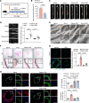
Figure 4. SLC44A2 knockout in VSMCs aggravates aortic aneurysm in Ang II–infused mice.
(A) Eight- to 10-week-old male Slc44a2WT and Slc44a2SMKO mice were infused with saline or Ang II (1,000 ng/kg/min) for 28 days by osmotic pumps. (B) The systolic blood pressure of Slc44a2WT and Slc44a2SMKO mice at 0, 7, 14, 21, and 28 days after osmotic pump implantation. n = 11–20. NS, no significance. (C) The incidence of aortic aneurysm in Ang II–infused mice. n = 20. (D) Representative morphology of aortas from saline- or Ang II–infused mice. Scale bars: 5 mm. n = 11–20. (E) Ultrasound images and inner diameter quantification of the suprarenal abdominal aorta. n = 11–20. (F) Electron microscopic images of the suprarenal abdominal aorta. Red arrowheads indicate elastin breaks. El, elastin; Nu, nucleus. Scale bars: 5 μm. n = 3. (G) H&E and EVG staining of the suprarenal abdominal aorta. Red arrowheads indicate elastin breaks. n = 5. (H) Immunofluorescence images of in situ zymography (DQ gelatin, green) in the suprarenal abdominal aorta. Scale bars: 200 μm. n = 5. (I) Immunofluorescent staining for OPN (red), ACTA2 (green), and staining with DAPI (blue) in the suprarenal abdominal aorta. Scale bars: 200 μm. n = 6. Differences were analyzed by 2-way ANOVA with mixed effects followed by Tukey’s multiple-comparison test (B), Fisher’s exact test (C), Kruskal-Wallis test followed by Dunn’s multiple-comparison test (E), or 1-way ANOVA followed by Tukey’s multiple-comparison test (H and I).
Figure 5
Figure 5. SLC44A2 activates TGF-β signaling to maintain the VSMC contractile phenotype via NRP1.
(A) Lysates from HASMCs were immunoprecipitated with anti-SLC44A2 antibody followed by mass spectrometry analysis to identify the proteins that interact with SLC44A2. The graph shows the TGF-β signaling–related proteins. iBAQ, intensity-based absolute quantification. (B) HASMCs were treated with Ang II (1 μM). Lysates were immunoprecipitated with anti-SLC44A2 antibody, and blotted with anti-NRP1 and anti-SLC44A2 antibodies. n = 5. (C) HASMCs were treated with Ang II. Lysates were immunoprecipitated with anti-NRP1 antibody, and blotted with anti-SLC44A2 and anti-NRP1 antibodies. n = 4. (D) Apoe–/– TaglnCre/+ mice were intravenously injected with lentivirus containing control vector or Slc44a2. Osmotic pumps were then implanted subcutaneously to infuse saline or Ang II. The interaction of SLC44A2 with NRP1 (red dots marked by arrowheads) in suprarenal abdominal aorta was detected by proximity ligation assay (PLA). Scale bars: 10 μm. n = 5. (EJ) HASMCs were infected with lentivirus containing empty vector or SLC44A2-encoding plasmids with or without siNRP1 transfection, and then treated with Ang II. (E) The TGF-β levels in culture medium was measured by ELISA. n = 5. (F) The levels of p-SMAD2 and p-SMAD3 were detected by Western blotting. n = 4. (G) The levels of VSMC synthetic and contractile markers were detected by Western blotting. n = 5. (H) Contraction of HASMCs grown in collagen discs was assessed and quantified by gel area. Scale bars: 5 mm. n = 5. (I) The activity of MMP2 and MMP9 in culture medium was measured by gel zymography. n = 6. (J) Immunofluorescence images of in situ zymography (DQ gelatin) in HASMCs. MMP activity was quantified by immunofluorescence intensity. Scale bars: 40 μm. n = 5. Differences were analyzed by Welch’s ANOVA followed by Tamhane’s T2 multiple-comparison test (D and I), 1-way ANOVA followed by Tukey’s multiple-comparison test (E, H, and J), or 1-way ANOVA followed by Tukey’s multiple-comparison test or Welch’s ANOVA followed by Tamhane’s T2 multiple-comparison test (G).
Figure 6
Figure 6. SLC44A2 mediates the activation of TGF-β by interacting with NRP1 and ITGB3.
(A) HEK293 cells were transfected with SLC44A2WT and plasmids encoding NRP1WT, NRP1ΔCUB, NRP1Δb1/b2, and NRP1ΔMAM. Lysates were immunoprecipitated with anti-HA antibody, and blotted with anti-His and anti-HA antibodies. n = 3. (B) HEK293 cells were transfected with NRP1WT and plasmids encoding SLC44A2WT, SLC44A2Δ55–232, SLC44A2Δ254–480, or SLC44A2Δ505–659. Lysates were immunoprecipitated with anti-His antibody, and blotted with anti-His and anti-HA antibodies. n = 3. (C) MASMCs from Slc44a2KO mice were infected with lentivirus containing empty vector or SLC44A2-encoding plasmids. The TGF-β level in culture medium was measured by ELISA. n = 5. (D) The interaction of SLC44A2 with ITGB3 in suprarenal abdominal aorta from Apoe–/– mice was detected by PLA. Scale bars: 10 μm. n = 3. (E) HEK293 cells were transfected with ITGB3WT and plasmids encoding SLC44A2WT, SLC44A2Δ55–232, SLC44A2Δ254–480, or SLC44A2Δ505–659. Lysates were immunoprecipitated with anti-FLAG antibody, and blotted with anti-FLAG and anti-HA antibodies. n = 3. (F) The interaction of NRP1 with ITGB3 in suprarenal abdominal aortas from Slc44a2WT and Slc44a2SMKO mice was detected by PLA. Scale bars: 10 μm. n = 5. (G) The interaction of NRP1 with ITGB3 was detected in siSLC44A2-transfected HASMCs by PLA. Scale bars: 20 μm. n = 3. (HK) HASMCs were infected with lentivirus containing empty vector or SLC44A2-encoding plasmids with or without siITGB3 transfection, and then treated with Ang II. (H) The TGF-β level in culture medium was measured by ELISA. n = 5. (I) p-SMAD2 and p-SMAD3 levels were detected by Western blotting. n = 3. (J) Immunofluorescence images and quantification of in situ zymography (DQ gelatin). Scale bars: 40 μm. n = 5. (K) VSMC synthetic and contractile markers were detected by qRT-PCR. n = 5. Differences were analyzed by 1-way ANOVA followed by Tukey’s multiple-comparison test (C, H, J, and K).
Figure 7
Figure 7. The transcription of SLC44A2 is regulated by RUNX1.
(A) Prediction of SLC44A2 promoter–binding transcription factors by JASPAR (https://jaspar.elixir.no/) and with the upstream 2,000 bp to downstream 100 bp region of SLC44A2 gene transcription initiation site set as the promoter region. Venn diagram of DEGs in murine (GSE17901 and GSE51229) and human (GSE7084) aortic aneurysm samples relative to normal controls from the NCBI GEO database. AA, aortic aneurysm; PPE, porcine pancreatic elastase. (B) HASMCs were transfected with siRUNX1 or siNC, and then treated with Ang II (1 μM, 24 hours). The levels of SLC44A2 and RUNX1 were detected by Western blotting. n = 5. (C) Western blot analysis of RUNX1 in the aortas of non-AAA and AAA individuals. n = 6. (D) RUNX1 mRNA level in the aortas of non-AAA and AAA individuals was detected by qRT-PCR. n = 6. (E) Relative luciferase activity in HEK293 cells transfected with luciferase reporter constructs containing SLC44A2 promoter truncations or its mutants along with pRL-TK (internal control plasmid) followed by transfection with RUNX1-encoding plasmid. n = 5. Differences were analyzed by 1-way ANOVA followed by Tukey’s multiple-comparison test (B), unpaired, 2-tailed Student’s t test (C and D), or unpaired, 2-tailed Student’s t test or Welch’s t test (E).
Figure 8
Figure 8. Administration of LEN relieves aortic aneurysm in mice.
(A) Eight- to 10-week-old male Apoe–/– mice were implanted subcutaneously with osmotic pumps to infuse saline or Ang II (1,000 ng/kg/min) with or without intragastric administration of LEN (20 mg/kg/day) for 28 days. (B) The incidence of aortic aneurysm in Ang II–infused Apoe–/– mice administered vehicle or LEN. n = 11. (C) Representative morphology of aortas from Ang II–infused Apoe–/– mice administered vehicle or LEN. Scale bars: 5 mm. n = 11. (D) Ultrasound images and inner diameter quantification of the suprarenal abdominal aorta. n = 9–11. (E) Electron microscopic images of the suprarenal abdominal aorta. Red arrowheads indicate elastin breaks. El, elastin; Nu, nucleus. Scale bars: 5 μm. n = 3. (F) H&E and EVG staining of the suprarenal abdominal aorta. Red arrowheads indicate elastin breaks. n = 5. (G) Immunofluorescence images of in situ zymography (DQ gelatin, green) in the suprarenal abdominal aorta. Scale bars: 200 μm. n = 5. (H) Immunofluorescent staining for OPN (red), ACTA2 (green), and staining with DAPI (blue) in the suprarenal abdominal aorta. Scale bars: 200 μm. n = 5. Differences were analyzed by Fisher’s exact test (B), Welch’s ANOVA followed by Tamhane’s T2 multiple-comparison test (D and G), or 1-way ANOVA followed by Tukey’s multiple-comparison test (H).
Figure 9
Figure 9. Proposed model for SLC44A2 as a therapeutic target in aortic aneurysm.
Lenalidomide promotes RUNX1-mediated transcription of SLC44A2. The upregulated SLC44A2 acts as a scaffolding protein to interact with NRP1 and ITGB3 to activate TGF-β/SMAD signaling, further promoting the expression of VSMC contractile genes and inhibiting the expression of VSMC synthetic genes to restrain the VSMC phenotypic switching in aortic aneurysm.

Comment in

  • SLC44A2-mediated phenotypic switch of vascular smooth muscle cells contributes to aortic aneurysm

References

    1. Bossone E, Eagle KA. Epidemiology and management of aortic disease: aortic aneurysms and acute aortic syndromes. Nat Rev Cardiol. 2021;18(5):331–348. doi: 10.1038/s41569-020-00472-6. - DOI - PubMed
    1. Pinard A, et al. Genetics of thoracic and abdominal aortic diseases. Circ Res. 2019;124(4):588–606. doi: 10.1161/CIRCRESAHA.118.312436. - DOI - PMC - PubMed
    1. Golledge J. Abdominal aortic aneurysm: update on pathogenesis and medical treatments. Nat Rev Cardiol. 2019;16(4):225–242. doi: 10.1038/s41569-018-0114-9. - DOI - PubMed
    1. Chakraborty R, et al. Histone Acetyltransferases p300 and CBP coordinate distinct chromatin remodeling programs in vascular smooth muscle plasticity. Circulation. 2022;145(23):1720–1737. doi: 10.1161/CIRCULATIONAHA.121.057599. - DOI - PubMed
    1. Cai D, et al. A novel mechanism underlying inflammatory smooth muscle phenotype in abdominal aortic aneurysm. Circ Res. 2021;129(10):e202–e214. doi: 10.1161/CIRCRESAHA.121.319374. - DOI - PMC - PubMed

MeSH terms