. 2024 Jul 2;121(27):e2403777121.
doi: 10.1073/pnas.2403777121. Epub 2024 Jun 25.

Parvalbumin gates chronic pain through the modulation of firing patterns in inhibitory neurons

Affiliations

Parvalbumin gates chronic pain through the modulation of firing patterns in inhibitory neurons

Haoyi Qiu et al. Proc Natl Acad Sci U S A. .

Abstract

Spinal cord dorsal horn inhibition is critical to the processing of sensory inputs, and its impairment leads to mechanical allodynia. How this decreased inhibition occurs and whether its restoration alleviates allodynic pain are poorly understood. Here, we show that a critical step in the loss of inhibitory tone is the change in the firing pattern of inhibitory parvalbumin (PV)-expressing neurons (PVNs). Our results show that PV, a calcium-binding protein, controls the firing activity of PVNs by enabling them to sustain high-frequency tonic firing patterns. Upon nerve injury, PVNs transition to adaptive firing and decrease their PV expression. Interestingly, decreased PV is necessary and sufficient for the development of mechanical allodynia and the transition of PVNs to adaptive firing. This transition of the firing pattern is due to the recruitment of calcium-activated potassium (SK) channels, and blocking them during chronic pain restores normal tonic firing and alleviates chronic pain. Our findings indicate that PV is essential for controlling the firing pattern of PVNs and for preventing allodynia. Developing approaches to manipulate these mechanisms may lead to different strategies for chronic pain relief.

Keywords: chronic pain; dorsal horn; parvalbumin.


Conflict of interest statement

Competing interests statement:The authors declare no competing interest.

Figures

Fig. 1.
Fig. 1.
PVN firing activity and PV expression are disrupted after nerve injury. (A) Whole-cell current-clamp representative traces of PVN response to step current injections in the lumbar DH of naive (Left) and 28 d post-nerve-injury (CCI 28 d) (Right) male mice. The same stimulation protocol is used for all subsequent electrophysiology traces unless otherwise stated. (B) Spike count (mean ± SEM) of PVN response to step current injections in naive and CCI 28 d mice (n = 28 cells from nine naive mice and n = 30 cells from 16 CCI mice) ****P-value < 0.0001, Two-way ANOVA, Šídák's multiple comparisons test. (C and D) Same as (A and B) in female mice. (n = 13 cells from four naive mice and n = 9 cells from 4 CCI mice.) **P-value = 0.0093, ***P-value = 0.0003, and ****P-value < 0.0001, two-way ANOVA, Šídák's multiple comparisons test. (E) Spike count (mean ± SEM) of PVN response to a 150 pA step current injection in naive, 2 d, 7 d, and 28 d CCI mice (n = 28 cells from nine naive mice, n = 9 cells from 4 CCI 4 d mice, n = 11 from 3 CCI 7 d mice, and n = 30 cells from 16 CCI 28 d mice) ***P-value =0.0002 and ****P-value < 0.0001, one-way ANOVA, Tukey’s multiple comparisons test. (F) Percentage of tonic firing (dark bar) and adaptive firing (light bar) cells at 150 pA step current injection in naive, 2 d, 7 d, and 28 d CCI male mice. ****P-value < 0.001, contingency test with Fisher’s exact probability test compares each time point with naive. (G) Pvalb promoter-driven tdTomato fluorescence in transverse DH sections of naive and CCI 28 d Pvalb-tdTomato male mice. (Scale bar, 100 µm.) (H) Reduced area of tdTomato-positive cells per spinal cord section (n = 33 sections in 3 CCI mice) on the ipsilateral side in CCI male mice. ****P-value < 0.0001, paired t test. No changes were observed in the area of tdTomato-positive cells in naive mice (n = 30 sections in three naive mice). (I) Mean (±SEM) Pvalb expression relative to β-actin in the dorsal ipsilateral quadrant of naive and CCI 28d mice (n = 4 mice per group). Results were normalized to Actb based on SI Appendix, Table S2. **P-value = 0.0021, unpaired t test. (J) Single-cell qPCR of Pvalb expression with respect to Gapdh in PVNs fromnaive and CCI 28 d mice (n = 13 cells from three naive mice and n = 10 cells from 3 CCI mice).*P-value = 0.049, unpaired t test.
Fig. 2.
Fig. 2.
PV expression is sufficient and necessary for preventing mechanical allodynia. (A and B) Paw withdrawal latency to heat (A) and nocifensive threshold to mechanical stimuli (B) of PV knockout (PV KO, n = 13) mice (light gray bar) and PV wild-type (PV WT, n = 11) mice (dark gray bar). ns, not statistically significant, unpaired t test. ***P-value < 0.001, unpaired t test. (C) Intraspinal delivery of a lentivirus carrying PV shRNA (n =10 mice) produces mechanical allodynia 8 wk postinjection (W8) compared to baseline (BL). Mice receiving scrambled shRNA (n =9) are unaffected. Two-way ANOVA, Šídák's multiple comparisons test. **P-value = 0.0088 and ***P-value < 0.001. (D and E) Spinal injection of AAV2/2-EF1α-DIO-WGA, allows for the expression of WGA in tdTomato+ PVNs (red, box i) (10/284 neurons), and their postsynaptic neurons (blue) which colocalize with PKCγ-expressing neurons (green, box ii) (50/284), or unidentified neurons (224/284). (Scale bar, 10 µm.) n = 11 sections from three mice. (F) Mean (±SEM) nocifensive responses in PV shRNA mice (shPV-W8) 20 min after an intrathecal injection of the control TAT peptide (red, n = 6) or PKCγ enzyme inhibitor, γV5-3 (100 pmol) (black, n = 6). *P-value = 0.0297, **P-value = 0.0022, ***P-value = 0.0002, and ****P-value < 0.0001, two-way ANOVA, Tukey's multiple comparisons test. (G and H) DH images (G) and quantification of tdTomato-positive cells (H) in Pvalb-tdTomato mice intraspinally injected with shPGC-1α (Bottom) or shControl adenovirus (Top) 4-d postinjection. (Scale bar, 100 µm.) (n = 32 sections from three shControl-treated mice and n = 22 section from shPGC-1α-treated mice) ****P-value < 0.0001 paired t test. (I) Same as (F) in shPGC-1α-injected mice (n = 8) 4-d postinjection (red) compared to shControl (blue, n = 6), and baseline (black, n = 14). Two-way ANOVA, Tukey's multiple comparisons test, *, # compares shPGC-1α to shControl and baseline, respectively. (J) AAV viral construct to overexpress PV in PVNs. (K) Neuroanatomical section of DH PVCre:tdTom neurons (red, Top) expressing the PV-WT sequence (blue arrowhead) visualized by a HA-tag (HA-IR; green, Bottom). (Scale bar, 10 µm.) (L) Mean (±SEM) of the number of nocifensive responses out of five von Frey filament stimulations (0.4 g) in mice injected with either the PV-ΔEF or the PV-WT sequence before nerve injury (CCI) **P-value = 0.0014, two-way ANOVA, Tukey’s multiple comparisons test. (n = 4 PV-WT and n = 4 PV-ΔEF). (M) Same as (L) in mice injected with the PV-ΔEF or the PV-WT sequence after CCI. n.s. nonstatistically significant, two-way ANOVA, Tukey's multiple comparisons test. (n = 5 PV-WT and n = 5 PV-ΔEF).
Fig. 3.
Fig. 3.
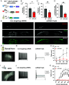
PV controls tonic firing of PVNs in the DH and hippocampus. (A) Schematic diagram of the technique to visualize shPvalb expressing PVNs for spinal cord slice electrophysiology (Materials and Methods). (B) Mean (±SEM) mechanically evoked response threshold as a percent of baseline in PVcre;RCF tdTomato male mice injected with shRNA-Pvalb (36.95 ± 6.0%, n = 7 mice, red, shPvalb) or nontargeting (64.23 ± 7.5%, n = 9 mice, gray, NT). *P-value = 0.0166, unpaired t test. (C) Same as in (B) in female mice (36.11 ± 5.4%, n = 6 shPvalb) (62.67 ± 4.5%, n = 5 NT) **P-value = 0.0053, unpaired t test. (D and E) Transverse DH sections of PVcre;RCF tdTOM mice injected with either NT (Left) or shPvalb (Right) (E). Anti-PV staining (green) showed that 21.79 ± 3.3% of tdTomato+ cells were PV-immunoreactivity (IR)-positive in the shPvalb-injected mice (n = 21 sections from three mice, red), whereas 71.33 ± 4.7% of tdTomato-positive cells were PV-IR-positive in the NT control (n = 13 sections from two mice, gray) (D). **P-value = 0.0028, unpaired t test. (Scale bar, 100 µm.) (F) Epifluorescence image of a spinal cord slice from a shPvalb injected PVcre;RCF tdTom mouse showing tdTomato-positive used for targeted electrophysiology recordings. (Scale bar, 100 µm.) (G and H) Whole-cell current clamp representative traces (G) and mean ± SEM of PVNs in response to step current injections (H) recorded in the DH of NT- (G, Left) and shPvalb-injected male and female mice (G, Right). (n = 6 cells from 4 NT-injected mice and n = 7 from 3 shPvalb-injected mice). *P-value = 0.0236 and **P-value = 0.0025, uncorrected Fisher’s LSD. (I) Same as (F) in the hippocampus (HPC) of the shPvalb-injected PVcre;RCF tdTom mouse. (Scale bar, 100 μm.) (J and K) Same as (G and H) in the hippocampus PVNs of NT- (J, Right) and shPvalb-injected mice (J, Right). (n = 4 cells from an NT-injected mouse and n = 4 cells from a shPvalb-injected mouse). ***P-value = 0.004 and ****P-value < 0.0001, two-way ANOVA, Šídák's multiple comparisons test.
Fig. 4.
Fig. 4.
Blocking SK channels of PVNs after nerve injury alleviates mechanical allodynia. (A–C) Whole-cell current clamp representative traces and mean ± SEM of PVNs spike count (C) in response to increasing step current injections recorded in the lumbar DH spinal cord of CCI male mice 0 min (Left) and after 15 min (Right) of cytoplasmic dialysis of internal solution containing 0.1 mM EGTA (A) or 2 mM EGTA (B). (n = 5 cells from 5 CCI mice for 0.1 mM EGTA and n = 5 cells from 3 CCI mice for 2 mM EGTA). *P-value = 0.0178 compares 0.1 mM EGTA (15 min) and 2 mM EGTA (15 min); #P-value = 0.0403 compares 2 mM EGTA (0 min) and 2 mM EGTA (15 min), two-way ANOVA, Dunnett’s multiple comparisons test. (D) Left, fluorescent in situ hybridization of transverse lumbar spinal DH sections from male mice (white outline) showing Pvalb (red), Vgat mRNA (Slc32a1, yellow), SK2 mRNA (Kcnn2, green), and SK3 mRNA (Kcnn3, cyan) colocalization. Right, zoomed-in section from the yellow box on the Left shows white arrows indicating inhibitory PVNs colocalizing with Kcnn2. (Scale bar, 100 µm, 50 µm.) (E) Quantification of (D) indicating inhibitory PVNs have a higher expression of Kcnn2 [273.5 ± 7.745 Intensity arbitrary units of measure (AUM)] than Kcnn3 (130.9 ± 2.722 Intensity AUM). Total of 684 VGAT+/PV+ cells counted in three mice, ****P-value < 0.0001 paired t test. (F and G) Whole-cell current clamp representative traces (F) and mean ± SEM of PVNs spike count in response to increasing step current injections (G) recorded in the lumbar DH spinal cord of naive male mice before (F, Left) and after bath application of 1-EBIO (0.3 mM) (F, Right) (n = 7 cells from three mice). Spike count of PVNs from CCI mice shown in the dotted line for comparison but was not used in statistics. **P-value = 0.0021, two-way ANOVA, Šídák's multiple comparisons test. (H and I) Same as (F and G) in PVNs of CCI male mice before (H, Left) and after bath application of apamin (200 nM) (I, Right). (n = 8 cells from 6 CCI mice). n.s. nonstatistically significant between naive PVNs and CCI + apamin. *P-value < 0.033 comparing naive and CCI PVNs, n.s. nonstatistically significant between naive PVNs and CCI + apamin. (J and K) Same as (F and G) in PVNs of CCI male mice before (J, Left) and after bath application of Lei-Dab7 (100 nM) (J, Right) (n = 8 cells from 4 CCI mice). *P-value < 0.033 comparing naive and CCI PVNs, n.s. nonstatistically significant between naive PVNs and CCI + Lei-Dab7. (L) Mechanical sensitivity timeline of downregulation of SK2 channels in PVNs after nerve injury. (M) Baseline mechanical sensitivity was measured before CCI surgery at week 0. After verifying mechanical sensitivity is present at 28 d post-CCI (week 4, W4, gray bars), mice were injected with either the NT control (n = 11) or the shKcnn2 (n = 8) and tested again 2 wk after injection (week 6, W6, blue bars) (Right). The downregulation of Kcnn2 was verified by RT-qPCR of cerebellum tissue in intracerebellar injected PVcre; RCF tdTomato mice (50.48 ± 6.1% NT, 33.32 ± 4.3% shKcnn2 normalized to Tbp, *P-value = 0.041, unpaired t test, n = 3 mice per condition). Both male and female mice were used.
Fig. 5.
Fig. 5.
General organization of PVN and PKCγ neuron relationship in normal and neuropathic pain conditions. Under naive conditions, PV in the PVNs chelates the free intracellular calcium ions, thereby inhibiting the activation of SK2 channels and preventing the influx of K+ ions. No PVN spike frequency adaption occurs which allows for inhibition of the polysynaptic pathway links between Aβ fibers to lamina I projection neurons. After nerve injury, a decrease in PV concentration results in an increase of the free intracellular calcium and activation of SK2 channels that allow for spike frequency adaptation of PVNs to appear. This adaption decreases their inhibition on PKCγ neurons enabling low-threshold inputs to drive lamina I projection neurons and causes mechanical allodynia.

References

    1. Jensen M. P., Chodroff M. J., Dworkin R. H., The impact of neuropathic pain on health-related quality of life: Review and implications. Neurology 68, 1178–1182 (2007). - PubMed
    1. Castro-Lopes J. M., Tavares I., Coimbra A., GABA decreases in the spinal cord dorsal horn after peripheral neurectomy. Brain Res. 620, 287–291 (1993). - PubMed
    1. Coull J. A. M., et al. , Trans-synaptic shift in anion gradient in spinal lamina I neurons as a mechanism of neuropathic pain. Nature 424, 938–942 (2003). - PubMed
    1. Moore K. A., et al. , Partial peripheral nerve injury promotes a selective loss of GABAergic inhibition in the superficial dorsal horn of the spinal cord. J. Neurosci. 22, 6724–6731 (2002). - PMC - PubMed
    1. Petitjean H., et al. , Dorsal horn parvalbumin neurons are gate-keepers of touch-evoked pain after nerve injury. Cell Rep. 13, 1246–1257 (2015). - PMC - PubMed

Substances

LinkOut - more resources