

Neonatal cerebral ultrasound: anatomical variants and age-related diseases
- PMID: 38918318
- PMCID: PMC11496415
- DOI: 10.1007/s40477-024-00914-8
Neonatal cerebral ultrasound: anatomical variants and age-related diseases
Abstract
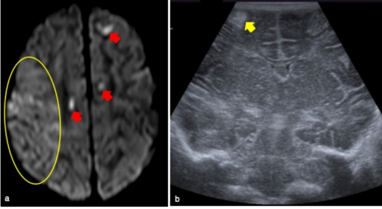
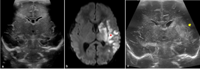
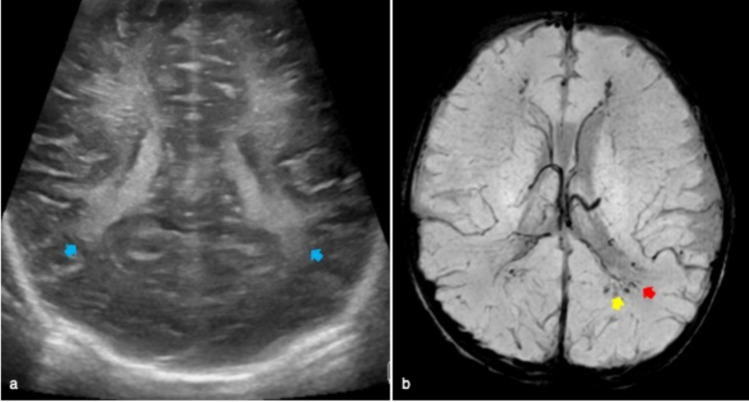
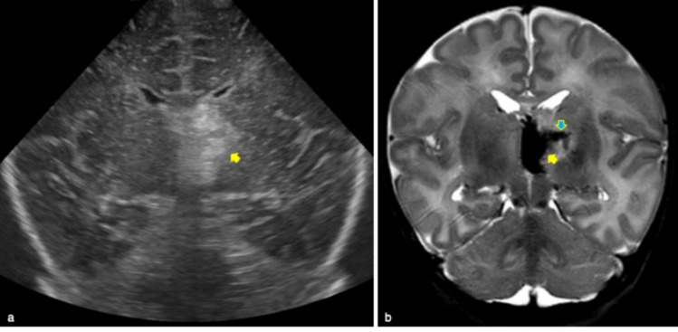
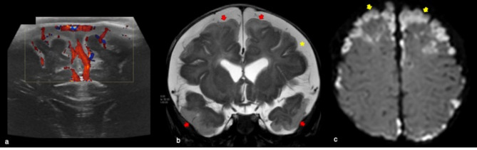
Cerebral ultrasound is a non-invasive imaging technique widely used for the assessment of brain anatomy and diseases in neonates and infants. Indeed, it allows a precise characterization of common variants such as cavum septum pellucidum or diseases like intraventricular hemorrhage. The aim of this pictorial review is to provide a comprehensive overview of the main ultrasound features of the most common cerebral anatomical variants and disorders detectable by cerebral ultrasound using an age-related approach which could support non-subspecialized radiologists.
Keywords: Anatomical variants; Brain disorders; Cerebral ultrasound; Infants.
© 2024. The Author(s).
Conflict of interest statement
The authors have no financial interests to disclose.
Figures


References
-
- Siegel, MJ (2018) Pediatric Sonography 5th Ed. Lippincott Williams & Wilkins (LWW)
-
- Winter TC, Kennedy AM, Byrne J, Woodward PJ (2010) The cavum septi pellucidi: why is it important? J Ultrasound Med 29:427–444. 10.7863/jum.2010.29.3.427 - PubMed
-
- Epelman M, Daneman A, Blaser SI, Ortiz-Neira C, Konen O, Jarrín J, Navarro OM (2006) Differential diagnosis of intracranial cystic lesions at head US: correlation with CT and MR imaging. Radiographics 26:173–196. 10.1148/rg.261055033 - PubMed
Publication types
MeSH terms
LinkOut - more resources
Full Text Sources
Medical
