. 2024 Jun 25;15(1):5360.
doi: 10.1038/s41467-024-49489-2.

N-terminal cysteine acetylation and oxidation patterns may define protein stability

Affiliations

N-terminal cysteine acetylation and oxidation patterns may define protein stability

Karen C Heathcote et al. Nat Commun. .

Abstract

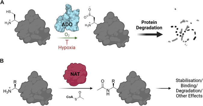
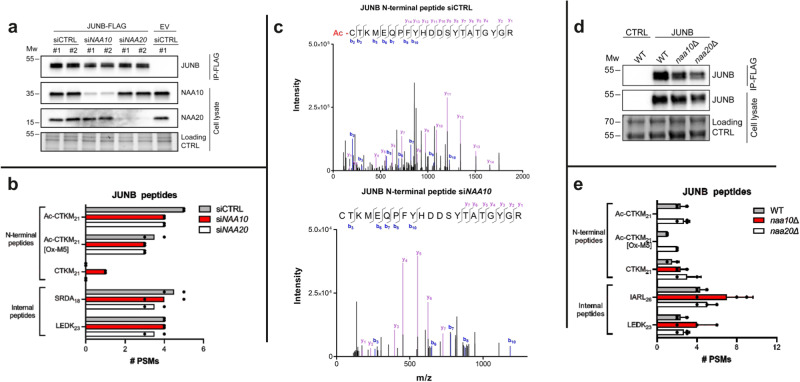
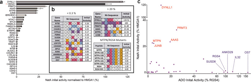
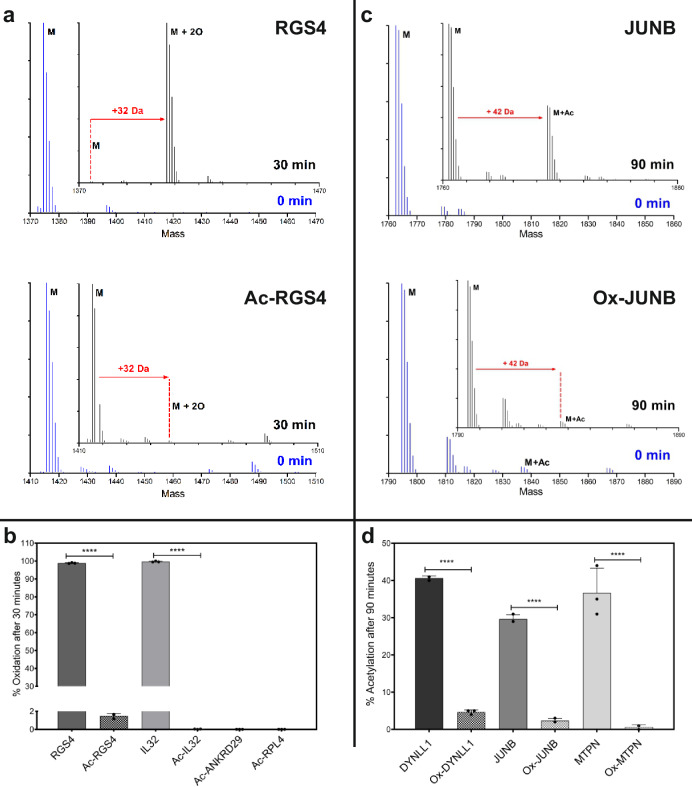
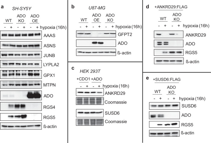
Oxygen homeostasis is maintained in plants and animals by O2-sensing enzymes initiating adaptive responses to low O2 (hypoxia). Recently, the O2-sensitive enzyme ADO was shown to initiate degradation of target proteins RGS4/5 and IL32 via the Cysteine/Arginine N-degron pathway. ADO functions by catalysing oxidation of N-terminal cysteine residues, but despite multiple proteins in the human proteome having an N-terminal cysteine, other endogenous ADO substrates have not yet been identified. This could be because alternative modifications of N-terminal cysteine residues, including acetylation, prevent ADO-catalysed oxidation. Here we investigate the relationship between ADO-catalysed oxidation and NatA-catalysed acetylation of a broad range of protein sequences with N-terminal cysteines. We present evidence that human NatA catalyses N-terminal cysteine acetylation in vitro and in vivo. We then show that sequences downstream of the N-terminal cysteine dictate whether this residue is oxidised or acetylated, with ADO preferring basic and aromatic amino acids and NatA preferring acidic or polar residues. In vitro, the two modifications appear to be mutually exclusive, suggesting that distinct pools of N-terminal cysteine proteins may be acetylated or oxidised. These results reveal the sequence determinants that contribute to N-terminal cysteine protein modifications, with implications for O2-dependent protein stability and the hypoxic response.


Conflict of interest statement

The authors declare no competing interests.

Figures

Fig. 1
Fig. 1. Oxidation and acetylation of protein N-termini.
A Cysteamine dioxygenase (ADO) can catalyse the oxidation of the N-terminal cysteine (Nt-Cys) of a target protein, incorporating two oxygen atoms from molecular oxygen,. This allows the protein to be recognised and so degraded by the Arg/N-degron pathway. ADO-catalysed Nt-Cys oxidation is significantly reduced in hypoxia. B N-terminal acetyltransferase enzymes (NATs) catalyse the acetylation of the N-terminal residue of a target protein, involving the transfer of an acetyl group from acetyl coenzyme A to the N-terminal amine. This can have a range of effects including stabilisation of the target protein, enhanced binding and degradation by the acetyl branch of the N-degron pathway. Created using Biorender (license agreement #YB26TL2XO9).
Fig. 2
Fig. 2. In vitro screening of Nt-Cys initiating peptide sequences from the human proteome with recombinant HsADO.
a Bar graphs show ADO-catalysed oxidation of 14-mer peptides representing the (Met-cleaved) Cys-initiating N-termini of potential ADO substrates (listed by gene name) after 1 min; activity was normalised to oxidation of RGS42-15 positive control peptide. Data are presented as mean ± standard deviation, n ≥ 3. b, c Sequences of peptides with which ADO was observed to have > 90% (b) or < 5% (v) activity relative to RGS42-15 positive control peptide. Amino acids colour coded according to side chain chemical properties: basic (H/K/R) = red; acidic (D/E) = blue; aromatic (F/W) = orange; polar (S/T) = purple. Source data are provided as a Source Data file.
Fig. 3
Fig. 3. In vitro testing of proposed ADO optimum substrate (OS) and anti-substrate (AS) peptides.
a Sequences of ADO optimum substrate (OS) peptides and initial activity of HsADO with OS peptides. Significant differences (p < 0.05) from 1-way ANOVA results summarised on graph: a = lower than OS7; b = lower than OS8; c = lower than OS3; d = lower than OS9 and OS10. Full 1-way ANOVA results and p-value ranges available in Supplementary Data 3. b Sequences of ADO anti-substrate (AS) peptides and initial activity of HsADO with AS peptides and RGS4 K3D/G. ASNS was included as a negative control for ADO activity. Significant differences (p < 0.05) from 1-way ANOVA results summarised on graph: a = lower than AS2; b = lower than AS6; c = lower than AS5; ASNS, RGS4 K3D/G and AS1-12 all lower than RGS4. Full 1-way ANOVA results available and p-value ranges in Supplementary Data 4. For (a, b), initial activity is turnover of peptide after 1-min incubation with HsADO normalised to that of RGS42-15 positive control peptide (%). Data represent mean ± standard deviation; n ≥ 3. Amino acids are colour coded according to chemical properties: basic (H/K/R) = red; acidic (D/E) = blue; aromatic (F/W) = orange; polar (S/T) = purple. Source data are provided as a Source Data file.
Fig. 4
Fig. 4. Regulation of potential ADO substrate sequences in Dual-Fluorescence Oxygen Reporter (DFOR) assay.
a Sequences of proteins selected from in vitro screening for testing in cells; including RAP2.12, a substrate of related Plant Cysteine Oxidase (PCO) enzymes. Amino acids are colour coded according to chemical properties: basic (H/K/R) = red; acidic (D/E) = blue; aromatic (F/W) = orange; polar (S/T) = purple. “in vitro Activity” values are relative to RGS42-15 (%). b, c SH-SY5Y cell lines stably expressing MCX13GFP:P2A:mCherry were (b) treated for 20 h with 2,2-dipyridyl (200 µM) or DMSO vehicle control and (c) incubated for 24 h in normoxia or hypoxia (1% O2). GFP/mCherry ratio was calculated using fluorescence measurements and normalised to the GFP:mCherry ratio obtained in cells expressing the positive control construct RGS4(1-15)-GFP:P2A:mCherry. Higher GFP/mCherry ratio indicates stabilisation of eGFP protein; increased stability after dipyridyl or hypoxia treatment is consistent with reduced ADO activity. Error bars show SEM. Statistical significance determined with 2-way ANOVA with Holm-Sidak multiple comparisons post-hoc test: ns (p > 0.05); *p ≤ 0.05; **p ≤ 0.01; ***p ≤ 0.001: ****p ≤ 0.0001. Data were collected across at least three biological repeats. Source data are provided as a Source Data file.
Fig. 5
Fig. 5. Validation of NatA-catalysed acetylation of Nt-Cys JunB.
a JUNB-FLAG was overexpressed in HAP1 ADO KO cells and treated with either siCTRL, siNAA10 or siNAA20. Cells treated with siCTRL as well as empty vector (EV) were included as controls. Anti-NAA10 and anti-NAA20 were used to verify successful knockdown and disrupted NatA/NatB complexes, whereas anti-JUNB was used to verify successful overexpression and IP. Protein gel image included as loading control. #1 and #2 represent two independent samples. b The number of peptide-spectrum matches (PSMs) identified by mass spectrometry for JUNB N-terminal peptides as well as two selected JUNB internal peptides. JUNB peptides designated XXXXn, where XXX are the first four amino acids of the peptide and n is the length of the peptide fragment. Data represent 2 independent biological samples; black dots represent each independent replicate. c Example MS/MS spectra for the N-terminal peptide of JUNB in its Nt-acetylated state from siCTRL treated cells and unacetylated state from siNAA10 treated cells showing bx (blue) and yx (purple) +1 ions with their respective mass/charge ratio and intensity. Free thiols in protein samples were alkylated prior to digestion and LC/MSMS, so Nt‑Cys bears a carbamidomethyl group adding 57 Da to the expected mass of b fragment ions. For expanded view see Supplementary Fig. 6. d S. cerevisiae strains WT, naa10∆ and naa20∆, with the last two lacking functional NatA and NatB, respectively, were transformed with a JUNB-FLAG plasmid or control plasmid. Protein gel image included as loading control. e The number of PSMs identified by mass spectrometry for JUNB N-terminal peptides as well as two selected JUNB internal peptides. JUNB peptides designated XXXXn, where XXXX are the first four amino acids of the peptide and n is the length of the peptide fragment. Data represent 3 independent biological samples ± standard deviation; black dots represent each independent replicate. MW units = kDa. Source data are provided as a Source Data file.
Fig. 6
Fig. 6. Investigation of NatA-catalysed acetylation of Nt-Cys sequences.
a [14C]-Acetyl incorporation into CX6RWGRPVGRRRRPVRVYP peptides (where CX6 is the 1st 7 amino acids of the Met-excised N-terminal sequence of protein of interest) after 5 min (200 µM peptide, 0.3 µM NatA, 200 µM Ac-CoA, 37 °C); activity is normalised to [14C]Acetyl incorporation into SESSSKSRWGRPVGRRRRPVRVYP (HMGA1) positive control peptide in the same assay (indicated with *). Data represent mean ± standard deviation; n = 4. b N-terminal sequences of peptides with which NatA was observed to have < 0.3% or > 20% activity compared to HMGA1 positive control peptide. MTPN peptide mutants varying the identity of the amino acid following the Nt-Cys were designed and tested (as in a) as well as RGS4 K3D. Amino acids are colour coded according to chemical properties: basic (H/K/R) = red; acidic (D/E) = blue; aromatic (F/W) = orange; polar (S/T) = purple. c Activity of NatA (a) plotted against activity of ADO (Fig. 2a) for Nt-Cys proteins of interest. Names of best NatA substrate peptides shown in red and names of best ADO substrate peptides shown in blue. Source data are provided as a Source Data file.
Fig. 7
Fig. 7. Nt-acetylation abrogates oxidation by ADO in vitro and vice versa.
a Deconvoluted MS spectra showing oxidation (32 Da increase) of RGS42-15 (RGS4) and N-terminally acetylated RGS42-15 (Ac-RGS4) before (blue, 0 min) and after (black, 30 min) incubation with HsADO at 37 °C (100 µM peptide, 0.1 µM ADO). b Oxidation of other acetylated ADO substrate peptides (Fig. 2a) compared to oxidation of RGS42-15 and IL322-15 peptides after 30 min (100 µM peptide, 0.1 µM HsADO, 37 °C). c Deconvoluted MS spectra showing acetylation (42 Da increase) of JUNB2-15 (JUNB) and N-terminally oxidised JUNB2-15 (Ox-JUNB) before (blue, 0 min) and after (black, 30 min) incubation with NatA at 37 °C (100 µM peptide, 0.15 µM NatA). d Acetylation of other oxidised NatA substrates peptides (Fig. 6a) compared to acetylation of non-oxidised NatA substrates peptides after 90 min (100 µM peptide, 0.15 µM NatA, 37 °C). In (b, d), data are presented as mean ± standard deviation, n = 3. Statistical significance determined using 2-way ANOVA with Bonferroni’s multiple comparison post-test: ****p ≤ 0.0001. Source data are provided as a Source Data file.
Fig. 8
Fig. 8. Regulation of putative ADO substrates human cells.
a, b WT, ADO KO or ADO overexpressing (OE) SH-SY5Y (a) or U87-MG (b) were exposed to hypoxia (1% O2) for 16 h, then protein samples were collected and analysed for the expression of various potential ADO substrates. RGS4 and RGS5 were used as positive controls. c HEK 293T cells were transiently co-transfected with cDNAs encoding ANKRD29 or SUSD6, alongside either CDO1 or ADO, and subjected to hypoxia as before. d, e SH-SY5Y cells stably expressing either ANKRD29:FLAG (d) or SUSD6:FLAG (e) were subjected to hypoxia for 16 h and the expression of both proteins was analysed using an anti-FLAG antibody. RGS5 expression was also analysed as a positive control. SUSD6 was detected at a much higher level relative to RGS5 and ANKRD29 in a stably polyclonal population. All blots (ae) are representative of at least 3 independent experiments. Source data (including relevant molecular weight markers) are provided as a Source Data file.

References

    1. Schofield CJ, Ratcliffe PJ. Oxygen sensing by HIF hydroxylases. Nat. Rev. Mol. Cell Biol. 2004;5:343–354. doi: 10.1038/nrm1366. - DOI - PubMed
    1. Epstein ACR, et al. C. elegans EGL-9 and mammalian homologs define a family of dioxygenases that regulate HIF by prolyl hydroxylation. Cell. 2001;107:43–54. doi: 10.1016/S0092-8674(01)00507-4. - DOI - PubMed
    1. Jaakkola P, et al. Targeting of HIF-α to the von Hippel-Lindau ubiquitylation complex by O2-regulated prolyl hydroxylation. Science. 2001;292:468–472. doi: 10.1126/science.1059796. - DOI - PubMed
    1. Ivan M, et al. HIFα targeted for VHL-mediated destruction by proline hydroxylation: implications for O2 sensing. Science. 2001;292:464–468. doi: 10.1126/science.1059817. - DOI - PubMed
    1. Kaelin WG, Ratcliffe PJ. Oxygen sensing by metazoans: the central role of the HIF hydroxylase pathway. Mol. Cell. 2008;30:393–402. doi: 10.1016/j.molcel.2008.04.009. - DOI - PubMed