. 2024 Jun 25;14(1):14610.
doi: 10.1038/s41598-024-65623-y.

Efficient and highly reproducible production of red blood cell-derived extracellular vesicle mimetics for the loading and delivery of RNA molecules

Affiliations

Efficient and highly reproducible production of red blood cell-derived extracellular vesicle mimetics for the loading and delivery of RNA molecules

Sara Biagiotti et al. Sci Rep. .

Abstract

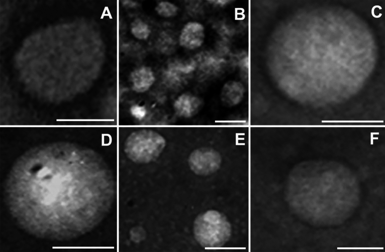
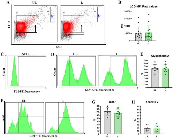
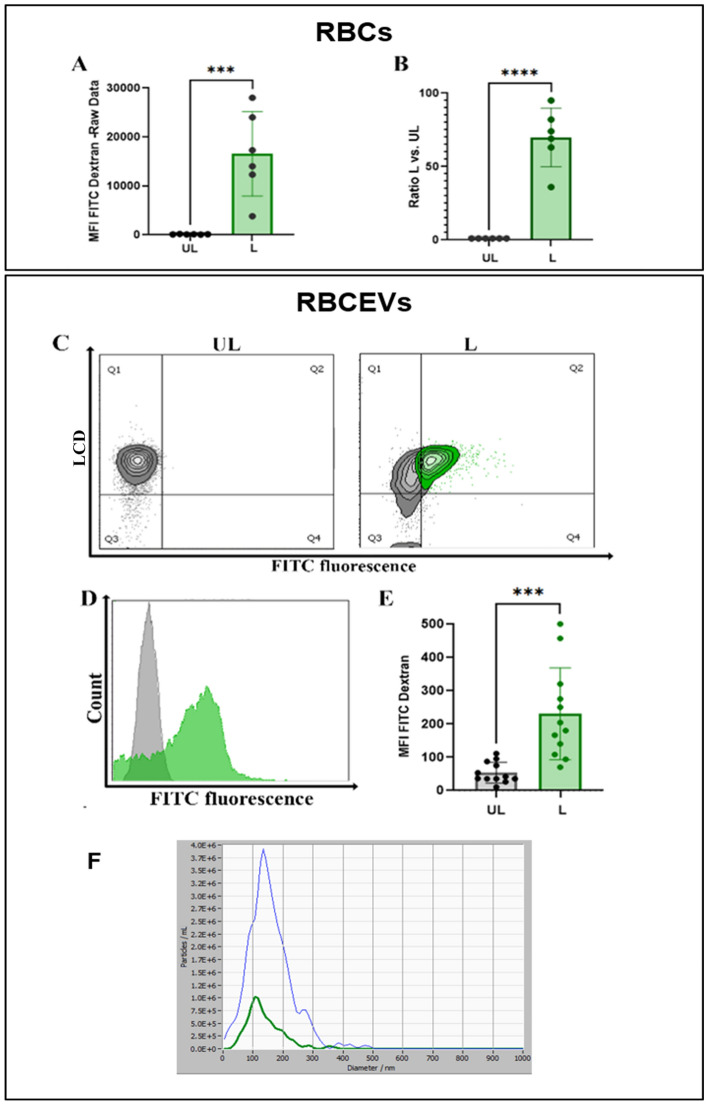
Extracellular vesicles (EVs) are promising natural nanocarriers for the delivery of therapeutic agents. As with any other kind of cell, red blood cells (RBCs) produce a limited number of EVs under physiological and pathological conditions. Thus, RBC-derived extracellular vesicles (RBCEVs) have been recently suggested as next-generation delivery systems for therapeutic purposes. In this paper, we show that thanks to their unique biological and physicochemical features, RBCs can be efficiently pre-loaded with several kinds of molecules and further used to generate RBCEVs. A physical vesiculation method, based on "soft extrusion", was developed, producing an extremely high yield of cargo-loaded RBCEV mimetics. The RBCEVs population has been deeply characterized according to the new guidelines MISEV2023, showing great homogeneity in terms of size, biological features, membrane architecture and cargo. In vitro preliminary results demonstrated that RBCEVs are abundantly internalized by cells and exert peculiar biological effects. Indeed, efficient loading and delivery of miR-210 by RBCEVs to HUVEC has been proven, as well as the inhibition of a known mRNA target. Of note, the bench-scale process can be scaled-up and translated into clinics. In conclusion, this investigation could open the way to a new biomimetic platform for RNA-based therapies and/or other therapeutic cargoes useful in several diseases.

Keywords: Drug delivery; EV engineering; Extracellular vesicles; RBC-derived extracellular vesicles; RNA therapy; miRNAs.


Conflict of interest statement

All the authors declare that the research was conducted in the absence of any commercial or financial relationships that could be construed as a potential conflict of interest. The authors also declare that some of the data presented herein have been used to fill a patent application (patent application number 102023000026244).

Figures

Figure 1
Figure 1
Physical characterization of RBCEVs. (A) Representative distribution plots of the UL RBCEVs diameter. (B) Representative distribution plots of the L RBCEVs diameter. (C) Histograms of the physical parameters of the distribution. Bar graphs showing inter-experiment variability over several replicates (n = 9) in terms of mode and mean of the RBCEVs size (particle diameter) and yield (particle number). As shown, no significant difference can be observed between UL and L samples for all the considered parameters, confirming the possibility of encapsulating cargoes inside RBCEVs (T-test (n = 9), P > 0.05). (D) Zeta Potential analyses of loaded RBCEVs at the end of their purification.
Figure 2
Figure 2
TEM analysis. Representative pictures of transmission electron microscopy analysis of negative stained RBCEVs. They were obtained at 20–50,000× magnification and represented the size distribution of the obtained RBCEVs (50–200 nm) that correlates quite well with the NTA characterization shown in Fig. 3. Scale bar = 100 nm.
Figure 3
Figure 3
Biological characterization of unloaded and loaded RBCEVs. (A) Dot plots of LCD fluorescent probe positivity and (B) bar graph of MFI values for both UL and L RBCEVs. The black arrow in panel (A) indicates the shift in fluorescence, highlighting the specific signal of the probe. In blue, enclosed by gate P3, Dako Cytocount beads are traceable. No statistical difference can be observed between UL and L positivity in graph (B). In the next panels, only LCD+ events are shown. In (C) the marker setting up to evaluate glycophorin A positivity is reported. In (D) UL and L RBCEVs glycophorin A profiles; (E) overall statistical evaluations for PE fluorescence- GYPA RBCEVs percentages. In (F) UL and L RBCEVs CD47 profiles. In (G), overall statistical evaluations for PE fluorescence- CD47 RBCEVs percentages. (H) Bar graphs show the low mean percentages of Annexin V positivity for both UL and L RBCEVs. No statistical difference can be found between L and UL samples. Unpaired T test by PRISM software was employed for statistical analyses.
Figure 4
Figure 4
Monitoring the production of RBCEVs loaded with Cascade Blue-dextran (10 kDa). (A) Dot plots showing vesicle number and size distribution using NTA fluorescence mode in L samples compared to UL as control. (B) Representative snapshots of the Cascade Blue positive particle acquisition in L samples. In (C), the bar graph of MFI Cascade blue values recorded by flow cytometry in UL and L RBCEVs is reported. A statistically higher value is shown for L samples compared to UL ones. Unpaired T test by PRISM software was employed for statistical analyses of flow cytometric evaluations (p-values *** < 0.001).
Figure 5
Figure 5
Monitoring the production of RBCEVs loaded with FITC-dextran (70 kDa). (A) Bar graph reporting green fluorescence MFI values for RBCs L compared to the relative UL controls. (B) Ratio calculated between L and UL samples. (C) Dot plot showing FITC-dextran positivity of LCD positive events in L RBCEVs compared to UL controls. (D) Representative histogram overlay for UL green fluorescent RBCEVs (grey histogram) and for L green fluorescent-FITC dextran (green histogram) RBCEVs evaluation. (E) Mean fluorescence intensity found in L RBCEVs compared to UL samples. (F) Analysis of loaded RBCEVs size distribution acquired in scattering and fluorescence mode, respectively. Unpaired T test by PRISM software was employed for statistical analyses (p-values *** < 0.001, **** < 0.0001).
Figure 6
Figure 6
PKH26-labeled RBCEVs uptake in HUVEC. (A) Histograms of PKH26 positivity in HUVEC untreated labeled and treated by labeled RBCEVs after 4 h and (B) labeled after 24 h. (C) Dot plots for assessing 7′ AAD positivity in order to detect cell death eventually induced in HUVEC by RBCEVs. (D) Bar graphs of 7′ AAD percentage of positivity in HUVEC treated or not with RBCEVs. Unpaired T test by PRISM software was employed for this statistical analysis. (E) Confocal images of PKH26 labeled RBCEVs and relative untreated controls. In red, PKH26 fluorophore signal, and in green, phalloidin-FITC that co-localizes into HUVEC. (F) Bar graphs reporting percentages of PKH26 positivity in HUVEC treated with labeled RBCEVs compared to untreated ones at 4 and 24 h. (G) Bar graphs reporting PHK26 MFI values in HUVEC treated by PKH26 labeled RBCEVs compared to the untreated ones at 4 and 24 h. One Way ANOVA, with Tukey multiple Comparison Tests, by PRISM Software, was employed for this statistical analysis (p-values ** < 0.05, *** < 0.001).
Figure 7
Figure 7
Production of miR-210-loaded RBCEVs and biological effect. (A) Absolute miR-210 concentrations have been calculated by a standard curve set up with synthetic RNA standards at 20–0.0002 nM. miR-210 concentration into loaded RBCEVs is compared to UL RBCEVs and to the starting concentration obtained into the mother RBCs (UL and L) before vesiculation. High loading efficiency can be appreciated. The amount of miR-210 found in UL RBCs and RBCEVs is due to the endogenous miRNA. Data are mean and SEM (n = 4). In (B), miR-210 relative quantification into HUVEC is reported. Relative quantification has been performed using U6 snRNA as a reference gene and UL sample as a control. miR-210 concentration found in HUVEC treated with L RBCEVs are compared to cells treated with UL RBCEVs and to cells transfected at different miRNA concentrations. Data are mean and SEM, n = 4 (Unpaired t-test; *two-tailed p-values < 0.05). In (C), the evaluation of the effect of miR210-loaded RBCEVs at the mRNA level is shown. Relative quantification of PTB1B mRNA has been performed using ATCB as reference gene and UL sample as control. PTP1B mRNA found in HUVEC treated with L RBCEVs is compared to cells treated with UL RBCEVs and to cells transfected at different miRNA concentrations. Data are mean and SEM, n = 4 (Unpaired t-test; *two-tailed p-values < 0.05). (D) Western blot of PTP1B in protein extracts from HUVEC treated with RBCEVs UL or L and transfected with different amounts of miR-210. Lanes 1–5: RBCEVs UL, RBCEVs L, miR-210 1 nM, 10 nM, and 50 nM plus Transit-2X. This figure has been cropped in order to report the most relevant results and the original blot is available in Supplementary Fig. S8. In (E), the evaluation of the effect of miR210-loaded RBCEVs at the protein level. Quantification of PTP1B band normalized to total proteins. Data are the mean and SEM, n = 4 (Unpaired t-test; p-values * < 0.05, *** < 0.001, **** < 0.0001). (F) Glycolytic and mitochondrial ATP production rates obtained in both miR210-transfected and RBCEVs-treated HUVEC (Unpaired t-test; p-values *** < 0.001).

References

    1. Yáñez-Mó M, et al. Biological properties of extracellular vesicles and their physiological functions. J. Extracell. Vesicles. 2015;4:27066. doi: 10.3402/jev.v4.27066. - DOI - PMC - PubMed
    1. Margolis L, Sadovsky Y. The biology of extracellular vesicles: The known unknowns. PLoS Biol. 2019;17:e300363. doi: 10.1371/journal.pbio.3000363. - DOI - PMC - PubMed
    1. Kalluri R, LeBleu VS. The biology, function, and biomedical applications of exosomes. Science. 2020;367:6478. doi: 10.1126/science.aau6977. - DOI - PMC - PubMed
    1. Chiangjong W, Netsirisawan P, Hongeng S, Chutipongtanate S. Red blood cell extracellular vesicle-based drug delivery: Challenges and opportunities. Front. Med. 2021;8:761362. doi: 10.3389/fmed.2021.761362. - DOI - PMC - PubMed
    1. Thangaraju K, Neerukonda SN, Katneni U, Buehler PW. Extracellular vesicles from red blood cells and their evolving roles in health, coagulopathy and therapy. Int. J. Mol. Sci. 2021;22:153. doi: 10.3390/ijms22010153. - DOI - PMC - PubMed