. 2024 Jun 11:15:1309846.
doi: 10.3389/fimmu.2024.1309846. eCollection 2024.

Acid ceramidase expression reduces IFNγ secretion by mouse CD4+ T cells and is crucial for maintaining B-cell numbers in mice

Affiliations

Acid ceramidase expression reduces IFNγ secretion by mouse CD4+ T cells and is crucial for maintaining B-cell numbers in mice

Putri Mandasari et al. Front Immunol. .

Abstract

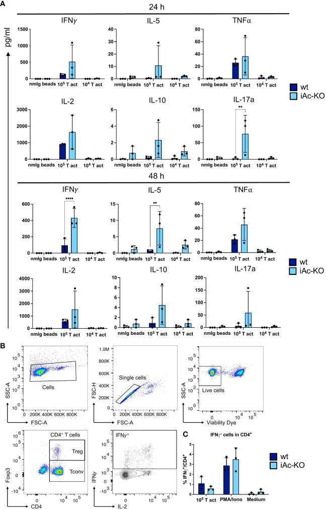
Acid ceramidase (Ac) is a lysosomal enzyme catalyzing the generation of sphingosine from ceramide, and Ac inhibitors are currently being investigated as potential cancer therapeutics. Yet, the role of the Ac in immune responses, particularly anti-viral immunity, is not fully understood. To investigate the impact of Ac expression on various leukocyte populations, we generated a tamoxifen-inducible global knockout mouse model for the Ac (iAc-KO). Following tamoxifen administration to healthy mice, we extracted primary and secondary lymphoid organs from iAc-KO and wild-type (wt) littermates and subsequently performed extensive flow cytometric marker analysis. In addition, we isolated CD4+ T cells from the spleen and lymph nodes for sphingolipid profiling and restimulated them in vitro with Dynabeads™ Mouse T-activator CD3/CD28. Intracellular cytokine expression (FACS staining) was analyzed and secreted cytokines detected in supernatants. To study cell-intrinsic effects, we established an in vitro model for iAc-KO in isolated CD4+ T and B cells. For CD4+ T cells of iAc-KO versus wt mice, we observed reduced Ac activity, an increased ceramide level, and enhanced secretion of IFNγ upon CD3/CD28 costimulation. Moreover, there was a marked reduction in B cell and plasma cell and blast numbers in iAc-KO compared to wt mice. To study cell-intrinsic effects and in line with the 3R principles, we established in vitro cell culture systems for iAc-KO in isolated B and CD4+ T cells. Our findings pinpoint to a key role of the Ac in mature B and antibody-secreting cells and in IFNγ secretion by CD4+ T cells.

Keywords: B cell survival; CD4+ T cell; acid ceramidase; cytokine secretion; in vitro culture assay; inducible knockout mice.


Conflict of interest statement

The authors declare that the research was conducted in the absence of any commercial or financial relationships that could be construed as a potential conflict of interest. The author(s) declared that they were an editorial board member of Frontiers, at the time of submission. This had no impact on the peer review process and the final decision.

Figures

Figure 1
Figure 1
Tamoxifen administration to iAc-KO in vivo led to efficient gene recombination and subsequently to ceramide accumulation and reduced Ac activity. (A) In vivo experimental procedure to induce the knockout of Ac. Tamoxifen was administered for four consecutive days per oral. Mice were then sacrificed on day 7 for analysis. (B) DNAs of purified CD4+ T cells from iAc-KO and wt littermates on day 7 were isolated and subsequent PCR to validate the Asah1 recombination (left panel). As comparison, genotyping results were shown to indicate non-excised band of Asah1 (right panel). (C) The levels of ceramides, sphingosine, and sphingomyelins of purified CD4+ T cells of iAc-KO and iAc-wt on day 7 were measured by HPLC-MS/MS (n=3). (D) Lysates of day 7 CD4+ T cells of iAc-KO and Ac-wt were measured for their Ac enzymatic activity using TLC-based assay (n=3). All depicted graphs show data from individual mice together with means ± SD. Statistical analysis was performed by two-way ANOVA with Sidak’s multiple comparison test or unpaired t-test (**p<0.01).
Figure 2
Figure 2
In vivo Ac deletion only had minor effects on subset composition of CD4+ and CD8+ T cells. (A) Representative dot plots showing the gating strategy for CD4+, CD8+ T cells, and their subsets. (B) Frequencies of CD4+ and CD8+ T cells together with their subsets in the spleen, lymph nodes, and thymus comparing iAc-KO to wt littermates on day 7 (n=3). Columns show each individual mice together with means ± SD. Two-way ANOVA followed by Sidak’s multiple comparison test was done for statistical analysis (*p<0.05, ***p<0.001).
Figure 3
Figure 3
CD4+ T-cell activation and proliferation were not altered in after Ac knock-out. (A) CD4+ T cells of iAc-KO and iAc-wt day 7 were restimulated with 105, 104 T-activator beads and normal mouse Ig (nmIg) beads for 24 and 48 h. In the left panel, representative flow cytometric dot plots as example from three independent experiments depict the frequencies of single positive for activation markers CD25 and CD69 among CD4+ T cells upon different stimulation for 2 days. The frequency of CD4+ T cells that are double positive for CD25 and CD69 comparing iAc-KO to wt littermates after 24 and 48 h stimulation is shown in the right panel (n=3). (B) Proliferation of CD4+ T cells from the same culture was monitored. Representative histograms displayed proliferation (marker, Ki-67) of CD4+ T cells of iAc-KO and iAc-wt upon 2 days stimulation with T-activator beads and nmIg beads as negative control. The bar graphs are summarized data of the proliferating CD4+ T cells of iAc-KO and iAc-wt upon 1 and 2 days stimulation (n=3). Data are depicted from individual mice together with means ± SD. Statistical analysis was performed by two-way ANOVA with Sidak’s multiple comparison test.
Figure 4
Figure 4
Ac-deficient CD4+ T cells secreted more upon optimal restimulation with 10e5 T-activator beads. (A) Supernatant from the 2 days stimulation culture of iAc-KO and iAc-wt CD4+ T cells were measured using bead-based assay and flow cytometry (n=3). (B) Flow cytometric gating strategy for IFNγ-producing cells among CD4+ T cells shown following intracellular cytokine staining. CD4+ T cells of iAc-KO and iAc-wt were stimulated with 105 T-activator beads for 4 h in the presence of brefeldin A to block the cytokine secretion. PMA/Iono and medium were used as positive and negative control, respectively. (C) Frequency of IFNγ-producing cells among CD4+ T cells were then measured by flow cytometry comparing iAc-KO and wt CD4+ T cells (n=3). Data are compiled from three mice per genotype and shown together with means ± SD. Two-way ANOVA with Sidak’s multiple comparison test was done to test for statistical significance (**p<0.01, ****p<0.0001).
Figure 5
Figure 5
The recombination of Ac gene (Asah1) could be induced in vitro and CD4+ T cells subsequently maintained in in vitro culture. (A) In vitro experimental setup for CD4+ T-cell pre-expansion and restimulation culture. Cells from the spleen and lymph nodes were treated with 4-OH-Tm for 1 h, which is followed by CD4+ purification. CD4+ T cells were then cultured in the presence of CD28-Superagonist (CD28-SA) and pan mouse Ig beads for 7 days. On day 7, beads were removed, and the cells were washed before seeded in only medium for another 3 days to induce steady state. Cells were harvested on day 10 and restimulated for 24 h and 48 h with T-activator beads and nmIg beads. (B) CD4+ T cells yield of iAc-KO and iAc-wt on day 0, 7, and 10 (n=3). (C) Representative gating strategy showing Treg and Tconv of iAc-KO and its wt littermates after 10 days of culture. (D) DNA of ex vivo spleen/LN cells treated with 4-OH-tamoxifen and day 10 CD4+ T cells of iAc-KO (and wt littermates) was isolated and used for PCR to validate Asah1 recombination after 10 days of culture (left panel). Lysates of day 10 CD4+ T cells from iAc-KO and iAc-wt were measured for its Ac activity (right panel). Data are shown as converted sphingosine of total lipid (n=3). All depicted graphs show data from individual mice together with means ± SD. Statistical analysis was performed by two-way ANOVA with Sidak’s multiple comparison test or unpaired t-test (*p<0.05).
Figure 6
Figure 6
Increase in IFNγ secretion by Ac-deficient CD4+ T cells was evident upon restimulation following in vitro pre-expansion. (A) Frequencies of double-positive CD25 and CD69 among CD4+ T cells from iAc-KO and iAc-wt representing activation (left panel) and proliferation using Ki-67 as marker (right panel) (n=3). On day 10, CD4+ T cells were restimulated with different concentration of T-activator beads. As negative control, nmIg beads were employed. (B) Cytokines were measured using supernatant originated from restimulation culture comparing Ac-deficient CD4+ T cells to wt littermates (n=4–6). (C) Cytokine mRNA expression profiles of wt and iAc-KO CD4+ T cells after restimulation with T-activator beads for 4 h at a 1:1 bead to cell ratio (culture triplicates; experiment was repeated with similar result) (D) Intracellular cytokine staining was performed with CD4+ T cells of iAc-KO and iAc-wt on day 10 of culture using different stimulation (T-activator beads, PMA/Iono, and medium) for 4 h in the presence of brefeldin (A) The frequency of IFNγ-producing cells among CD4+ T cells upon different stimulation is summarized in the graph (n=2). All depicted data show means ± SD. Two-way ANOVA with Sidak’s multiple comparison test was done to test for statistical significance (**p<0.01, ****p<0.0001).
Figure 7
Figure 7
Global Ac ablation resulted in B-cell number reduction in vivo and in vitro. (A) Representative flow cytometric gating strategy for B cells and its subsets on day 7 after tamoxifen in vivo treatment. (B) Absolute cell number of B cells and their subsets comparing iAc-KO and iAc-wt in spleen and bone marrow ex vivo on day 7 (n=3). (C) DNA of iAc-KO and iAc-wt B cells after 4 days of culture were employed to confirm the recombination via genomic PCR (left panel). Ac enzymatic activity was also measured from the isolated B cells on day 4 and shown as representative (right panel). (D) In vitro B-cell culture setup (left panel). Cells from the spleen were treated with 4-OH-tamoxifen for 1 h and subsequently purified for B cells. B cells were cultured for 4 days before analyzed. Representative absolute cell number of B cells and mature B cells was determined on day 4 of culture (right panel) (n=3 replicates). Experiment was repeated and showed same results. Data are depicted in means ± SD. Two-way ANOVA with Sidak’s multiple comparison test or unpaired t-test were performed for statistics (*p<0.05, **p<0.01).

Similar articles

References

    1. Thudichum JLW. A Treatise on the Chemical Constitution of the Brain: Archon Books. (London: Baillière, Tindall and Cox; ) (1962).
    1. Hannun YA, Obeid LM. Sphingolipids and their metabolism in physiology and disease. Nat Rev Mol Cell Biol. (2018) 19:175–91. doi: 10.1038/nrm.2017.107 - DOI - PMC - PubMed
    1. Maceyka M, Spiegel S. Sphingolipid metabolites in inflammatory disease. Nature. (2014) 510:58–67. doi: 10.1038/nature13475 - DOI - PMC - PubMed
    1. Hannun YA, Obeid LM. Many ceramides. J Biol Chem. (2011) 286:27855–62. doi: 10.1074/jbc.R111.254359 - DOI - PMC - PubMed
    1. Zeidan YH, Jenkins RW, Korman JB, Liu X, Obeid LM, Norris JS, et al. . Molecular targeting of acid ceramidase: implications to cancer therapy. Curr Drug Targets. (2008) 9:653–61. doi: 10.2174/138945008785132358 - DOI - PMC - PubMed