. 2024 Aug;55(8):2139-2150.
doi: 10.1161/STROKEAHA.124.046400. Epub 2024 Jun 26.

Circadian-Dependent Intermittent Fasting Influences Ischemic Tolerance and Dendritic Spine Remodeling

Affiliations

Circadian-Dependent Intermittent Fasting Influences Ischemic Tolerance and Dendritic Spine Remodeling

Soomin Jeong et al. Stroke. 2024 Aug.

Abstract

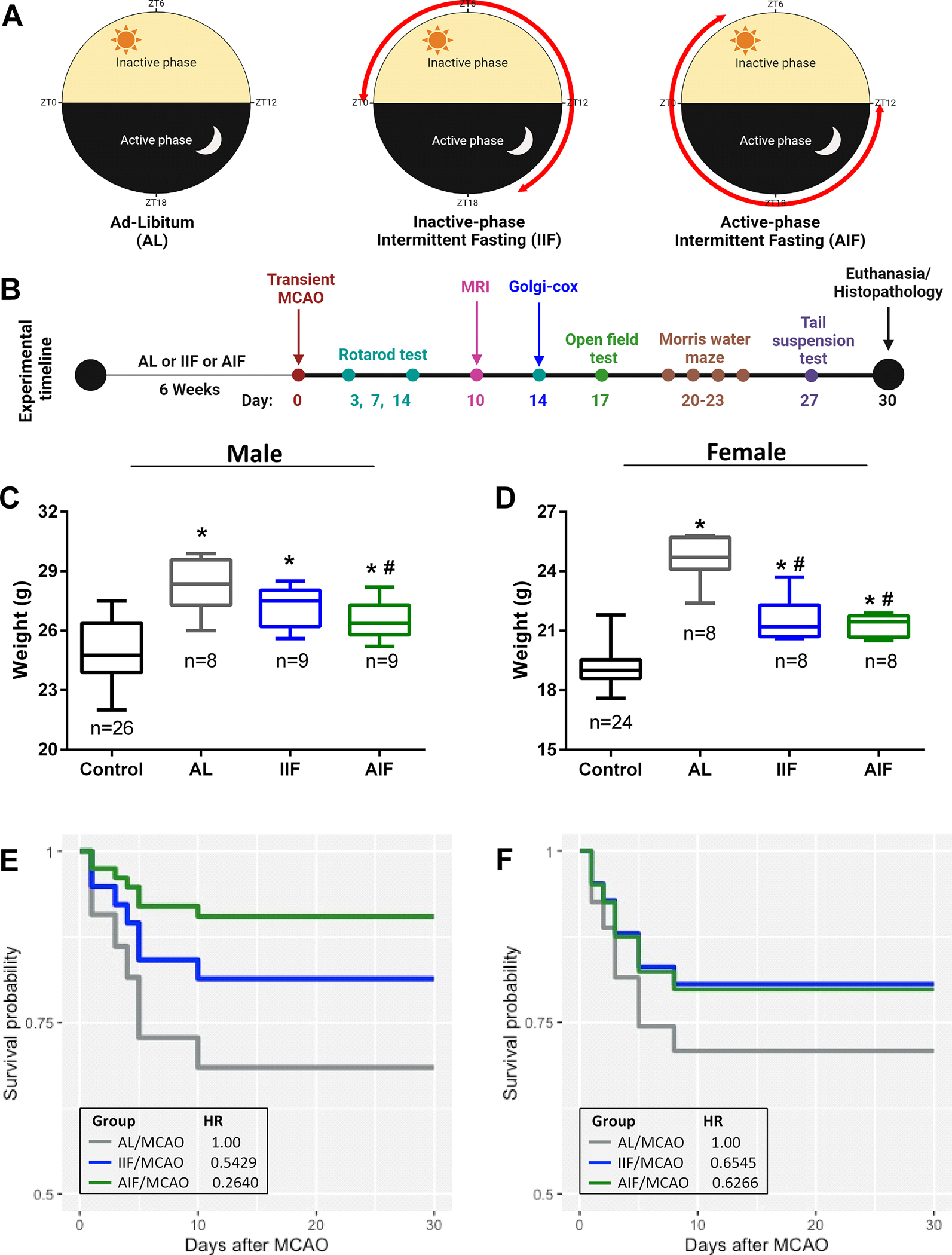
Background: Preconditioning by intermittent fasting is linked to improved cognition and motor function, and enhanced recovery after stroke. Although the duration of fasting was shown to elicit different levels of neuroprotection after ischemic stroke, the impact of time of fasting with respect to the circadian cycles remains unexplored.

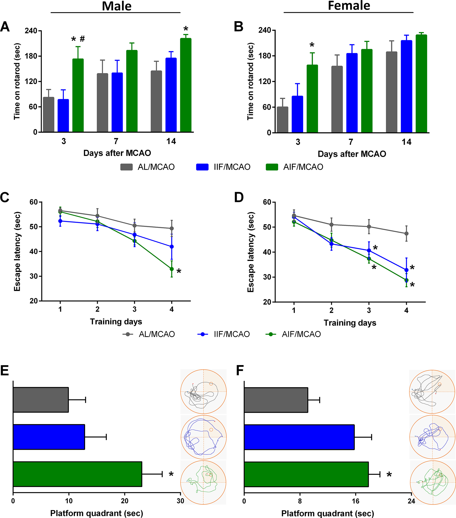
Methods: Cohorts of mice were subjected to a daily 16-hour fast, either during the dark phase (active-phase intermittent fasting) or the light phase (inactive-phase intermittent fasting) or were fed ad libitum. Following a 6-week dietary regimen, mice were subjected to transient focal cerebral ischemia and underwent behavioral functional assessment. Brain samples were collected for RNA sequencing and histopathologic analyses.

Results: Active-phase intermittent fasting cohort exhibited better poststroke motor and cognitive recovery as well as reduced infarction, in contrast to inactive-phase intermittent fasting cohort, when compared with ad libitum cohort. In addition, protection of dendritic spine density/morphology and increased expression of postsynaptic density protein-95 were observed in the active-phase intermittent fasting.

Conclusions: These findings indicate that the time of daily fasting is an important factor in inducing ischemic tolerance by intermittent fasting.

Keywords: cerebrovascular diseases; circadian rhythm; dendritic spines; intermittent fasting; postsynaptic density protein-95; structural plasticity.


Conflict of interest statement

None.

Figures

Fig. 1:
Fig. 1:. Intermittent fasting, regardless of circadian rhythms, reduces weight gain during the fasting period and mortality during the acute phase after stroke.
Randomly selected mice were subjected to ad-libitum (AL) feeding, inactive-phase intermittent fasting (IIF), or active-phase intermittent fasting (AIF) for 6 weeks. The red arrow represents the fasting time (A). All three groups of mice were subjected to 1h transient MCAO after 6 weeks of feeding paradigms and then tested for motor function (rotarod test on days 3, 7, and 14 of reperfusion), infarct volume (T2-MRI on day 10 of reperfusion), anxiety-like behavior (open-field test on day 17 of reperfusion), cognition (Morris water maze test between days 20 and 23 of reperfusion), and depression-like behavior (tail suspension test on day 27 of reperfusion). Mice were euthanized on day 30 for histopathologic assessment of atrophy volume (cresyl violet staining) and white matter damage (luxol fast blue staining) (B). A cohort of 26 male and 24 female mice were weighed before starting the feeding regimens (control). They were divided into AL, IIF and AIF groups and weighed again after 6 weeks of dietary regimens (C and D). The hazard ratio (HR), indicative of post-stroke survival probability, was measured by the Univariate Cox proportional hazard model (E and F). Data are mean ± SD of n=8–9/group. *p<0.05 compared with control and #p<0.05 compared with AL by one-way ANOVA followed by Tukey’s test (C-D).
Fig. 2:
Fig. 2:. Post-stroke motor and cognitive functional recovery is sensitive to circadian-dependent feeding in both sexes.
Post-stroke motor function recovery estimated by rotarod test on days 3, 7 and 14 of reperfusion (A and B). Cognitive function was assessed by Morris water maze test between days 20 and 23 after transient MCAO. 2 blocks of the test were performed on each training day and escape latency was measured (C and D). In the probe test on the last training day of Morris water maze, the time spent in the platform quadrant was measured. Track plots from representative mice of each cohort are shown next to the graphs (E and F). Data are mean ± SEM of n=7–9/group. *p<0.05 compared with AL/MCAO and #p<0.05 compared with IIF/MCAO by one-way ANOVA (A-B, E-F) or two-way repeated-measures ANOVA (C-D) followed by Tukey’s test.
Fig. 3:
Fig. 3:. AIF reduces gray and white matter damage after focal ischemia in both sexes.
T2-weighted MRI images were analyzed for infarct volume on day 10 of reperfusion (A). Sectioned brain samples on day 30 of reperfusion were stained with the cresyl violet method to measure atrophy volume (B). Thickness of the medial corpus callosum was estimated in luxol fast blue stained brain section (coordinate +0.5 mm from bregma). The values were normalized to the sham-operated control (C). Data are mean ± SD of n=7–8/group. &p<0.05 compared with sham, *p<0.05 compared with AL/MCAO, and #p<0.05 compared with IIF/MCAO by one-way ANOVA followed by Tukey’s test.
Fig. 4:
Fig. 4:. Effect of feeding regimens on pre- and post-stroke gene expression profile in peri-infarct cortex.
Level of cortical mRNA expression from cohorts of mice subjected to 6 weeks of AL, IIF or AIF (A), followed by 1h transient MCAO and euthanized at 1 day of reperfusion (B) was conducted by RNA-seq (n=4/group). Top view of the 3D volcano plot (generated using the volcano3D Rstudio package) of the expression of all transcripts analyzed between the three groups. Through one-way ANOVA, among the transcripts where the mean values of three groups are not equal, the cases in which the means of one group is greater than the means of the other two groups are displayed in the following colors (AL and AL/MCAO: dark gray, IIF and IIF/MCAO: blue, AIF and AIF/MCAO: green). The remaining transcripts are shown in light gray (A and B). Gene ontology (GO) analyses for the biological process of 769 upregulated transcripts in AIF compared with AL and IIF (C) and of 637 upregulated transcripts in the AIF/MCAO cohort compared with AL/MCAO cohort and IIF/MCAO cohort (D) were shown with bubble plots. Gene counts and p-values are indicated by circle size and circle color, respectively. Bubble plots were generated in Rstudio followed by analysis of GO pathways by DAVID Bioinformatics Resources, (C and D).
Fig. 5:
Fig. 5:. AIF enriches the postsynaptic compartment-related transcripts and promotes the post-stroke survival of dendritic spines.
In respect of location in the synapse, upregulated transcripts in AIF (769) and AIF/MCAO (637) were analyzed with Synaptic Gene Ontology (A and B). Spine density and morphology were analyzed in the Golgi-Cox stained brain sections collected at 6 weeks after IF, as well as on 14 days of reperfusion following MCAO, in all three cohorts. Peri-infarct cortex area (black rectangle) was imaged and infarct was shown with red dotted line. Scale bar, 500μm (C). Dendritic segments from each group were magnified. Pink, blue, green and yellow arrows indicate stubby, mushroom, thin, and filopodia protrusions, respectively. Scale bar, 5μm (D). Spine density in MCAO cohorts was compared with respective diet-matched controls (E). The number of spine subtypes was compared between after IF and after MCAO cohorts for each feeding paradigm (F). Data are means ± SD (E) or means ± SEM (F) of n=3/group (1–3 segments/5–6 cells/group). *p<0.05 using Mann-Whitney t-test.
Fig. 6:
Fig. 6:. AIF increases PSD95-dependent structural plasticity following stroke.
mRNA and protein levels of PSD95 were quantified by real-time PCR and dot blot from the brain samples on day 14, respectively (A and B). Fluorescent images immunostained with postsynaptic density protein 95 (PSD95; red) and microtubule-associated protein 2 (MAP2; green) were generated on sectioned brain samples collected on day 14 following focal ischemia. Scale bar, 20μm (C). PSD95 expression was measured by integrated density (2 ROIs/sample) (D). Data are mean ± SD of n=4/group. *p<0.05 compared with AL/MCAO and #p<0.05 compared with IIF/MCAO by one-way ANOVA followed by Tukey’s test.

References

    1. Vemuganti R, Arumugam TV. Much ado about eating: Intermittent fasting and post-stroke neuroprotection. J Cereb Blood Flow Metab. 2021;41:1791–1793. doi: 10.1177/0271678X211009362 - DOI - PMC - PubMed
    1. St-Onge MP, Ard J, Baskin ML, Chiuve SE, Johnson HM, Kris-Etherton P, Varady K. Meal Timing and Frequency: Implications for Cardiovascular Disease Prevention: A Scientific Statement From the American Heart Association. Circulation. 2017;135:e96–e121. doi: 10.1161/cir.0000000000000476 - DOI - PMC - PubMed
    1. Mattson MP, Moehl K, Ghena N, Schmaedick M, Cheng A. Intermittent metabolic switching, neuroplasticity and brain health. Nature reviews Neuroscience. 2018;19:63–80. doi: 10.1038/nrn.2017.156 - DOI - PMC - PubMed
    1. Lee J, Duan W, Mattson MP. Evidence that brain-derived neurotrophic factor is required for basal neurogenesis and mediates, in part, the enhancement of neurogenesis by dietary restriction in the hippocampus of adult mice. Journal of neurochemistry. 2002;82:1367–1375. doi: 10.1046/j.1471-4159.2002.01085.x - DOI - PubMed
    1. Singh R, Lakhanpal D, Kumar S, Sharma S, Kataria H, Kaur M, Kaur G. Late-onset intermittent fasting dietary restriction as a potential intervention to retard age-associated brain function impairments in male rats. Age (Dordrecht, Netherlands). 2012;34:917–933. doi: 10.1007/s11357-011-9289-2 - DOI - PMC - PubMed

Publication types