

Analysis of morphology and symmetry of the root canal system of incisors, premolars and mandibular molars using CBCT
- PMID: 38920123
- PMCID: PMC11212214
- DOI: 10.54589/aol.37/1/25
Analysis of morphology and symmetry of the root canal system of incisors, premolars and mandibular molars using CBCT
Abstract
Knowledge of root canal internal anatomy and its variations is important forproper endodontic treatment. It is therefore necessary to investigate morphological aspects among different dental groups in the same patient to define the best protocol for the case.
Aim: To evaluate the morphology and symmetry of homologous incisors, premolars and mandibular molars using cone beam computed tomography (CBCT).
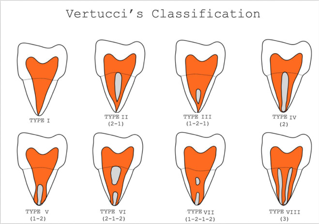
Materials and method: Descriptive statistical analysis was performed for the frequency of categorical variables, and a chi-square test or Fisher 's exact test was used to test whether gender and side were associated with number of roots, number of canals, and Vertucci's classification. Forty-five CBCT scans were evaluated, and 444 mandibular teeth were analyzed. The number of roots, number of canals, classification of the canals in each root according to Vertucci and presence of a symmetrical relationship between pairs of posterior teeth were analyzed.
Results: The resuls showed that 74% of mandibular central incisors had type I root canal, 26% of mandibular lateral incisors had type I and, with a significant difference in the number of canals between males and females (p < 0.05). In mandibular first premolars, 70.5% had type I; and in mandibular second premolars, 98.5% had type I. Mandibular first molars had two roots in 98% of the cases. Second mandibular molars had two roots in 92.5% of the cases, one root in 6%, and three roots in 1.5%. Symmetry between central incisors was higher in females than in males.
Conclusión: Teeth of the same group can have different morphologies in the same patient.
0 conhecimento da anatomia interna e suas variagoes anatómicas é fator importante para o adequado tratamento endodóntico. Portanto, é necessário investigar esses aspectos morfológicos entre diferentes grupos dentários de um mesmo paciente para definir o melhor protocolo para o caso.
Objetivo: Avaliar a morfologia e simetria de incisivos, pré-molares e molares inferiores homólogos por meio de tomografia computadorizada de feixe cónico (TCFC).
Materiais e método: Foi realizada análise estatística descri-tiva para a frequéncia das variáveis categóricas e foi utilizado o teste do qui-quadrado ou teste exato de Fisher para testar a relagao entre sexo e lado em comparagao com número de raízes, número de canais e classificagao de Vertucci. Quarenta e cinco TCFC foram avaliadas e 444 dentes inferiores foram analisados. Foram considerados: o número de raízes, o número de canais, o tipo dos canais acordo com a classificagao de Vertucci e a presenga de relagao simétrica entre pares de dentes posteriores.
Resultados: Os resultados mostraram que 74% dos incisivos centrais inferiores tinham um canal radicular tipo 1 e 26% tinham dois canais; 73% dos incisivos laterais inferiores, 26%oeram do tipo I, tinham um canal e 27% tinham dois canais, com diferenga significativa no número de canais entre os grupos masculino e feminino (p < 0,05). Nos primeiros pré-molares inferiores, tipo I, um canal foi detectado em 70,5% e dois canais em 29,5%; nos segundos pré-molares inferiores, tipo I, um único canal foi detectado em 98,5%. O primeiro molar inferior foi observado com duas raízes em 98% e tres raízes em 2%o. O segundo molar inferior tinha duas raízes em 92,5% dos casos, uma raiz em 6% e tres raízes em 1,5%. A simetria foi maior nas mulheres em comparagao aos homens nos incisivos centrais.
Conclusão: Pode-se concluir que dentes de um mesmo grupo podem apresentar morfologias diferentes no mesmo paciente.
Keywords: anatomy; endodontics; permanent teeth.
Copyright© 2024 Sociedad Argentina de Investigación Odontológica.
Conflict of interest statement
The authors declare no potential conflicts of interest regarding the research, authorship, and/or publication of this article.
Figures
Similar articles
-
The association between complex root canal morphology of mandibular anteriors and distolingual roots in mandibular first molars in a Turkish population.BMC Oral Health. 2025 Jul 16;25(1):1174. doi: 10.1186/s12903-025-06515-z. BMC Oral Health. 2025. PMID: 40670962 Free PMC article.
-
Anatomical Evaluation of Root and Root Canal Morphology of Permanent Mandibular Dentition among the Saudi Arabian Population: A Systematic Review.Biomed Res Int. 2022 Aug 2;2022:2400314. doi: 10.1155/2022/2400314. eCollection 2022. Biomed Res Int. 2022. PMID: 35958809 Free PMC article.
-
Assessment of Root Canal Anatomy of Mandibular Permanent Incisors in a Sample of Yemeni Population.Int J Dent. 2025 Aug 2;2025:2973236. doi: 10.1155/ijod/2973236. eCollection 2025. Int J Dent. 2025. PMID: 40786530 Free PMC article.
-
Root anatomy and canal configuration of primary molars: a systemic review and meta-analysis.Eur Arch Paediatr Dent. 2025 Jun;26(3):409-421. doi: 10.1007/s40368-025-01011-y. Epub 2025 May 5. Eur Arch Paediatr Dent. 2025. PMID: 40325295
-
Variations in root canal morphology of permanent incisors and canines among Asian population: A systematic review and meta-analysis.J Oral Biosci. 2021 Dec;63(4):337-350. doi: 10.1016/j.job.2021.09.004. Epub 2021 Sep 20. J Oral Biosci. 2021. PMID: 34547454
References
-
- Vertucci FJ. Root canal morphology and its relationship to endodontic procedures. Endod Topics [Internet]. 2005 Mar;10(1):3-29. https://doi.org/10.1111/j.1601-1546.2005.00129.x
- Vertucci FJ. Root canal morphology and its relationship to endodontic procedures. Endod Topics [Internet] 2005 Mar;10(1):3–29. doi: 10.1111/j.1601-1546.2005.00129.x. - DOI
-
- Vertucci FJ. Root canal anatomy of the human permanent teeth. Oral Surg Oral Med Oral Pathol. 1984 Nov;58(5):589-99. https://doi.org/10.1016/0030-4220(84)90085-9 - PubMed
- Vertucci FJ. Root canal anatomy of the human permanent teeth. Oral Surg Oral Med Oral Pathol. 1984 Nov;58(5):589–99. doi: 10.1016/0030-4220(84)90085-9. - DOI - PubMed
-
- Plotino G, Tocci L, Grande NM, Testarelli L, Messineo D, Ciotti M, et al. Symmetry of root and root canal morphology of maxillary and mandibular molars in a white population: A cone-beam computed tomography study in vivo. J Endod. 2013;39(12):1545-8. https://doi.org/10.1016/j.joen.2013.09.012 - PubMed
- Plotino G, Tocci L, Grande NM, Testarelli L, Messineo D, Ciotti M, et al. Symmetry of root and root canal morphology of maxillary and mandibular molars in a white population: A cone-beam computed tomography study in vivo. J Endod. 2013;39(12):1545–8. doi: 10.1016/j.joen.2013.09.012. - DOI - PubMed
-
- Leoni GB, Versiani MA, Pécora JD, Damiao de Sousa-Neto M. Micro-Computed Tomographic Analysis of the Root Canal Morphology of Mandibular Incisors. J Endod. 2014 May;40(5):710-6. https://doi.org/10.1016/j.joen.2013.09.003 - PubMed
- Leoni GB, Versiani MA, Pécora JD, Damiao de Sousa-Neto M. Micro-Computed Tomographic Analysis of the Root Canal Morphology of Mandibular Incisors. J Endod. 2014 May;40(5):710–6. doi: 10.1016/j.joen.2013.09.003. - DOI - PubMed
-
- Tang Y, Wu Y, Pei F, Liu C, Qiu Y, Yang T, et al. A micro-computed tomographic analysis of the root canal systems in the permanent mandibular incisors in a Chinese population. BMC Oral Health. 2023 Mar 8;23(1): 129. https://doi.org/10.1186/s12903-023-02830-5 - PMC - PubMed
- Tang Y, Wu Y, Pei F, Liu C, Qiu Y, Yang T, et al. A micro-computed tomographic analysis of the root canal systems in the permanent mandibular incisors in a Chinese population. BMC Oral Health. 2023 Mar 8;23(1):129. doi: 10.1186/s12903-023-02830-5. - DOI - PMC - PubMed
