. 2024 Jun 7;13(12):995.
doi: 10.3390/cells13120995.

Elevated NLRP3 Inflammasome Activation Is Associated with Motor Neuron Degeneration in ALS

Affiliations

Elevated NLRP3 Inflammasome Activation Is Associated with Motor Neuron Degeneration in ALS

Hilal Cihankaya et al. Cells. .

Abstract

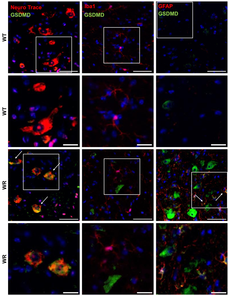
Amyotrophic lateral sclerosis (ALS) is a fatal neurodegenerative disease characterized by motor neuron degeneration in the central nervous system. Recent research has increasingly linked the activation of nucleotide oligomerization domain-like receptor protein 3 (NLRP3) inflammasome to ALS pathogenesis. NLRP3 activation triggers Caspase 1 (CASP 1) auto-activation, leading to the cleavage of Gasdermin D (GSDMD) and pore formation on the cellular membrane. This process facilitates cytokine secretion and ultimately results in pyroptotic cell death, highlighting the complex interplay of inflammation and neurodegeneration in ALS. This study aimed to characterize the NLRP3 inflammasome components and their colocalization with cellular markers using the wobbler mouse as an ALS animal model. Firstly, we checked the levels of miR-223-3p because of its association with NLRP3 inflammasome activity. The wobbler mice showed an increased expression of miR-223-3p in the ventral horn, spinal cord, and cerebellum tissues. Next, increased levels of NLRP3, pro-CASP 1, cleaved CASP 1 (c-CASP 1), full-length GSDMD, and cleaved GDSMD revealed NLRP3 inflammasome activation in wobbler spinal cords, but not in the cerebellum. Furthermore, we investigated the colocalization of the aforementioned proteins with neurons, microglia, and astrocyte markers in the spinal cord tissue. Evidently, the wobbler mice displayed microgliosis, astrogliosis, and motor neuron degeneration in this tissue. Additionally, we showed the upregulation of protein levels and the colocalization of NLRP3, c-CASP1, and GSDMD in neurons, as well as in microglia and astrocytes. Overall, this study demonstrated the involvement of NLRP3 inflammasome activation and pyroptotic cell death in the spinal cord tissue of wobbler mice, which could further exacerbate the motor neuron degeneration and neuroinflammation in this ALS mouse model.

Keywords: NLRP3 inflammasome; amyotrophic lateral sclerosis; miR-223-3p; motor neuron degeneration; pyroptotic cell death; wobbler mouse.


Conflict of interest statement

The authors declare no conflicts of interest.

Figures

Figure 1
Figure 1
Wobbler mice demonstrated elevated levels of miR-223-3p in laser-microdissected ventral horn, cervical spinal cord, and cerebellum. (A) Image of cresyl-violet-stained spinal cord section. Gray matter can be seen as dark purple, and white matter can be seen as light purple/pink. Blunt-ended lines show ventral and dorsal horns. Arrows show individual motor neurons. Scale: 400 µm. Close-up image of cresyl-violet-stained gray matter of ventral horn (B) before and (C) after LMD. Scale: 200 µm. RT-qPCR analysis of miR-223-3p using samples from (D) laser-microdissected ventral horn, (E) cervical spinal cord, and (F) cerebellum of p40 wild-type and wobbler mice. The differences in the expression levels were calculated using the 2−ΔΔCt method with endogenous normalization to UniSp6. Data are presented as mean ± SD, n = 4–8. *** p < 0.001; **** p < 0.0001.
Figure 2
Figure 2
Increased levels of NLRP3, pro-CASP 1, c-CASP 1, GSDMD-FL, C-GSDMD, IL-1β, and IL-18 proteins revealed NLRP3 inflammasome activation and thus pyroptotic cell death in wobbler spinal cords, but not in the cerebellum, despite increased NLRP3 expression. (A) Western blot image and semi-quantitative analyses of (B) NLRP3, (C) pro-CASP 1, (D) c-CASP 1, (E) GSDMD-FL, (F) C-GSDMD, (G) IL-1β, and (H) IL-18 in cervical spinal cord tissues of p40 wild-type and wobbler mice. (I) Western blot image and quantitative analyses of (J) NLRP3, (K) pro-CASP 1, (L) c-CASP 1, and (M) C-GSDMD in cerebellum tissues of p40 wild-type and wobbler mice. Calnexin or actin was used as a loading control. Data are presented as mean ± SD, n varies between 4 and 12 per group. ns: not significant. * p < 0.05; *** p < 0.001; **** p < 0.0001.
Figure 3
Figure 3
Representative images of double immunofluorescence staining for NLRP3 with NeuN, Iba1, or GFAP in the cervical spinal cord tissues of p40 wild-type and wobbler mice. NLRP3 was predominantly expressed in wobbler NeuN+ cells (motor neuronal cells), as well as in Iba1+ (microglia) and GFAP+ (astrocytes), which were in close proximity to motor neurons. Arrows indicate double-labeled cells. White frames indicate the areas of magnification. Scale: 50 µm; scale in the close-up images: 20 µm.
Figure 4
Figure 4
Representative images of double immunofluorescence staining for CASP 1 with NeuN, Iba1, or GFAP in the cervical spinal cord tissues of p40 wild-type and wobbler mice. CASP 1 was predominantly expressed in wobbler NeuN+ (motor neuronal cells), as well as in Iba1+ (microglia) and GFAP+ (astrocytes), which were in close proximity to motor neurons. Arrows indicate double-labeled cells. White frames indicate the areas of magnification. Scale: 50 µm; scale in the close-up images: 20 µm.
Figure 5
Figure 5
Representative images of double immunofluorescence staining for GSDMD with Neuro Trace, Iba1, or GFAP in the cervical spinal cord tissues of p40 wild-type and wobbler mice. GSDMD was predominantly expressed in wobbler NeuN+ (motor neuronal cells), as well as in GFAP+ (astrocytes), which were in close proximity to motor neurons. Arrows indicate double-labeled cells. White frames indicate the areas of magnification. Scale: 50 µm; scale in the close-up images: 20 µm.
Figure 6
Figure 6
The statistical mean intensity analysis of (A) NeuN, (B) Iba1, (C) GFAP, (D) NLRP3, (E) CASP 1, and (F) GSDMD in the spinal cord of p40 wild-type and wobbler mice. Data are presented as mean ± SD, n is at least 30 per group. ** p < 0.01; *** p < 0.001; **** p < 0.0001.
Figure 7
Figure 7
Activation of NLRP3 inflammasome and pyroptotic cell death in motor neurons of the wobbler spinal cord. During the priming step, DAMP and PAMP molecules activate several different receptors such as TLR4, IL-1R, TNFR, and RAGE. This activation leads to ubiquitination and thereby degradation of IκB, the inhibitor of NFκB. As a consequence, P50 and RelA subunits of NFκB can be translocated into the nucleus, where they can upregulate the expression of inflammasome genes such as Nlrp3, pro-Il-1b, and pro-Il-18. (A) Increasing levels of miR-223-3p has been shown in the lasered ventral horn and cervical spinal cord of the wobbler mouse. However, it is unclear whether miR-223-3p targets NLRP3 or an alternative protein(s) in the NFκB translocation pathway in wobbler mice. (B) Elevated levels of ROS and (C) mitochondrial fragmentation in the spinal cord of the wobbler motor neurons trigger (D) NLRP3 inflammasome formation, leading to (E) auto-cleavage of pro-CASP 1 into c-CASP 1. On one hand, c-CASP 1 is able to cleave pro-IL-1β and pro-IL-18 into their mature forms, and on the other hand, it can cleave GSDMD protein. N-GSDMD subunits can translocate into the cellular membrane and form pores, which cause membrane rupture and eventually (F) pyroptotic motor neuronal death. Through these pores, mature cytokines can be released, leading to (G) inflammation for the neighboring cells. This image was created with BioRender.com.

References

    1. Wolfson C., Gauvin D.E., Ishola F., Oskoui M. Global Prevalence and Incidence of Amyotrophic Lateral Sclerosis: A Systematic Review. Neurology. 2023;101:E613–E623. doi: 10.1212/WNL.0000000000207474. - DOI - PMC - PubMed
    1. Ferraiuolo L., Kirby J., Grierson A.J., Shaw P.J. Molecular Pathways of Motor Neuron Injury in Amyotrophic Lateral Sclerosis. Nat. Rev. Neurol. 2011;7:616–630. doi: 10.1038/nrneurol.2011.152. - DOI - PubMed
    1. Bruijn L.I., Miller T.M., Cleveland D.W. Unraveling the Mechanisms Involved in Motor Neuron Degeneration in ALS. Annu. Rev. Neurosci. 2004;27:723–749. doi: 10.1146/ANNUREV.NEURO.27.070203.144244. - DOI - PubMed
    1. Falconer D.S. Wobbler (Wr) Mouse News Lett. 1956;15:23–29.
    1. Moser J.M., Bigini P., Schmitt-John T. The Wobbler Mouse, an ALS Animal Model. Mol. Genet. Genom. 2013;288:207–229. doi: 10.1007/s00438-013-0741-0. - DOI - PMC - PubMed

Publication types

MeSH terms

LinkOut - more resources