

Morphological Clues of Acute Monocytic Leukemia in COVID-19-Induced Transient Leukoerythroblastic Reaction with Monocytosis
- PMID: 38921181
- PMCID: PMC11203109
- DOI: 10.3390/hematolrep16020033
Morphological Clues of Acute Monocytic Leukemia in COVID-19-Induced Transient Leukoerythroblastic Reaction with Monocytosis
Abstract
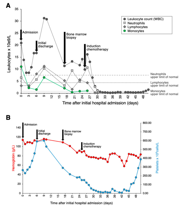
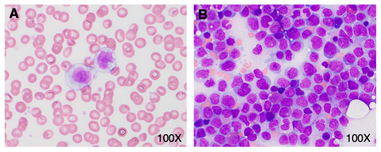
Viral infections, including those caused by COVID-19, can produce striking morphologic changes in peripheral blood. Distinguishing between reactive changes and abnormal morphology of monocytes remains particularly difficult, with low consensus rates reported amongst hematopathologists. Here, we report a patient who developed transient monocytosis of 11.06 × 10⁹/L with 32% promonocytes and 1% blasts during hospitalization that was secondary to severe COVID-19 infection. Three days later, the clinical status of the patient improved and the WBC had decreased to 8.47 × 10⁹/L with 2.2 × 10⁹/L monocytes. Flow cytometry studies did not reveal immunophenotypic findings specific for an overt malignant population. At no time during admission did the patient develop cytopenia(s), and she was discharged upon clinical improvement. However, the peripheral blood sample containing promonocytes was sent for molecular testing with an extended next-generation sequencing myeloid panel and was positive for pathogenic NPM1 Type A and DNMT3A R882H mutations. Subsequently, despite an essentially normal complete blood count, the patient underwent a bone marrow assessment that showed acute myeloid leukemia with 77% promonocytes. This case emphasizes the critical importance of a full work up to exclude acute leukemia when classical promonocyte morphology is encountered in the peripheral blood. Promonocytes are not a part of the reactive changes associated with COVID-19 and remain specific to myeloid neoplasia.
Keywords: COVID-19; acute myeloid leukemia; monocytes; promonocytes.
Conflict of interest statement
The authors declare no conflicts of interest.
Figures
Similar articles
-
Concordance among hematopathologists in classifying blasts plus promonocytes: A bone marrow pathology group study.Int J Lab Hematol. 2020 Aug;42(4):418-422. doi: 10.1111/ijlh.13212. Epub 2020 Apr 16. Int J Lab Hematol. 2020. PMID: 32297416
-
Classification of Monocytes, Promonocytes and Monoblasts Using Deep Neural Network Models: An Area of Unmet Need in Diagnostic Hematopathology.J Clin Med. 2021 May 24;10(11):2264. doi: 10.3390/jcm10112264. J Clin Med. 2021. PMID: 34073699 Free PMC article.
-
Flow cytometric analysis of different CD14 epitopes can help identify immature monocytic populations.Am J Clin Pathol. 2005 Dec;124(6):930-6. Am J Clin Pathol. 2005. PMID: 16416743
-
Clinical and Molecular Approach to Adult-Onset, Neoplastic Monocytosis.Curr Hematol Malig Rep. 2021 Jun;16(3):276-285. doi: 10.1007/s11899-021-00632-6. Epub 2021 Apr 22. Curr Hematol Malig Rep. 2021. PMID: 33890194 Review.
-
Acute myeloid leukemia: 2013 update on risk-stratification and management.Am J Hematol. 2013 Apr;88(4):318-27. doi: 10.1002/ajh.23404. Am J Hematol. 2013. PMID: 23526416 Review.
References
-
- Salib C., Khattar P., Cheng J., Teruya-Feldstein J. Atypical Peripheral Blood Cell Morphology in COVID-19 (Sars-CoV-2) Patients from Mount Sinai Health System in New York City. Blood. 2020;136:26–27. doi: 10.1182/blood-2020-142581. - DOI
-
- Bain B.J. Blood Cells: A Practical Guide. 5th ed. John Wiley & Sons; West Sussex, UK: 2014.
Publication types
LinkOut - more resources
Full Text Sources
