. 2024 May 21;9(6):308.
doi: 10.3390/biomimetics9060308.

Synthesis of Chitosan and Ferric-Ion (Fe3+)-Doped Brushite Mineral Cancellous Bone Scaffolds

Affiliations

Synthesis of Chitosan and Ferric-Ion (Fe3+)-Doped Brushite Mineral Cancellous Bone Scaffolds

Lemiha Yildizbakan et al. Biomimetics (Basel). .

Abstract

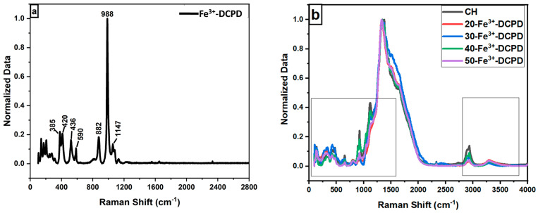
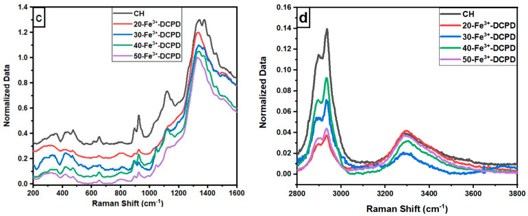
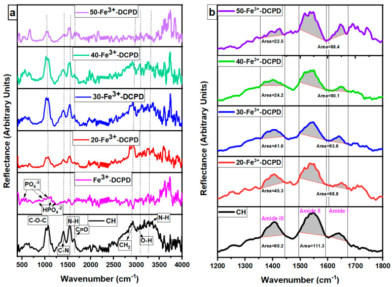
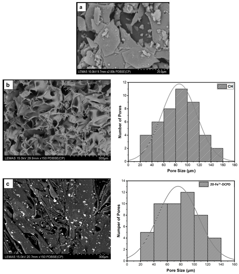
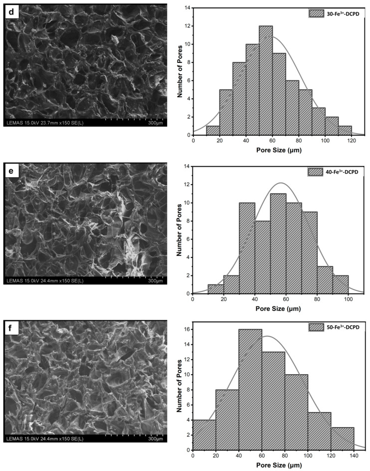
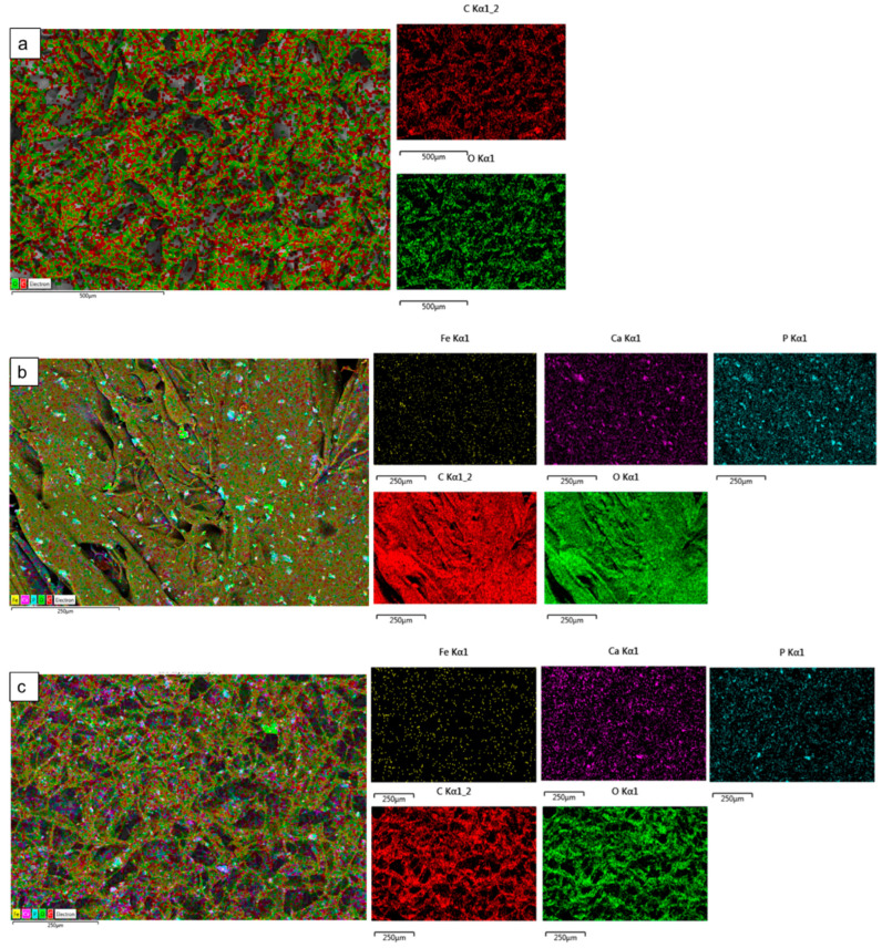
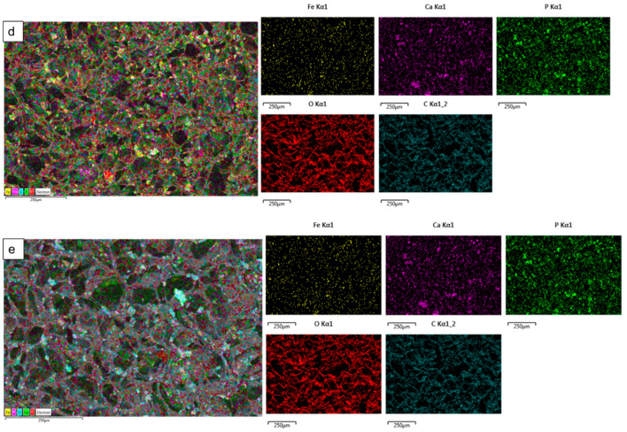
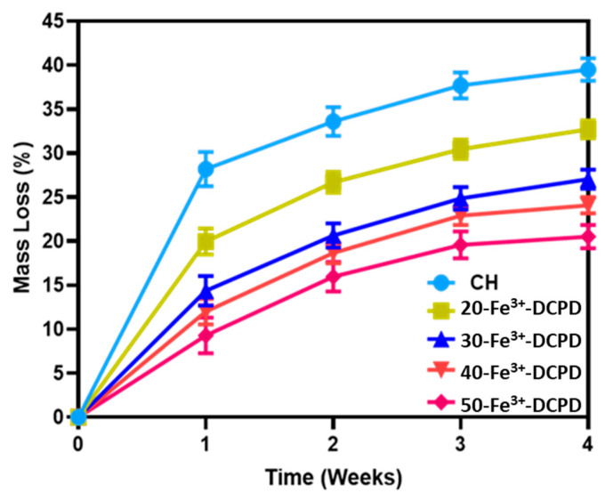
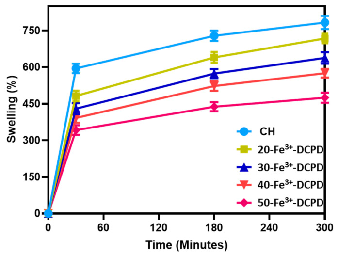
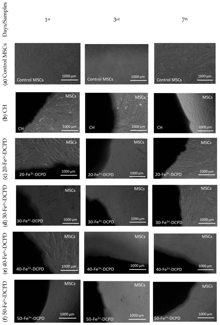
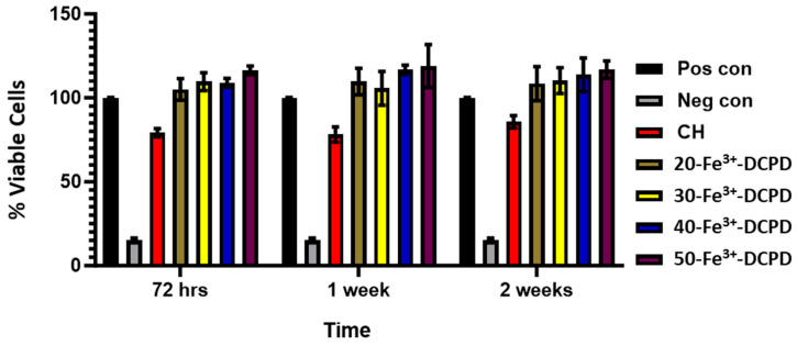
Biodegradable scaffolds are needed to repair bone defects. To promote the resorption of scaffolds, a large surface area is required to encourage neo-osteogenesis. Herein, we describe the synthesis and freeze-drying methodologies of ferric-ion (Fe3+) doped Dicalcium Phosphate Dihydrate mineral (DCPD), also known as brushite, which has been known to favour the in situ condition for osteogenesis. In this investigation, the role of chitosan during the synthesis of DCPD was explored to enhance the antimicrobial, scaffold pore distribution, and mechanical properties post freeze-drying. During the synthesis of DCPD, the calcium nitrate solution was hydrolysed with a predetermined stoichiometric concentration of ammonium phosphate. During the hydrolysis reaction, 10 (mol)% iron (Fe3+) nitrate (Fe(NO3)3) was incorporated, and the DCPD minerals were precipitated (Fe3+-DCPD). Chitosan stir-mixed with Fe3+-DCPD minerals was freeze-dried to create scaffolds. The structural, microstructural, and mechanical properties of freeze-dried materials were characterized.

Keywords: Fe3+-doped brushite (dicalcium phosphate dihydrate); bone tissue engineering; chitosan; mechanical properties; scaffold.


Conflict of interest statement

The authors declare no conflict of interest.

Figures

Figure 1
Figure 1
Overview illustration of the freeze-dried scaffolds synthesis process.
Figure 2
Figure 2
Normalised Raman Spectra for the following: (a) Fe-Dicalcium Phosphate Dihydrate mineral (Fe3+-DCPD mineral) powder; (b) comparison of the various Fe3+-DCPD mineral concentrations (0, 20, 30, 40, and 50 (wt)%) loaded chitosan fabricated freeze-dried scaffolds in the range of 100 to 4000 cm−1; (c) 200 to 2800 cm−1 region with 0.2 offsets; and (d) 2800 to 3800 cm−1.
Figure 2
Figure 2
Normalised Raman Spectra for the following: (a) Fe-Dicalcium Phosphate Dihydrate mineral (Fe3+-DCPD mineral) powder; (b) comparison of the various Fe3+-DCPD mineral concentrations (0, 20, 30, 40, and 50 (wt)%) loaded chitosan fabricated freeze-dried scaffolds in the range of 100 to 4000 cm−1; (c) 200 to 2800 cm−1 region with 0.2 offsets; and (d) 2800 to 3800 cm−1.
Figure 3
Figure 3
Fabricated freeze-dried chitosan scaffolds were compared with different concentrations of the 10 (mol)% Iron (III) nitrate doped Dicalcium Phosphate Dihydrate (Fe3+-DCPD) mineral (CH, 20, 30, 40, and 50-Fe3+-DCPD). (a) Data were acquired using the Vertex 70 FTIR spectrometer in attenuated total reflection (ATR) mode, between 400 and 4000 cm−1, with a resolution of 4 cm−1, (b) Amide I, II, and III peak comparison.
Figure 4
Figure 4
Normalised X-ray diffraction data, (a) Experimental XRD spectra for synthesised Fe-DCPD mineral, mineral-free CH, and different amounts of Fe3+-DCPD-doped chitosan (20, 30, 40, and 50-Fe3+-DCPD) freeze-dried scaffolds, (● corresponds to miller indices (020), (040), (−112), (−231), and (080)). (b) DCPD reference spectra (JCPDS), and (c) crystallite size comparison. ‘■’ corresponds to the Bragg 2θ diffraction peaks of Fe3+-DCPD with miller indices (0 2 0), (0 4 0), (−1 1 2), (−2 3 1), and (0 8 0), respectively.
Figure 5
Figure 5
The Hitachi SU8230 SEM image results and corresponding pore size distribution comparisons of (a) 10 (mol)% iron (III) nitrate doped Dicalcium Phosphate Dihydrate (Fe3+-DCPD) mineral, (b) freeze-dried chitosan (CS), (c) 20-Fe3+-DCPD, (d) 30-Fe3+-DCPD, (e) 40-Fe3+-DCPD, and (f) 50-Fe3+-DCPD.
Figure 5
Figure 5
The Hitachi SU8230 SEM image results and corresponding pore size distribution comparisons of (a) 10 (mol)% iron (III) nitrate doped Dicalcium Phosphate Dihydrate (Fe3+-DCPD) mineral, (b) freeze-dried chitosan (CS), (c) 20-Fe3+-DCPD, (d) 30-Fe3+-DCPD, (e) 40-Fe3+-DCPD, and (f) 50-Fe3+-DCPD.
Figure 6
Figure 6
The Hitachi SU8230 SEM with a dispersive energy X-ray (EDX) detector was used to analyse the surface elemental characterization of freeze-dried chitosan scaffolds without Fe3+-DCPD mineral and with various Fe3+-DCPD mineral concentrations (20, 30, 40, and 50 (wt)%). Freeze-dried (a) Chitosan, (b) 20-Fe3+-DCPD, (c) 30-Fe3+-DCPD, (d) 40-Fe3+-DCPD, and (e) 50-Fe3+-DCPD scaffold. Each colour represents different types of elements in the sample.
Figure 6
Figure 6
The Hitachi SU8230 SEM with a dispersive energy X-ray (EDX) detector was used to analyse the surface elemental characterization of freeze-dried chitosan scaffolds without Fe3+-DCPD mineral and with various Fe3+-DCPD mineral concentrations (20, 30, 40, and 50 (wt)%). Freeze-dried (a) Chitosan, (b) 20-Fe3+-DCPD, (c) 30-Fe3+-DCPD, (d) 40-Fe3+-DCPD, and (e) 50-Fe3+-DCPD scaffold. Each colour represents different types of elements in the sample.
Figure 7
Figure 7
Mineral-free and different concentration Fe3+-DCPD mineral embedded chitosan freeze-dried scaffolds degradation results (CH, 20, 30, 40, and 50-Fe3+-DCPD) when they are dissolved in phosphate saline buffer (pH 7.4) at 37 °C. Over 4 weeks, the studies were performed in triplicate. The error bars demonstrate each group’s standard deviation (SD) ± mean, n = 3.
Figure 8
Figure 8
The swelling kinetics of mineral-free chitosan (CH), 20, 30, 40 and 50-Fe3+-DCPD freeze-dried scaffolds immersed in phosphate saline buffer (pH 7.4) at physiological temperature 37 °C. Experiments were carried out in triplicates for each of the fabricated scaffolds. The error bars represent the mean ± standard deviation (SD) for n = 3 in each group.
Figure 9
Figure 9
Direct cytotoxicity. Images of chitosan freeze-dried scaffolds with varied Fe3+-DCPD concentrations (0, 20, 30, 40, and 50 (wt)%) were obtained at ×4 on the first, third, and seventh days. (a) Control MSCs were bone marrow mesenchymal stem cells (MSCs) without scaffolds, (b) mineral-free CH scaffold, (c) 20-Fe3+-DCPD, (d) 30-Fe3+-DCPD, (e) 40-Fe3+-DCPD, and (f) 50-Fe3+-DCPD.
Figure 10
Figure 10
Cytotoxicity by XTT assay test on MSCs exposed to extracts taken from freeze-dried chitosan scaffolds and containing varying concentrations of Fe3+-DCPD minerals (20 (wt)% (20-Fe3+-DCPD), 30 (wt)% (30-Fe3+-DCPD), 40 (wt)% (40-Fe3+-DCPD), and 50 (wt)% (50-Fe3+-DCPD). The error bars represent the mean ± SD (n = 2 in each group).
Figure 11
Figure 11
Cell proliferation was assessed using the XTT assay on MSCs exposed to freeze-dried scaffold extracts (CH, 20-Fe3+-DCPD, 30-Fe3+-DCPD, 40-Fe3+-DCPD, and 50-Fe3+-DCPD). The fabricated scaffold extracts were seeded 500 cells/well. The error bars represent the mean ± SD (n = 2 in each group). There was no significant difference between freeze-dried scaffolds at all time points.

Similar articles

Cited by

References

    1. Bezstarosti H., Metsemakers W.J., van Lieshout E.M.M., Voskamp L.W., Kortram K., McNally M.A., Marais L.C., Verhofstad M.H.J. Management of critical-sized bone defects in the treatment of fracture-related infection: A systematic review and pooled analysis. Arch. Orthop. Trauma Surg. 2021;141:1215–1230. doi: 10.1007/s00402-020-03525-0. - DOI - PMC - PubMed
    1. Martin R.B., Burr D.B., Sharkey N.A., Fyhrie D.P. Skeletal Tissue Mechanics. Springer; New York, NY, USA: 1998.
    1. Wang W., Yeung K.W.K. Bone grafts and biomaterials substitutes for bone defect repair: A review. Bioact. Mater. 2017;2:224–247. doi: 10.1016/j.bioactmat.2017.05.007. - DOI - PMC - PubMed
    1. Kao S.T., Scott D.D. A Review of Bone Substitutes. Oral Maxillofac. Surg. Clin. N. Am. 2007;19:513–521. doi: 10.1016/j.coms.2007.06.002. - DOI - PubMed
    1. Zárate-Kalfópulos B., Reyes-Sánchez A. Bone grafts in orthopedic surgery. Cir. Cir. 2006;74:217–222. - PubMed

LinkOut - more resources