. 2024 May 30;10(6):391.
doi: 10.3390/jof10060391.

Mitochondrial Protease Oct1p Regulates Mitochondrial Homeostasis and Influences Pathogenicity through Affecting Hyphal Growth and Biofilm Formation Activities in Candida albicans

Affiliations

Mitochondrial Protease Oct1p Regulates Mitochondrial Homeostasis and Influences Pathogenicity through Affecting Hyphal Growth and Biofilm Formation Activities in Candida albicans

Xiaoxiao Zhu et al. J Fungi (Basel). .

Abstract

Mitochondria, as the core metabolic organelles, play a crucial role in aerobic respiration/biosynthesis in fungi. Numerous studies have demonstrated a close relationship between mitochondria and Candida albicans virulence and drug resistance. Here, we report an octapeptide-aminopeptidase located in the mitochondrial matrix named Oct1p. Its homolog in the model fungus Saccharomyces cerevisiae is one of the key proteins in maintaining mitochondrial respiration and protein stability. In this study, we utilized evolutionary tree analysis, gene knockout experiments, mitochondrial function detection, and other methods to demonstrate the impact of Oct1p on the mitochondrial function of C. albicans. Furthermore, through transcriptome analysis, real-time quantitative PCR, and morphological observation, we discovered that the absence of Oct1p results in functional abnormalities in C. albicans, affecting hyphal growth, cell adhesion, and biofilm formation. Finally, the in vivo results of the infection of Galleria mellonella larvae and vulvovaginal candidiasis in mice indicate that the loss of Oct1p led to the decreased virulence of C. albicans. In conclusion, this study provides a solid theoretical foundation for treating Candida diseases, developing new targeted drugs, and serves as a valuable reference for investigating the connection between mitochondria and virulence in other pathogenic fungi.

Keywords: Candida albicans; Oct1p; hypha formation; mitochondria; virulence.


Conflict of interest statement

We declare that we have no conflicts of interest.

Figures

Figure 1
Figure 1
Deletion of OCT1 reduces C. albicans mitochondrial function. (A) Mitochondrial morphology under 579 nm excitation was observed. The concentration of C. albicans suspension was adjusted with PBS to 1.0 × 10⁶ cells/mL for each strain, and then 200 μM Mito-tracker Red dye solution was added to the suspension at the ratio of 1:1000 and incubated at 30℃ in the dark for 30 min. Scale bar: 2 μm. (B) A total of 100 μL of 1.0 × 10⁷ cells/mL of each C. albicans liquid and an equal volume of BacTiter-GloTM reaction solution were added to an opaque 96-well plate and incubated for 5 min at room temperature after oscillating and mixing, and the ATP content of each sample was detected by a multifunctional enzyme marker. (C) Mitochondrial membrane potential (MMP) was assessed using the JC-1 assay kit and flow cytometry (Ex/Em of 595/488 nm). (D) The average red/green fluorescence intensity was recorded for each sample. The transition of JC-1 dye from red to green fluorescence was used to easily detect the decrease in MMP, after which MMP was determined as the ratio of red to green fluorescence. (E) ROS levels of WT, oct1∆/∆, and oct1∆/∆:OCT1 were detected using a DCFH-DA probe and visualized using a microscope (Ex/Em of 488/525 nm). Scale bar: 20 μm. (F) The fluorograms of each strain were quantitatively analyzed using Image J software. (ns, not significant; **, p < 0.01; ***, p < 0.001; and ****, p < 0.0001).
Figure 1
Figure 1
Deletion of OCT1 reduces C. albicans mitochondrial function. (A) Mitochondrial morphology under 579 nm excitation was observed. The concentration of C. albicans suspension was adjusted with PBS to 1.0 × 10⁶ cells/mL for each strain, and then 200 μM Mito-tracker Red dye solution was added to the suspension at the ratio of 1:1000 and incubated at 30℃ in the dark for 30 min. Scale bar: 2 μm. (B) A total of 100 μL of 1.0 × 10⁷ cells/mL of each C. albicans liquid and an equal volume of BacTiter-GloTM reaction solution were added to an opaque 96-well plate and incubated for 5 min at room temperature after oscillating and mixing, and the ATP content of each sample was detected by a multifunctional enzyme marker. (C) Mitochondrial membrane potential (MMP) was assessed using the JC-1 assay kit and flow cytometry (Ex/Em of 595/488 nm). (D) The average red/green fluorescence intensity was recorded for each sample. The transition of JC-1 dye from red to green fluorescence was used to easily detect the decrease in MMP, after which MMP was determined as the ratio of red to green fluorescence. (E) ROS levels of WT, oct1∆/∆, and oct1∆/∆:OCT1 were detected using a DCFH-DA probe and visualized using a microscope (Ex/Em of 488/525 nm). Scale bar: 20 μm. (F) The fluorograms of each strain were quantitatively analyzed using Image J software. (ns, not significant; **, p < 0.01; ***, p < 0.001; and ****, p < 0.0001).
Figure 2
Figure 2
Oct1p affects C. albicans growth ability. (A) Each strain was incubated overnight in YPD, then washed with PBS and serially diluted, and different concentrations of WT, oct1∆/∆, and oct1∆/∆:OCT1 (5 μL 106 cells/mL to 102 cells/mL) were spotted on YPD solid medium containing 2% dextrose as a carbon source and 3% glycerol and anhydrous ethanol mixture as a carbon source and YPEG solid medium with 3% glycerol and anhydrous ethanol mixture as a carbon source. The plates were then incubated at 30 °C, 35 °C, and 37 °C for 2 days and then photographed. (B) Each strain was incubated in YPD overnight, then diluted to 500 cells/mL with sterile PBS, and 100 μL was spread on YPD solid medium containing 2% glucose as a carbon source and YPEG solid medium with a 3% mixture of glycerol and anhydrous ethanol as a carbon source, respectively, and then incubated for 2 days at 30 °C, 35 °C, and 37 °C, respectively, and then measured by using a vernier caliper, the size of single colonies, and recorded. (C) Each strain was incubated at 200 mL with an initial concentration of OD600 = 0.04 on a shaker at 30 °C, 200 rpm for 24 h, and OD600 was measured at 2 h intervals; the data were recorded, and growth curves were plotted. (D) Doubling time of C. albicans in the exponential growth phase. (E) Each strain was incubated overnight in YPD, then washed with sterile PBS and serially diluted. Different concentrations of WT, oct1∆/∆, and oct1∆/∆:OCT1 (5 μL 106 cells/mL to 102 cells/mL) were spotted on YPD, fluconazole containing 2 μg/mL, 0.04% SDS, and amphotericin B medium at 0.05 μg/mL, and the plates were photographed after incubation at 30 °C for 2 days. (ns, not significant; *, p < 0.05; **, p < 0.01; ****, p < 0.0001).
Figure 3
Figure 3
RNA-seq analysis reveals the potential functions of Oct1p. (A) Genes (DEGs) significantly differentially expressed in oct1∆/∆. Expression difference multiples |log2fold change| > 1 and false discovery rate p-value < 0.05. (B) Major distribution of oct1∆/∆ down-regulated differential genes in GO function enrichment analysis (no distinction between molecular function, cellular components, and biological processes). (C) Primary distribution of oct1∆/∆ up-regulated differential genes in GO functional enrichment analyses (no distinction between molecular function, cellular components, and biological processes). (D) Heat map of up- and down-regulated genes associated with biofilm formation of the KEGG category. (E) Transcript levels of genes associated with C. albicans hyphal growth, adhesion in WT, and oct1∆/∆ strains were verified by qRT-PCR in genome-wide transcriptional data. (ns, not significant; *, p < 0.05; **, p < 0.01; ***, p < 0.001; and ****, p < 0.0001).
Figure 4
Figure 4
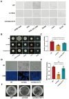
Deficiency of Oct1p attenuates C. albicans hyphal formation, biofilm formation, and adhesion. (A) Each strain was incubated in YPD overnight, then washed twice with sterile PBS, and incubated in six different liquid mycelium induction media: RPMI-1640, YPD, YPD + 10% FBS, Lee’s, Spider, and SLAD. The concentration of each solution was adjusted to 1.0 × 10⁶ cells/mL, and 2 mL of each solution was added into 6-well plates at 37 °C for 4 h. The formation of liquid mycelium was observed under a microscope. Scale bar: 4 μm. (B) Each strain was incubated in YPD overnight, and then the concentration of each strain was adjusted to 1.0 × 10⁵ cells/mL after being washed twice with sterile PBS, and 5 μL was spotted on five different solid mycelium-inducing media (YPD, YPD + 10% FBS, Lee’s, Spider, and SLAD) and incubated in a 37 °C incubator. The results were observed and photographed for recording. Scale bar: 4 mm. (C) XTT reduction assay to detect the in vitro biofilm formation ability of WT, oct1∆/∆, and oct1∆/∆:OCT1. (D) Each strain was cultured in YPD overnight, then washed twice with sterile PBS, and then the concentration of each bacterial solution was adjusted to 1.0 × 10⁶ cells/mL with RPMI-1640 liquid medium, incubated at 37 °C for 24 h, and then observed under a fluorescence microscope. After incubation at 37 °C for 24 h, the C. albicans were stained with Calcium fluorescent white for 5 min and then observed under a fluorescence microscope. Scale bar: 40 μm. (E) The fluorograms of each strain were quantitatively analyzed using Image J software. (F) WT, oct1∆/∆, and oct1∆/OCT1 in vitro adhesion capacity assay. (ns, not significant; *, p < 0.05; **, p < 0.01).
Figure 5
Figure 5
The absence of Oct1p reduces the infective ability of C. albicans in G. mellonella and mice. (A) The concentration of each C. albicans liquid was adjusted to 8.0 × 10⁶ cells/mL with PBS, and 10 μL of each C. albicans liquid was introduced and injected into the left or right hypophysis of G. mellonella larvae using a pointed microinjection needle, and then placed in the incubator at 37 °C after making the molds and incubated, and then the deaths of G. mellonella were observed and recorded every day. The experiment lasted for 14 days. (B) Fungal load in the G. mellonella at 48 h after the injection of each fungal solution into the larvae of the G. mellonella. (C) Localized state of the mouse vagina, histopathological changes in the vaginal mucosa, and the morphology of C. albicans in vaginal lavage fluid 7 days after infection with vulvovaginal candidiasis. Scale bar: 100 μm. (D) Fungal load in the vaginal lavage fluid of mice 7 days after infection with vulvovaginal candidiasis. (E) The method of ELISA was used to detect the levels of IL-6, TNF-α, and IL-10 in mouse vaginal tissues. (F) The method of qRT-PCR was used to detect the expression of IL-6, TNF-α, and IL-10 genes in mouse vaginal tissues. (ns, not significant; *, p < 0.05; **, p < 0.01; ***, p < 0.001; and ****, p < 0.0001).
Figure 6
Figure 6
Diagram of the working mode of Oct1p in C. albicans. The upward or downward arrow indicates an increase or decrease in the corresponding parameter.

Similar articles

Cited by

References

    1. Lane N., Martin W. The energetics of genome complexity. Nature. 2010;467:929–934. doi: 10.1038/nature09486. - DOI - PubMed
    1. Zeng L.B., Huang Y.C., Tan J.J., Peng J., Hu N.Y., Liu Q., Cao Y.L., Zhang Y.P., Chen J.Z., Huang X.T. QCR7 affects the virulence of Candida albicans and the uptake of multiple carbon sources present in different host niches. Front. Cell. Infect. Microbiol. 2023;13:1136698. doi: 10.3389/fcimb.2023.1136698. - DOI - PMC - PubMed
    1. Tsui C., Kong E.F., Jabra-Rizk M.A. Pathogenesis of Candida albicans biofilm. Pathog. Dis. 2016;74:ftw018. doi: 10.1093/femspd/ftw018. - DOI - PMC - PubMed
    1. Calderone R., Li D.M., Traven A. System-level impact of mitochondria on fungal virulence: To metabolism and beyond. FEMS Yeast Res. 2015;15:fov027. doi: 10.1093/femsyr/fov027. - DOI - PMC - PubMed
    1. Shingu-Vazquez M., Traven A. Mitochondria and Fungal Pathogenesis: Drug Tolerance, Virulence, and Potential for Antifungal Therapy. Eukaryot. Cell. 2011;10:1376–1383. doi: 10.1128/EC.05184-11. - DOI - PMC - PubMed

LinkOut - more resources