. 2024 May 21;14(6):118.
doi: 10.3390/membranes14060118.

Cryptotanshinone-Induced Permeabilization of Model Phospholipid Membranes: A Biophysical Study

Affiliations

Cryptotanshinone-Induced Permeabilization of Model Phospholipid Membranes: A Biophysical Study

Julia Ortiz et al. Membranes (Basel). .

Abstract

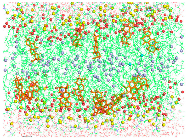
The Danshen terpenoid cryptotanshinone (CPT) is gaining enormous interest in light of its various outstanding biological activities. Among those, CPT has been shown to interact with cell membranes and, for instance, to have antibacterial activity. Several works have shown that CPT alone, or in combination with other drugs, can effectively act as an antibiotic against various infectious bacteria. Some authors have related the mechanism underlying this action to CPT-membrane interaction. This work shows that CPT readily partitions into phosphatidylcholine membranes, but there is a limiting capacity of accommodation of ca. 1 mol CPT to 3 mol phospholipid. The addition of CPT to unilamellar liposomes composed of 1-palmitoyl-2-oleoylphosphatidylcholine (POPC) causes membrane permeabilization, as shown by fluorescent probe leakage. This process has been kinetically studied, as well as its modulation by incorporation of phosphatidylethanolamine or phosphatidylglycerol, as a model for pathogenic cell membranes. The thermotropic behavior of 1,2-dimyristoylphosphatidylcholine (DMPC) model membranes is weakly affected by CPT, but the terpenoid causes significant dehydration of the polar region of the bilayer and weak disordering of the acyl chain palisade, as observed in Fourier-transform infrared spectroscopy (FTIR) results. Small-angle X-ray scattering (SAXS) shows that CPT increases DMPC bilayer thickness, which could be due to localization near the phospholipid/water interface. Molecular dynamics (MD) simulations show that the lateral diffusion coefficient of the phospholipid increases with the presence of CPT. CPT extends from the polar head region to the center of the bilayer, being centered between the carbonyl groups and the unsaturated region of the POPC, where there is greater overlap. Interestingly, the free energy profiles of a water molecule crossing the lipid membrane show that the POPC membrane becomes more permeable in the presence of CPT. In summary, our results show that CPT perturbs the physicochemical properties of the phospholipid membrane and compromises its barrier function, which could be of relevance to explain part of its antimicrobial or anticancer activities.

Keywords: cryptotanshinone; membrane permeabilization; molecular dynamics; phospholipid membranes.


Conflict of interest statement

The authors declare no conflicts of interest.

Figures

Figure 1
Figure 1
The chemical structure of cryptotanshinone.
Figure 2
Figure 2
Partition of CPT into POPC membranes. The actual molar ratio of CPT in the membrane (CPTmem/POPC), determined as described in Materials and Methods, is plotted against the total initial CPT to POPC molar ratio (CPTtot/POPC). Data correspond to the mean of three independent experiments ± SD (error bars).
Figure 3
Figure 3
Content leakage curves for CPT-induced permeabilization of POPC LUV. The concentration of POPC was kept constant at 20 µM. Numbers on the curves indicate the molar ratio of total CPT to phospholipid in the cuvette (CPTtot/POPC).
Figure 4
Figure 4
The influence of membrane composition on CPT-induced leakage. Experiments were conducted under the same conditions as in Figure 3, for liposomes composed of pure POPC (black), POPC/POPG (5:1.7, mol/mol) (blue), and POPC/POPE (5:1.7, mol/mol) (red), at two different CPT concentrations, as indicated.
Figure 5
Figure 5
High-sensitivity DSC heating thermograms for mixtures of CPT with DMPC. CPTtot/DMPC molar ratios are indicated on the curves. Scans were carried out at 60 °C h−1.
Figure 6
Figure 6
The effect of CPT on the region of the DMPC acyl chains and polar headgroups, determined by FTIR. Top: the effect on the maximum frequency of the νCH2 symmetric stretching band. Bottom: the effect on the maximum frequency of the νCO stretching band. Plots correspond to pure DMPC (blue), and CPTtot/DMPC (mol/mol) 0.1 (red) and 0.25 (green). Spectra were collected from 10 to 34 °C, every 2 °C. Data correspond to the mean of three independent repetitions ± SD (error bars).
Figure 7
Figure 7
Effect of CPT on the SAXS profiles of DMPC. Diffractograms correspond to pure DMPC (red) and CPTtot/DMPC 0.1 (mol/mol) (blue), at 7 °C (A), 18 °C (B), and 34 °C (C).
Figure 8
Figure 8
Area per lipid vs. simulated time for all simulated systems: POPC at 298 K (black), POPC + CPT at 298 K (red), DMPC at 312 K (blue), and DMPC + CPT at 312 K (green).
Figure 9
Figure 9
Mass density profiles along the z-axis of the simulation box of the simulated system of POPC + CPT. POPC phosphorus atoms are in green, CPT in red, POPC terminal methyl of sn-1 chains in blue, and POPC carbonyl groups in black. Curves are symmetrized around the center of the bilayer.
Figure 10
Figure 10
Final snapshot of the simulation box of POPC + CPT membranes. Water molecules are shown in red lines, CPT in orange sticks, POPC atoms in green lines, POPC carbonyl groups in red spheres, POPC methyl terminals of sn-1 chains in blue spheres, and phosphorous atoms in yellow spheres.
Figure 11
Figure 11
Free energy profiles for a water molecule crossing a POPC membrane (black line) and a POPC + CPT membrane (red line). Profiles are assumed to be symmetric across the bilayer center.

Similar articles

Cited by

References

    1. Jiang Z., Gao W., Huang L. Tanshinones, Critical Pharmacological Components in Salvia miltiorrhiza. Front. Pharmacol. 2019;10:439237. doi: 10.3389/FPHAR.2019.00202. - DOI - PMC - PubMed
    1. Ke L., Zhong C., Chen Z., Zheng Z., Li S., Chen B., Wu Q., Yao H. Tanshinone I: Pharmacological activities, molecular mechanisms against diseases and future perspectives. Phytomedicine. 2023;110:154632. doi: 10.1016/J.PHYMED.2022.154632. - DOI - PubMed
    1. Gao H., Huang L., Ding F., Yang K., Feng Y., Tang H., Xu Q.M., Feng J., Yang S. Simultaneous purification of dihydrotanshinone, tanshinone I, cryptotanshinone, and tanshinone IIA from Salvia miltiorrhiza and their anti-inflammatory activities investigation. Sci. Rep. 2018;8:8460. doi: 10.1038/s41598-018-26828-0. - DOI - PMC - PubMed
    1. Wu Y.R.Y.H., Wu Y.R.Y.H., Li B., Yan Z.Y. Cryptotanshinone: A review of its pharmacology activities and molecular mechanisms. Fitoterapia. 2020;145:104633. doi: 10.1016/j.fitote.2020.104633. - DOI - PubMed
    1. Ashrafizadeh M., Zarrabi A., Orouei S., Saberifar S., Salami S., Hushmandi K., Najafi M. Recent advances and future directions in anti-tumor activity of cryptotanshinone: A mechanistic review. Phyther. Res. 2021;35:155–179. doi: 10.1002/ptr.6815. - DOI - PubMed

LinkOut - more resources